Aortic regurgitation echocardiography: Difference between revisions
m (Robot: Automated text replacement (-msbeih@perfuse.org +msbeih@wikidoc.org, -psingh@perfuse.org +psingh13579@gmail.com, -agovi@perfuse.org +agovi@wikidoc.org, -rgudetti@perfuse.org +ravitheja.g@gmail.com, -lbiller@perfuse.org +lbiller@wikidoc.org,...) |
Esther Lee (talk | contribs) No edit summary |
||
| Line 40: | Line 40: | ||
{{#ev:youtube|3W_YX_GbpK8}} | {{#ev:youtube|3W_YX_GbpK8}} | ||
==2008 ACC/AHA Guidelines for Management of Patients | ==2008 and incorporated 2006 ACC/AHA Guidelines incorporated into the 2006 guidelines for the management of patients with valvular heart disease <ref name="pmid18820172">{{cite journal |author=Bonow RO, Carabello BA, Chatterjee K, ''et al.'' |title=2008 Focused update incorporated into the ACC/AHA 2006 guidelines for the management of patients with valvular heart disease: a report of the American College of Cardiology/American Heart Association Task Force on Practice Guidelines (Writing Committee to Revise the 1998 Guidelines for the Management of Patients With Valvular Heart Disease): endorsed by the Society of Cardiovascular Anesthesiologists, Society for Cardiovascular Angiography and Interventions, and Society of Thoracic Surgeons |journal=Circulation |volume=118 |issue=15 |pages=e523–661 |year=2008 |month=October |pmid=18820172 |doi=10.1161/CIRCULATIONAHA.108.190748 |url=}}</ref>== | ||
===Echocardiography (DO NOT EDIT) <ref name="pmid18820172">{{cite journal |author=Bonow RO, Carabello BA, Chatterjee K, ''et al.'' |title=2008 Focused update incorporated into the ACC/AHA 2006 guidelines for the management of patients with valvular heart disease: a report of the American College of Cardiology/American Heart Association Task Force on Practice Guidelines (Writing Committee to Revise the 1998 Guidelines for the Management of Patients With Valvular Heart Disease): endorsed by the Society of Cardiovascular Anesthesiologists, Society for Cardiovascular Angiography and Interventions, and Society of Thoracic Surgeons |journal=Circulation |volume=118 |issue=15 |pages=e523–661 |year=2008 |month=October |pmid=18820172 |doi=10.1161/CIRCULATIONAHA.108.190748 |url=}}</ref>=== | |||
{|class="wikitable" | {|class="wikitable" | ||
| Line 46: | Line 48: | ||
| colspan="1" style="text-align:center; background:LightGreen"|[[ACC AHA guidelines classification scheme#Classification of Recommendations|Class I]] | | colspan="1" style="text-align:center; background:LightGreen"|[[ACC AHA guidelines classification scheme#Classification of Recommendations|Class I]] | ||
|- | |- | ||
| bgcolor="LightGreen"|<nowiki>"</nowiki>'''1.''' [[Echocardiography]] is indicated to confirm the presence and severity of acute or chronic [[AR]]. ([[ACC AHA guidelines classification scheme#Level of Evidence|Level of Evidence: B]])<nowiki>"</nowiki> | | bgcolor="LightGreen"|<nowiki>"</nowiki>'''1.''' [[Echocardiography]] is indicated to confirm the presence and severity of acute or chronic [[AR]]. ''([[ACC AHA guidelines classification scheme#Level of Evidence|Level of Evidence: B]])''<nowiki>"</nowiki> | ||
|- | |- | ||
| bgcolor="LightGreen"|<nowiki>"</nowiki>'''2.''' [[Echocardiography]] is indicated for diagnosis and assessment of the cause of chronic [[AR]] (including valve morphology and aortic root size and morphology) and for assessment of [[LV hypertrophy]], dimension (or volume), and [[systolic]] function. ([[ACC AHA guidelines classification scheme#Level of Evidence|Level of Evidence: B]])<nowiki>"</nowiki> | | bgcolor="LightGreen"|<nowiki>"</nowiki>'''2.''' [[Echocardiography]] is indicated for diagnosis and assessment of the cause of chronic [[AR]] (including valve morphology and aortic root size and morphology) and for assessment of [[LV hypertrophy]], dimension (or volume), and [[systolic]] function. ''([[ACC AHA guidelines classification scheme#Level of Evidence|Level of Evidence: B]])''<nowiki>"</nowiki> | ||
|- | |- | ||
| bgcolor="LightGreen"|<nowiki>"</nowiki>'''3.''' [[Echocardiography]] is indicated in patients with an enlarged [[aortic root]] to assess [[regurgitation]] and the severity of aortic dilatation. ([[ACC AHA guidelines classification scheme#Level of Evidence|Level of Evidence: B]])<nowiki>"</nowiki> | | bgcolor="LightGreen"|<nowiki>"</nowiki>'''3.''' [[Echocardiography]] is indicated in patients with an enlarged [[aortic root]] to assess [[regurgitation]] and the severity of aortic dilatation. ''([[ACC AHA guidelines classification scheme#Level of Evidence|Level of Evidence: B]])''<nowiki>"</nowiki> | ||
|- | |- | ||
| bgcolor="LightGreen"|<nowiki>"</nowiki>'''4.''' [[Echocardiography]] is indicated for the periodic re-evaluation of [[LV]] size and function in asymptomatic patients with severe [[AR]]. ([[ACC AHA guidelines classification scheme#Level of Evidence|Level of Evidence: B]])<nowiki>"</nowiki> | | bgcolor="LightGreen"|<nowiki>"</nowiki>'''4.''' [[Echocardiography]] is indicated for the periodic re-evaluation of [[LV]] size and function in asymptomatic patients with severe [[AR]]. ''([[ACC AHA guidelines classification scheme#Level of Evidence|Level of Evidence: B]])''<nowiki>"</nowiki> | ||
|- | |- | ||
| bgcolor="LightGreen"|<nowiki>"</nowiki>'''5.''' [[Radionuclide angiography]] or [[magnetic resonance imaging]] is indicated for the initial and serial assessment of [[LV]] volume and function at rest in patients with [[AR]] and suboptimal echocardiograms. ([[ACC AHA guidelines classification scheme#Level of Evidence|Level of Evidence: B]])<nowiki>"</nowiki> | | bgcolor="LightGreen"|<nowiki>"</nowiki>'''5.''' [[Radionuclide angiography]] or [[magnetic resonance imaging]] is indicated for the initial and serial assessment of [[LV]] volume and function at rest in patients with [[AR]] and suboptimal echocardiograms. ''([[ACC AHA guidelines classification scheme#Level of Evidence|Level of Evidence: B]])''<nowiki>"</nowiki> | ||
|- | |- | ||
| bgcolor="LightGreen"|<nowiki>"</nowiki>'''6.''' [[Echocardiography]] is indicated to re-evaluate mild, moderate, or severe [[AR]] in patients with new or changing symptoms. ([[ACC AHA guidelines classification scheme#Level of Evidence|Level of Evidence: B]])<nowiki>"</nowiki> | | bgcolor="LightGreen"|<nowiki>"</nowiki>'''6.''' [[Echocardiography]] is indicated to re-evaluate mild, moderate, or severe [[AR]] in patients with new or changing symptoms. ''([[ACC AHA guidelines classification scheme#Level of Evidence|Level of Evidence: B]])''<nowiki>"</nowiki> | ||
|} | |} | ||
==Sources== | ==Sources== | ||
*ACC/AHA | *2008 and incorporated 2006 ACC/AHA Guidelines incorporated into the 2006 guidelines for the management of patients with valvular heart disease <ref name="pmid18820172">{{cite journal |author=Bonow RO, Carabello BA, Chatterjee K, ''et al.'' |title=2008 Focused update incorporated into the ACC/AHA 2006 guidelines for the management of patients with valvular heart disease: a report of the American College of Cardiology/American Heart Association Task Force on Practice Guidelines (Writing Committee to Revise the 1998 Guidelines for the Management of Patients With Valvular Heart Disease): endorsed by the Society of Cardiovascular Anesthesiologists, Society for Cardiovascular Angiography and Interventions, and Society of Thoracic Surgeons |journal=Circulation |volume=118 |issue=15 |pages=e523–661 |year=2008 |month=October |pmid=18820172 |doi=10.1161/CIRCULATIONAHA.108.190748 |url=}}</ref> | ||
==References== | ==References== | ||
Revision as of 18:30, 7 November 2012
|
Aortic Regurgitation Microchapters |
|
Diagnosis |
|---|
|
Treatment |
|
Acute Aortic regurgitation |
|
Chronic Aortic regurgitation |
|
Special Scenarios |
|
Case Studies |
|
Aortic regurgitation echocardiography On the Web |
|
American Roentgen Ray Society Images of Aortic regurgitation echocardiography |
|
Risk calculators and risk factors for Aortic regurgitation echocardiography |
Editor-In-Chief: C. Michael Gibson, M.S., M.D. [1]; Associate Editor-In-Chief: Cafer Zorkun, M.D., Ph.D. [2], Varun Kumar, M.B.B.S., Lakshmi Gopalakrishnan, M.B.B.S., Mohammed A. Sbeih, M.D. [3]
Overview
The echocardiogram is the single most useful diagnostic imaging study in the diagnosis and ongoing surveillance of the severity of aortic insufficiency. Echocardiography allows for serial assessment of left ventricular volumes which can be critical in determining the timing of aortic valve replacement. Aortic valve replacement should be performed if the LVEF is ≤ 55% or if left ventricular end-systolic dimension is >55 mm.
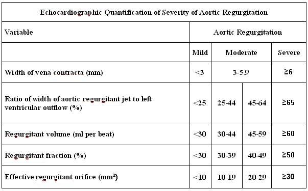
Echocardiographic Findings in Severe Aortic Insufficiency
The echocardiographic findings in severe aortic regurgitation include:
- An AI color jet dimension > 60 percent of the left ventricular outflow tract (LVOT) diameter (may not be true if the jet is eccentric)
- The pressure half-time of the regurgitant jet is < 250 msec
- Early termination of the mitral inflow (due to an increase in LV pressure as a result of the AI)
- Early diastolic flow reversal in the descending aorta.
- Regurgitant volume > 60 ml
- Regurgitant fraction > 55 percent
 |
Characteristics of aortic insufficiency are demonstrated by:
- Increased duration between E and A peaks
- Fluttering of the anterior mitral valve leaflet due to AI jet turbulence
Severe Aortic Insufficiency Video 1
{{#ev:googlevideo|4226733785410043550}}
Severe Aortic Insufficiency Video 2
{{#ev:googlevideo|3075471538892457393}}
Moderate Aortic Insufficiency (Color Doppler)
{{#ev:youtube|zMD3o0L5SlA}} {{#ev:youtube|3W_YX_GbpK8}}
2008 and incorporated 2006 ACC/AHA Guidelines incorporated into the 2006 guidelines for the management of patients with valvular heart disease [2]
Echocardiography (DO NOT EDIT) [2]
| Class I |
| "1. Echocardiography is indicated to confirm the presence and severity of acute or chronic AR. (Level of Evidence: B)" |
| "2. Echocardiography is indicated for diagnosis and assessment of the cause of chronic AR (including valve morphology and aortic root size and morphology) and for assessment of LV hypertrophy, dimension (or volume), and systolic function. (Level of Evidence: B)" |
| "3. Echocardiography is indicated in patients with an enlarged aortic root to assess regurgitation and the severity of aortic dilatation. (Level of Evidence: B)" |
| "4. Echocardiography is indicated for the periodic re-evaluation of LV size and function in asymptomatic patients with severe AR. (Level of Evidence: B)" |
| "5. Radionuclide angiography or magnetic resonance imaging is indicated for the initial and serial assessment of LV volume and function at rest in patients with AR and suboptimal echocardiograms. (Level of Evidence: B)" |
| "6. Echocardiography is indicated to re-evaluate mild, moderate, or severe AR in patients with new or changing symptoms. (Level of Evidence: B)" |
Sources
- 2008 and incorporated 2006 ACC/AHA Guidelines incorporated into the 2006 guidelines for the management of patients with valvular heart disease [2]
References
- ↑ Zoghbi WA, Enriquez-Sarano M, Foster E, Grayburn PA, Kraft CD, Levine RA, Nihoyannopoulos P, Otto CM, Quinones MA, Rakowski H, Stewart WJ, Waggoner A, Weissman NJ (2003). "Recommendations for evaluation of the severity of native valvular regurgitation with two-dimensional and Doppler echocardiography". Journal of the American Society of Echocardiography : Official Publication of the American Society of Echocardiography. 16 (7): 777–802. doi:10.1016/S0894-7317(03)00335-3. PMID 12835667. Retrieved 2011-03-02. Unknown parameter
|month=ignored (help) - ↑ 2.0 2.1 2.2 Bonow RO, Carabello BA, Chatterjee K; et al. (2008). "2008 Focused update incorporated into the ACC/AHA 2006 guidelines for the management of patients with valvular heart disease: a report of the American College of Cardiology/American Heart Association Task Force on Practice Guidelines (Writing Committee to Revise the 1998 Guidelines for the Management of Patients With Valvular Heart Disease): endorsed by the Society of Cardiovascular Anesthesiologists, Society for Cardiovascular Angiography and Interventions, and Society of Thoracic Surgeons". Circulation. 118 (15): e523–661. doi:10.1161/CIRCULATIONAHA.108.190748. PMID 18820172. Unknown parameter
|month=ignored (help)
