Angiolymphoid hyperplasia with eosinophilia: Difference between revisions
No edit summary |
|||
| Line 8: | Line 8: | ||
Angiolymphoid hyperplasia with eosinophilia <ref>Rapini, Ronald P.; Bolognia, Jean L.; Jorizzo, Joseph L. (2007). Dermatology: 2-Volume Set. St. Louis: Mosby. ISBN 1-4160-2999-0.</ref> is defined as red-brown, dome-shaped, dermal [[papule]] or [[nodule]], present in the head or neck, specifically around the ears and on the scalp.<ref name="Andrews">James, William; Berger, Timothy; Elston, Dirk (2005). ''Andrews' Diseases of the Skin: Clinical Dermatology''. (10th ed.). Saunders. ISBN 0-7216-2921-0.</ref> | Angiolymphoid hyperplasia with eosinophilia <ref>Rapini, Ronald P.; Bolognia, Jean L.; Jorizzo, Joseph L. (2007). Dermatology: 2-Volume Set. St. Louis: Mosby. ISBN 1-4160-2999-0.</ref> is defined as red-brown, dome-shaped, dermal [[papule]] or [[nodule]], present in the head or neck, specifically around the ears and on the scalp.<ref name="Andrews">James, William; Berger, Timothy; Elston, Dirk (2005). ''Andrews' Diseases of the Skin: Clinical Dermatology''. (10th ed.). Saunders. ISBN 0-7216-2921-0.</ref> | ||
==Historical Perspective== | ==Historical Perspective== | ||
*Angiolymphoid hyperplasia with eosinophilia was first discovered by G. C. | *Angiolymphoid hyperplasia with eosinophilia was first discovered by G. C. Wells and I. W. Whimster, two british physicians , in 1969.<ref name="pmid5763634">{{cite journal| author=Wells GC, Whimster IW| title=Subcutaneous angiolymphoid hyperplasia with eosinophilia. | journal=Br J Dermatol | year= 1969 | volume= 81 | issue= 1 | pages= 1-14 | pmid=5763634 | doi= | pmc= | url=http://www.ncbi.nlm.nih.gov/entrez/eutils/elink.fcgi?dbfrom=pubmed&tool=sumsearch.org/cite&retmode=ref&cmd=prlinks&id=5763634 }} </ref> | ||
==Pathophysiology== | ==Pathophysiology== | ||
*The pathogenesis of angiolymphoid hyperplasia with eosinophilia is characterized by red to brown papules or nodules dislocated in the dermis or subcutaneous tissue. | *The pathogenesis of angiolymphoid hyperplasia with eosinophilia is characterized by red to brown papules or nodules dislocated in the dermis or subcutaneous tissue. | ||
| Line 15: | Line 15: | ||
*On gross pathology, smooth-surface red to brown papules or nodules on the head, neck, trunk, extremities, genitalia, lips and oral mucosa; extracutaneous involvement are characteristic findings of angiolymphoid hyperplasia with eosinophilia. | *On gross pathology, smooth-surface red to brown papules or nodules on the head, neck, trunk, extremities, genitalia, lips and oral mucosa; extracutaneous involvement are characteristic findings of angiolymphoid hyperplasia with eosinophilia. | ||
*On microscopic histopathological analysis, florid vascular proliferation with atypical endothelial cells surrounded by a lymphocytic and eosinophilic infiltrate are characteristic findings of angiolymphoid hyperplasia with eosinophilia. | *On microscopic histopathological analysis, florid vascular proliferation with atypical endothelial cells surrounded by a lymphocytic and eosinophilic infiltrate are characteristic findings of angiolymphoid hyperplasia with eosinophilia. | ||
==Causes== | ==Causes== | ||
* Angiolymphoid hyperplasia with eosinophilia may be caused by either allergic reactions, traumas, or autoimmune disorders. | * Angiolymphoid hyperplasia with eosinophilia may be caused by either allergic reactions, traumas, or autoimmune disorders. | ||
==Differentiating Angiolymphoid hyperplasia with eosinophilia from other Diseases== | ==Differentiating Angiolymphoid hyperplasia with eosinophilia from other Diseases== | ||
*Angiolymphoid hyperplasia with eosinophilia must be differentiated from other diseases that cause swelling of face and neck, such as: | *Angiolymphoid hyperplasia with eosinophilia must be differentiated from other diseases that cause swelling of face and neck, such as: | ||
| Line 26: | Line 24: | ||
:*Cutaneous lymphoma | :*Cutaneous lymphoma | ||
:*Sarcoidosis | :*Sarcoidosis | ||
==Epidemiology and Demographics== | ==Epidemiology and Demographics== | ||
* The prevalence of [disease name] is approximately [number or range] per 100,000 individuals worldwide. | * The prevalence of [disease name] is approximately [number or range] per 100,000 individuals worldwide. | ||
| Line 36: | Line 33: | ||
===Race=== | ===Race=== | ||
*Angiolymphoid hyperplasia with eosinophilia usually affects predominently individuals of the Asian race. | *Angiolymphoid hyperplasia with eosinophilia usually affects predominently individuals of the Asian race. | ||
==Risk Factors== | ==Risk Factors== | ||
*Common risk factors in the development of angiolymphoid hyperplasia with eosinophilia are allergic reactions, traumas or autoimmune disorders. | *Common risk factors in the development of angiolymphoid hyperplasia with eosinophilia are allergic reactions, traumas or autoimmune disorders. | ||
== Natural History, Complications and Prognosis== | == Natural History, Complications and Prognosis== | ||
*The majority of patients with angiolymphoid hyperplasia with eosinophilia remain asymptomatic for several years. | *The majority of patients with angiolymphoid hyperplasia with eosinophilia remain asymptomatic for several years. | ||
*Common complications of angiolymphoid hyperplasia with eosinophilia include pruritis, pulsation, and spontaneous bleeding. | *Common complications of angiolymphoid hyperplasia with eosinophilia include pruritis, pulsation, and spontaneous bleeding. | ||
* One-third of cases of angiolymphoid hyperplasia with eosinophilia recur when incompletely excised. | * One-third of cases of angiolymphoid hyperplasia with eosinophilia recur when incompletely excised. | ||
== Diagnosis == | == Diagnosis == | ||
=== Symptoms === | === Symptoms === | ||
| Line 51: | Line 45: | ||
*Physical examination may be remarkable for: | *Physical examination may be remarkable for: | ||
:*Dome-shaped, smooth-surfaced papules or expanding nodule or group of nodules | :*Dome-shaped, smooth-surfaced papules or expanding nodule or group of nodules | ||
====Ear==== | |||
<gallery> | |||
Image:Epithelioid hemangioma01.jpg|Epithelioid hemangioma. <SMALL><SMALL>''[http://www.atlasdermatologico.com.br/ Adapted from Dermatology Atlas.]''<ref name="Dermatology Atlas">{{Cite | |||
Image:Epithelioid hemangioma02.jpg|Epithelioid hemangioma. <SMALL><SMALL>''[http://www.atlasdermatologico.com.br/ Adapted from Dermatology Atlas.]''<ref name="Dermatology Atlas">{{Cite | |||
Image:Epithelioid hemangioma03.jpg|Epithelioid hemangioma. <SMALL><SMALL>''[http://www.atlasdermatologico.com.br/ Adapted from Dermatology Atlas.]''<ref name="Dermatology Atlas">{{Cite | |||
Image:Epithelioid hemangioma04.jpg|Epithelioid hemangioma. <SMALL><SMALL>''[http://www.atlasdermatologico.com.br/ Adapted from Dermatology Atlas.]''<ref name="Dermatology Atlas">{{Cite | |||
Image:Epithelioid hemangioma05.jpg|Epithelioid hemangioma. <SMALL><SMALL>''[http://www.atlasdermatologico.com.br/ Adapted from Dermatology Atlas.]''<ref name="Dermatology Atlas">{{Cite | |||
Image:Epithelioid hemangioma06.jpg|Epithelioid hemangioma. <SMALL><SMALL>''[http://www.atlasdermatologico.com.br/ Adapted from Dermatology Atlas.]''<ref name="Dermatology Atlas">{{Cite | |||
Image:Epithelioid hemangioma07.jpg|Epithelioid hemangioma. <SMALL><SMALL>''[http://www.atlasdermatologico.com.br/ Adapted from Dermatology Atlas.]''<ref name="Dermatology Atlas">{{Cite | |||
Image:Epithelioid hemangioma08.jpg|Epithelioid hemangioma. <SMALL><SMALL>''[http://www.atlasdermatologico.com.br/ Adapted from Dermatology Atlas.]''<ref name="Dermatology Atlas">{{Cite | |||
Image:Epithelioid hemangioma09.jpg|Epithelioid hemangioma. <SMALL><SMALL>''[http://www.atlasdermatologico.com.br/ Adapted from Dermatology Atlas.]''<ref name="Dermatology Atlas">{{Cite | |||
Image:Epithelioid hemangioma10.jpg|Epithelioid hemangioma. <SMALL><SMALL>''[http://www.atlasdermatologico.com.br/ Adapted from Dermatology Atlas.]''<ref name="Dermatology Atlas">{{Cite | |||
</gallery> | |||
=== Laboratory Findings === | === Laboratory Findings === | ||
*A eosinophilia in CBC is charecteristic of angiolymphoid hyperplasia with eosinophilia. | *A eosinophilia in CBC is charecteristic of angiolymphoid hyperplasia with eosinophilia. | ||
| Line 57: | Line 64: | ||
*Angiolymphoid hyperplasia with eosinophilia may also be diagnosed using biopsy. | *Angiolymphoid hyperplasia with eosinophilia may also be diagnosed using biopsy. | ||
*Findings on biopsy include proliferation of small blood vessels with endothelial cells having a cobblestone appearance, a perivascular and interstitial infiltrate of lymphocytes and eosinophils. | *Findings on biopsy include proliferation of small blood vessels with endothelial cells having a cobblestone appearance, a perivascular and interstitial infiltrate of lymphocytes and eosinophils. | ||
== Treatment == | == Treatment == | ||
=== Medical Therapy === | === Medical Therapy === | ||
*The mainstay of therapy for angiolymphoid hyperplasia with eosinophilia is intralesional corticoid therapy and imiquimod. | *The mainstay of therapy for angiolymphoid hyperplasia with eosinophilia is intralesional corticoid therapy and imiquimod. | ||
*Imiquimod acts by induction of the production of interferon-α and induction or inhibition of certain cytokines, mainly interleukin-5.<ref name="EstevesBarbalho2015">{{cite journal|last1=Esteves|first1=Paola|last2=Barbalho|first2=Marcella|last3=Lima|first3=Tiago|last4=Quintella|first4=Leonardo|last5=Niemeyer-Corbellini|first5=Jo�o Paulo|last6=Ramos-e-Silva|first6=Marcia|title=Angiolymphoid Hyperplasia with Eosinophilia: A Case Report|journal=Case Reports in Dermatology|volume=7|issue=2|year=2015|pages=113–116|issn=1662-6567|doi=10.1159/000381843}}</ref> | *Imiquimod acts by induction of the production of interferon-α and induction or inhibition of certain cytokines, mainly interleukin-5.<ref name="EstevesBarbalho2015">{{cite journal|last1=Esteves|first1=Paola|last2=Barbalho|first2=Marcella|last3=Lima|first3=Tiago|last4=Quintella|first4=Leonardo|last5=Niemeyer-Corbellini|first5=Jo�o Paulo|last6=Ramos-e-Silva|first6=Marcia|title=Angiolymphoid Hyperplasia with Eosinophilia: A Case Report|journal=Case Reports in Dermatology|volume=7|issue=2|year=2015|pages=113–116|issn=1662-6567|doi=10.1159/000381843}}</ref> | ||
=== Surgery === | === Surgery === | ||
*Surgery is the mainstay of therapy for angiolymphoid hyperplasia with eosinophilia. | *Surgery is the mainstay of therapy for angiolymphoid hyperplasia with eosinophilia. | ||
| Line 68: | Line 73: | ||
* Other treatment options include radiotherapy, curettage, shave excision with electrodessication, cryotherapy, corticosteroids (topical, systemic or intralesional preparation), and laser therapy using Continuous wave carbon dioxide and argon lasers.<ref name="pmid21487464">{{cite journal| author=Lembo S, Balato A, Cirillo T, Balato N| title=A Long-Term Follow-Up of Angiolymphoid Hyperplasia with Eosinophilia Treated by Corticosteroids: When a Traditional Therapy is Still Up-to-Date. | journal=Case Rep Dermatol | year= 2011 | volume= 3 | issue= 1 | pages= 64-7 | pmid=21487464 | doi=10.1159/000323182 | pmc=PMC3073756 | url=http://www.ncbi.nlm.nih.gov/entrez/eutils/elink.fcgi?dbfrom=pubmed&tool=sumsearch.org/cite&retmode=ref&cmd=prlinks&id=21487464 }} </ref> | * Other treatment options include radiotherapy, curettage, shave excision with electrodessication, cryotherapy, corticosteroids (topical, systemic or intralesional preparation), and laser therapy using Continuous wave carbon dioxide and argon lasers.<ref name="pmid21487464">{{cite journal| author=Lembo S, Balato A, Cirillo T, Balato N| title=A Long-Term Follow-Up of Angiolymphoid Hyperplasia with Eosinophilia Treated by Corticosteroids: When a Traditional Therapy is Still Up-to-Date. | journal=Case Rep Dermatol | year= 2011 | volume= 3 | issue= 1 | pages= 64-7 | pmid=21487464 | doi=10.1159/000323182 | pmc=PMC3073756 | url=http://www.ncbi.nlm.nih.gov/entrez/eutils/elink.fcgi?dbfrom=pubmed&tool=sumsearch.org/cite&retmode=ref&cmd=prlinks&id=21487464 }} </ref> | ||
=== Prevention === | === Prevention === | ||
*There are no primary preventive measures available for angiolymphoid hyperplasia with eosinophilia | *There are no primary preventive measures available for angiolymphoid hyperplasia with eosinophilia | ||
==References== | ==References== | ||
{{reflist|2}} | {{reflist|2}} | ||
[[Category:Disease]] | [[Category:Disease]] | ||
[[Category:Hematology]] | [[Category:Hematology]] | ||
Revision as of 20:20, 9 May 2016
Editor-In-Chief: C. Michael Gibson, M.S., M.D. [1];Associate Editor(s)-in-Chief: Kiran Singh, M.D. [2] Ammu Susheela, M.D. [3]
Synonyms and Keywords: Epithelioid hemangioma; Histiocytoid hemangioma; Inflammatory angiomatous nodule; Intravenous atypical vascular proliferation; Papular angioplasia; Inflammatory arteriovenous hemangioma; Pseudopyogenic granuloma; ALHE; EH
Overview
Angiolymphoid hyperplasia with eosinophilia [1] is defined as red-brown, dome-shaped, dermal papule or nodule, present in the head or neck, specifically around the ears and on the scalp.[2]
Historical Perspective
- Angiolymphoid hyperplasia with eosinophilia was first discovered by G. C. Wells and I. W. Whimster, two british physicians , in 1969.[3]
Pathophysiology
- The pathogenesis of angiolymphoid hyperplasia with eosinophilia is characterized by red to brown papules or nodules dislocated in the dermis or subcutaneous tissue.
- Angiolymphoid hyperplasia with eosinophilia (ALHE) is a rare and idiopathic vascular disorder.
- Angiolymphoid hyperplasia with eosinophilia (ALHE) is an uncommon benign lesion, primarily occurring in the head and neck.
- On gross pathology, smooth-surface red to brown papules or nodules on the head, neck, trunk, extremities, genitalia, lips and oral mucosa; extracutaneous involvement are characteristic findings of angiolymphoid hyperplasia with eosinophilia.
- On microscopic histopathological analysis, florid vascular proliferation with atypical endothelial cells surrounded by a lymphocytic and eosinophilic infiltrate are characteristic findings of angiolymphoid hyperplasia with eosinophilia.
Causes
- Angiolymphoid hyperplasia with eosinophilia may be caused by either allergic reactions, traumas, or autoimmune disorders.
Differentiating Angiolymphoid hyperplasia with eosinophilia from other Diseases
- Angiolymphoid hyperplasia with eosinophilia must be differentiated from other diseases that cause swelling of face and neck, such as:
- Kimura’s disease
- Acial granuloma
- Insect bite reaction
- Cutaneous lymphoma
- Sarcoidosis
Epidemiology and Demographics
- The prevalence of [disease name] is approximately [number or range] per 100,000 individuals worldwide.
- In [year], the incidence of [disease name] was estimated to be [number or range] cases per 100,000 individuals in [location].
Age
- Angiolymphoid hyperplasia with eosinophilia is more commonly observed among young patients.
Gender
- Females are more commonly affected with angiolymphoid hyperplasia with eosinophilia than males.
Race
- Angiolymphoid hyperplasia with eosinophilia usually affects predominently individuals of the Asian race.
Risk Factors
- Common risk factors in the development of angiolymphoid hyperplasia with eosinophilia are allergic reactions, traumas or autoimmune disorders.
Natural History, Complications and Prognosis
- The majority of patients with angiolymphoid hyperplasia with eosinophilia remain asymptomatic for several years.
- Common complications of angiolymphoid hyperplasia with eosinophilia include pruritis, pulsation, and spontaneous bleeding.
- One-third of cases of angiolymphoid hyperplasia with eosinophilia recur when incompletely excised.
Diagnosis
Symptoms
- Angiolymphoid hyperplasia with eosinophilia lesions may be asymptomatic, painful or pruriginous.
Physical Examination
- Physical examination may be remarkable for:
- Dome-shaped, smooth-surfaced papules or expanding nodule or group of nodules
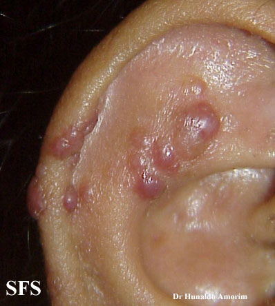
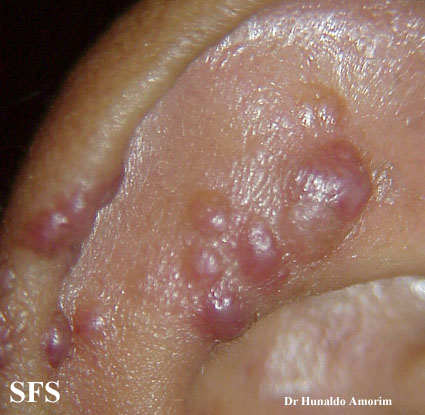
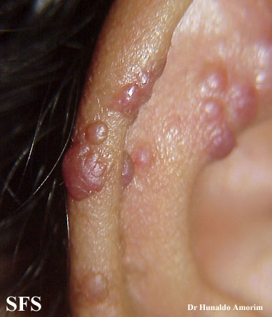
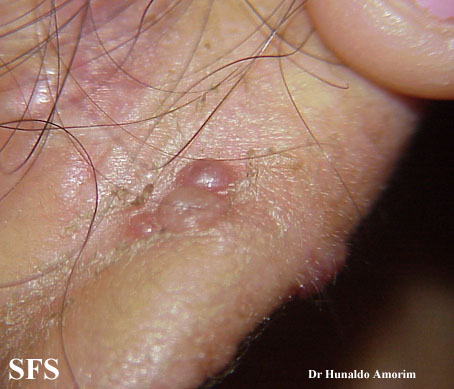
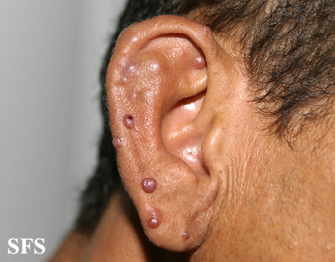
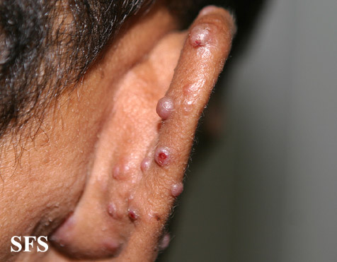
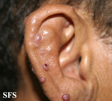
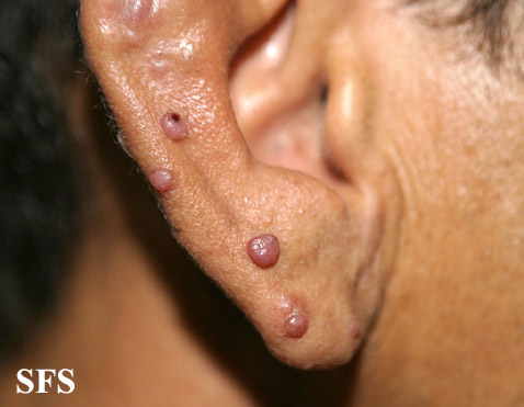
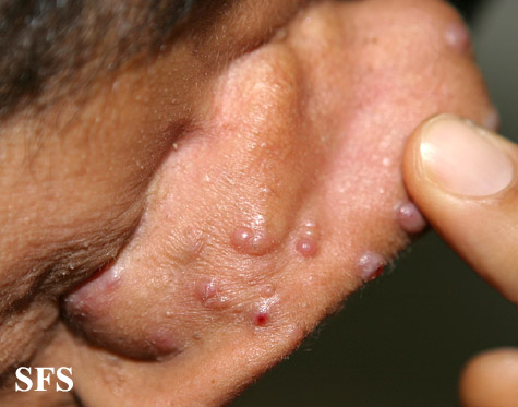
Ear
-
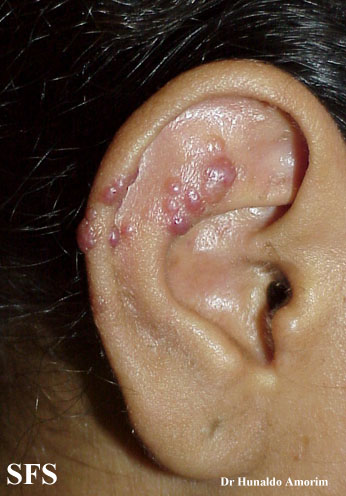
Epithelioid hemangioma. Adapted from Dermatology Atlas.<ref name="Dermatology Atlas">{{Cite
-
Epithelioid hemangioma. Adapted from Dermatology Atlas.<ref name="Dermatology Atlas">{{Cite
-
Epithelioid hemangioma. Adapted from Dermatology Atlas.<ref name="Dermatology Atlas">{{Cite
-
Epithelioid hemangioma. Adapted from Dermatology Atlas.<ref name="Dermatology Atlas">{{Cite
-
Epithelioid hemangioma. Adapted from Dermatology Atlas.<ref name="Dermatology Atlas">{{Cite
-
Epithelioid hemangioma. Adapted from Dermatology Atlas.<ref name="Dermatology Atlas">{{Cite
-
Epithelioid hemangioma. Adapted from Dermatology Atlas.<ref name="Dermatology Atlas">{{Cite
-
Epithelioid hemangioma. Adapted from Dermatology Atlas.<ref name="Dermatology Atlas">{{Cite
-
Epithelioid hemangioma. Adapted from Dermatology Atlas.<ref name="Dermatology Atlas">{{Cite
-
Epithelioid hemangioma. Adapted from Dermatology Atlas.<ref name="Dermatology Atlas">{{Cite


Laboratory Findings
- A eosinophilia in CBC is charecteristic of angiolymphoid hyperplasia with eosinophilia.
- Eosinophils in urinalysis is also a feature.
Other Diagnostic Studies
- Angiolymphoid hyperplasia with eosinophilia may also be diagnosed using biopsy.
- Findings on biopsy include proliferation of small blood vessels with endothelial cells having a cobblestone appearance, a perivascular and interstitial infiltrate of lymphocytes and eosinophils.
Treatment
Medical Therapy
- The mainstay of therapy for angiolymphoid hyperplasia with eosinophilia is intralesional corticoid therapy and imiquimod.
- Imiquimod acts by induction of the production of interferon-α and induction or inhibition of certain cytokines, mainly interleukin-5.[4]
Surgery
- Surgery is the mainstay of therapy for angiolymphoid hyperplasia with eosinophilia.
- Mohs micrographic surgery with complete margin examination is performed.
- Other treatment options include radiotherapy, curettage, shave excision with electrodessication, cryotherapy, corticosteroids (topical, systemic or intralesional preparation), and laser therapy using Continuous wave carbon dioxide and argon lasers.[5]
Prevention
- There are no primary preventive measures available for angiolymphoid hyperplasia with eosinophilia
References
- ↑ Rapini, Ronald P.; Bolognia, Jean L.; Jorizzo, Joseph L. (2007). Dermatology: 2-Volume Set. St. Louis: Mosby. ISBN 1-4160-2999-0.
- ↑ James, William; Berger, Timothy; Elston, Dirk (2005). Andrews' Diseases of the Skin: Clinical Dermatology. (10th ed.). Saunders. ISBN 0-7216-2921-0.
- ↑ Wells GC, Whimster IW (1969). "Subcutaneous angiolymphoid hyperplasia with eosinophilia". Br J Dermatol. 81 (1): 1–14. PMID 5763634.
- ↑ Esteves, Paola; Barbalho, Marcella; Lima, Tiago; Quintella, Leonardo; Niemeyer-Corbellini, Jo�o Paulo; Ramos-e-Silva, Marcia (2015). "Angiolymphoid Hyperplasia with Eosinophilia: A Case Report". Case Reports in Dermatology. 7 (2): 113–116. doi:10.1159/000381843. ISSN 1662-6567. replacement character in
|first5=at position 3 (help) - ↑ Lembo S, Balato A, Cirillo T, Balato N (2011). "A Long-Term Follow-Up of Angiolymphoid Hyperplasia with Eosinophilia Treated by Corticosteroids: When a Traditional Therapy is Still Up-to-Date". Case Rep Dermatol. 3 (1): 64–7. doi:10.1159/000323182. PMC 3073756. PMID 21487464.