Angiolymphoid hyperplasia with eosinophilia: Difference between revisions
(→Gender) |
No edit summary |
||
| (34 intermediate revisions by the same user not shown) | |||
| Line 3: | Line 3: | ||
{{CMG}};{{AE}} {{KS}} {{Ammu}} | {{CMG}};{{AE}} {{KS}} {{Ammu}} | ||
'''''Synonyms and Keywords:''''' Epithelioid hemangioma; Histiocytoid hemangioma; Inflammatory angiomatous nodule; Intravenous atypical vascular proliferation; Papular angioplasia; Inflammatory arteriovenous hemangioma; Pseudopyogenic granuloma; ALHE | '''''Synonyms and Keywords:''''' Epithelioid hemangioma; Histiocytoid hemangioma; Inflammatory angiomatous nodule; Intravenous atypical vascular proliferation; Papular angioplasia; Inflammatory arteriovenous hemangioma; Pseudopyogenic granuloma; ALHE; EH | ||
==Overview== | ==Overview== | ||
Angiolymphoid hyperplasia with eosinophilia <ref>Rapini, Ronald P.; Bolognia, Jean L.; Jorizzo, Joseph L. (2007). Dermatology: 2-Volume Set. St. Louis: Mosby. ISBN 1-4160-2999-0.</ref> is defined as red-brown, dome-shaped, dermal [[papule]] or [[nodule]], present in the | Angiolymphoid hyperplasia with eosinophilia was first discovered by G. C. Wells and I. W. Whimster, two British physicians, in 1969.<ref name="pmid5763634">{{cite journal| author=Wells GC, Whimster IW| title=Subcutaneous angiolymphoid hyperplasia with eosinophilia. | journal=Br J Dermatol | year= 1969 | volume= 81 | issue= 1 | pages= 1-14 | pmid=5763634 | doi= | pmc= | url=http://www.ncbi.nlm.nih.gov/entrez/eutils/elink.fcgi?dbfrom=pubmed&tool=sumsearch.org/cite&retmode=ref&cmd=prlinks&id=5763634 }} </ref>Angiolymphoid hyperplasia with eosinophilia <ref>Rapini, Ronald P.; Bolognia, Jean L.; Jorizzo, Joseph L. (2007). Dermatology: 2-Volume Set. St. Louis: Mosby. ISBN 1-4160-2999-0.</ref> is defined as red-brown, dome-shaped, dermal [[papule]] or [[nodule]], present in the around the ears, scalp, neck, trunk, extremities, genitalia, lips and oral mucosa.<ref name="Andrews">James, William; Berger, Timothy; Elston, Dirk (2005). ''Andrews' Diseases of the Skin: Clinical Dermatology''. (10th ed.). Saunders. ISBN 0-7216-2921-0.</ref>On gross pathology, smooth-surface red to brown papules or nodules are characteristic findings of angiolymphoid hyperplasia with eosinophilia. On microscopic histopathological analysis, florid vascular proliferation with atypical endothelial cells surrounded by a lymphocytic and eosinophilic infiltrate are characteristic findings of angiolymphoid hyperplasia with eosinophilia. Angiolymphoid hyperplasia with eosinophilia may be caused by either allergic reactions, traumas, or autoimmune disorders. Angiolymphoid hyperplasia with eosinophilia must be differentiated from other diseases that cause swelling of face and neck, such as [[Kimura disease]], acial granuloma, insect bite reaction, [[Lymphoma|cutaneous lymphoma]], [[sarcoidosis]]. The prevalence of angiolymphoid hyperplasia with eosinophilia is unknown as it is an extremely rare disease. Angiolymphoid hyperplasia with eosinophilia is more commonly observed among young patients. Females are more commonly affected with angiolymphoid hyperplasia with eosinophilia than males. Angiolymphoid hyperplasia with eosinophilia usually affects predominently individuals of the Asian race. Common complications of angiolymphoid hyperplasia with eosinophilia include [[pruritis]], pulsation, and spontaneous bleeding. Findings on [[biopsy]] include proliferation of small blood vessels with endothelial cells having a cobblestone appearance, a perivascular and interstitial infiltrate of lymphocytes and [[eosinophils]]. [[Surgery]] is the mainstay of therapy for angiolymphoid hyperplasia with eosinophilia. | ||
==Historical Perspective== | ==Historical Perspective== | ||
* | *Angiolymphoid hyperplasia with eosinophilia was first discovered by G. C. Wells and I. W. Whimster, two British physicians, in 1969.<ref name="pmid5763634">{{cite journal| author=Wells GC, Whimster IW| title=Subcutaneous angiolymphoid hyperplasia with eosinophilia. | journal=Br J Dermatol | year= 1969 | volume= 81 | issue= 1 | pages= 1-14 | pmid=5763634 | doi= | pmc= | url=http://www.ncbi.nlm.nih.gov/entrez/eutils/elink.fcgi?dbfrom=pubmed&tool=sumsearch.org/cite&retmode=ref&cmd=prlinks&id=5763634 }} </ref> | ||
== | |||
==Pathophysiology== | ==Pathophysiology== | ||
*Angiolymphoid hyperplasia with eosinophilia <ref>Rapini, Ronald P.; Bolognia, Jean L.; Jorizzo, Joseph L. (2007). Dermatology: 2-Volume Set. St. Louis: Mosby. ISBN 1-4160-2999-0.</ref> is defined as red-brown, dome-shaped, dermal [[papule]] or [[nodule]], present in the head or neck, specifically around the ears and on the scalp.<ref name="Andrews">James, William; Berger, Timothy; Elston, Dirk (2005). ''Andrews' Diseases of the Skin: Clinical Dermatology''. (10th ed.). Saunders. ISBN 0-7216-2921-0.</ref> | |||
*The pathogenesis of angiolymphoid hyperplasia with eosinophilia is characterized by red to brown papules or nodules dislocated in the dermis or subcutaneous tissue. | *The pathogenesis of angiolymphoid hyperplasia with eosinophilia is characterized by red to brown papules or nodules dislocated in the dermis or subcutaneous tissue. | ||
* Angiolymphoid hyperplasia with eosinophilia | * Angiolymphoid hyperplasia with eosinophilia is a rare and idiopathic vascular disorder. | ||
* | *On gross pathology, smooth-surface red to brown papules or nodules on the head, neck, trunk, extremities, genitalia, lips and oral mucosa are characteristic findings of angiolymphoid hyperplasia with eosinophilia. | ||
characteristic findings of | |||
*On microscopic histopathological analysis, florid vascular proliferation with atypical endothelial cells surrounded by a lymphocytic and eosinophilic infiltrate are characteristic findings of angiolymphoid hyperplasia with eosinophilia. | *On microscopic histopathological analysis, florid vascular proliferation with atypical endothelial cells surrounded by a lymphocytic and eosinophilic infiltrate are characteristic findings of angiolymphoid hyperplasia with eosinophilia. | ||
==Causes== | ==Causes== | ||
* Angiolymphoid hyperplasia with eosinophilia may be caused by either allergic reactions, traumas, or autoimmune disorders. | * Angiolymphoid hyperplasia with eosinophilia may be caused by either allergic reactions, traumas, or autoimmune disorders. | ||
==Differentiating Angiolymphoid hyperplasia with eosinophilia from other Diseases== | ==Differentiating Angiolymphoid hyperplasia with eosinophilia from other Diseases== | ||
*Angiolymphoid hyperplasia with eosinophilia must be differentiated from other diseases that cause swelling of face and neck, such as: | *Angiolymphoid hyperplasia with eosinophilia must be differentiated from other diseases that cause swelling of face and neck, such as: | ||
:* | :*[[Kimura disease]] | ||
:*Acial granuloma | |||
:*Insect bite reaction | |||
:*[[Lymphoma|Cutaneous lymphoma]] | |||
:*[[Sarcoidosis]] | |||
==Epidemiology and Demographics== | ==Epidemiology and Demographics== | ||
* The prevalence of | * The prevalence of angiolymphoid hyperplasia with eosinophilia is unknown as it is an extremely rare disease. | ||
===Age=== | ===Age=== | ||
* | *Angiolymphoid hyperplasia with eosinophilia is more commonly observed among young patients. | ||
===Gender=== | ===Gender=== | ||
*Females are more commonly affected with angiolymphoid hyperplasia with eosinophilia than males. | *Females are more commonly affected with angiolymphoid hyperplasia with eosinophilia than males. | ||
===Race=== | ===Race=== | ||
*Angiolymphoid hyperplasia with eosinophilia usually affects predominently individuals of the Asian race. | *Angiolymphoid hyperplasia with eosinophilia usually affects predominently individuals of the Asian race. | ||
==Risk Factors== | ==Risk Factors== | ||
*Common risk factors in the development of angiolymphoid hyperplasia with eosinophilia are allergic reactions, traumas or autoimmune disorders. | *Common risk factors in the development of angiolymphoid hyperplasia with eosinophilia are allergic reactions, traumas or autoimmune disorders. | ||
== Natural History, Complications and Prognosis== | == Natural History, Complications and Prognosis== | ||
*The majority of patients with | *The majority of patients with angiolymphoid hyperplasia with eosinophilia remain asymptomatic for several years. | ||
*Common complications of angiolymphoid hyperplasia with eosinophilia include pruritis, pulsation, and spontaneous bleeding. | |||
* One-third of cases of angiolymphoid hyperplasia with eosinophilia recur when incompletely excised. | |||
*Common complications of | |||
* | |||
== Diagnosis == | == Diagnosis == | ||
=== Symptoms === | === Symptoms === | ||
* | * Angiolymphoid hyperplasia with eosinophilia lesions may be asymptomatic, painful or pruriginous. | ||
=== Physical Examination === | === Physical Examination === | ||
*Physical examination may be remarkable for: | *Physical examination may be remarkable for: | ||
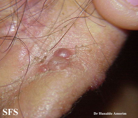
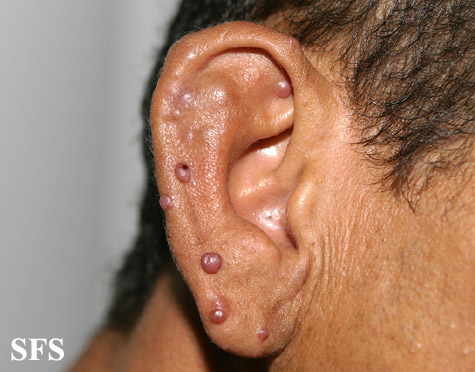
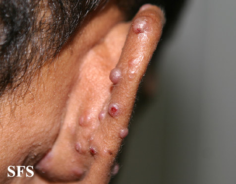
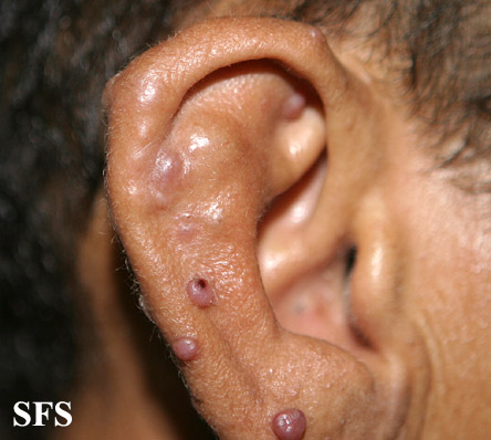
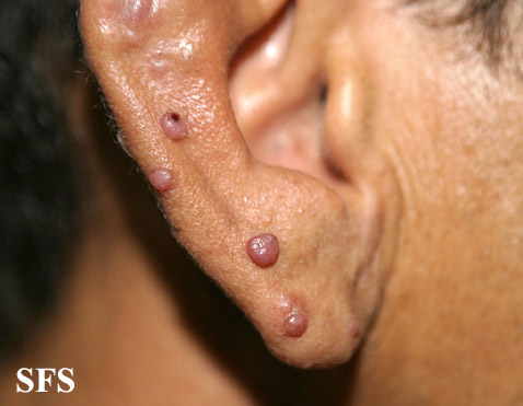
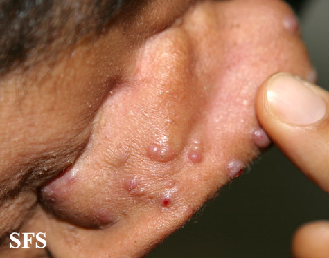
:* | :*Dome-shaped, smooth-surfaced papules or expanding nodule or group of nodules | ||
====Ear==== | ====Ear==== | ||
<gallery> | <gallery> | ||
| Line 149: | Line 56: | ||
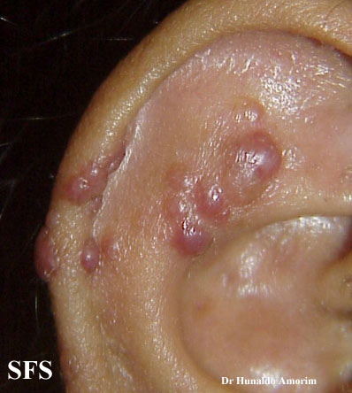
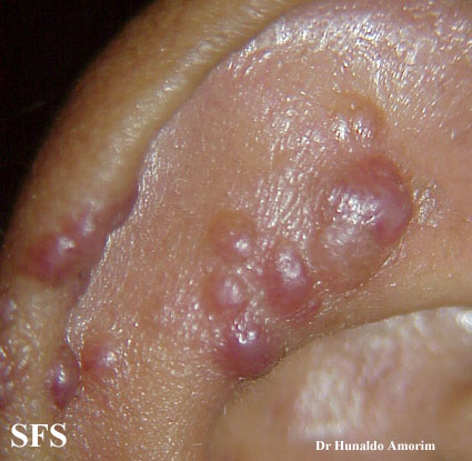
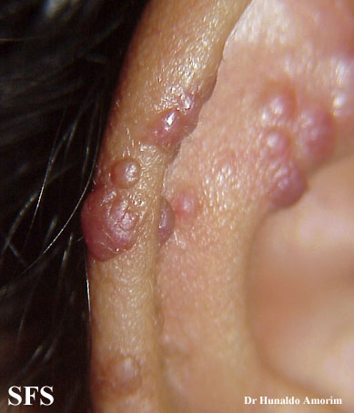
Image:Epithelioid hemangioma09.jpg|Epithelioid hemangioma. <SMALL><SMALL>''[http://www.atlasdermatologico.com.br/ Adapted from Dermatology Atlas.]''<ref name="Dermatology Atlas">{{Cite | Image:Epithelioid hemangioma09.jpg|Epithelioid hemangioma. <SMALL><SMALL>''[http://www.atlasdermatologico.com.br/ Adapted from Dermatology Atlas.]''<ref name="Dermatology Atlas">{{Cite | ||
Image:Epithelioid hemangioma10.jpg|Epithelioid hemangioma. <SMALL><SMALL>''[http://www.atlasdermatologico.com.br/ Adapted from Dermatology Atlas.]''<ref name="Dermatology Atlas">{{Cite | Image:Epithelioid hemangioma10.jpg|Epithelioid hemangioma. <SMALL><SMALL>''[http://www.atlasdermatologico.com.br/ Adapted from Dermatology Atlas.]''<ref name="Dermatology Atlas">{{Cite | ||
</gallery> | </gallery> | ||
=== Laboratory Findings === | |||
*A eosinophilia in [[complete blood count]] is characteristic of angiolymphoid hyperplasia with eosinophilia. | |||
*[[Eosinophils]] in [[urinalysis]] is also a feature. | |||
=== Other Diagnostic Studies === | |||
*Angiolymphoid hyperplasia with eosinophilia may also be diagnosed using [[biopsy]]. | |||
*Findings on [[biopsy]] include proliferation of small blood vessels with endothelial cells having a cobblestone appearance, a perivascular and interstitial infiltrate of lymphocytes and [[eosinophils]]. | |||
== Treatment == | |||
=== Medical Therapy === | |||
*The mainstay of therapy for angiolymphoid hyperplasia with eosinophilia is intralesional corticoid therapy and [[imiquimod]]. | |||
*[[Imiquimod]] acts by induction of the production of interferon-α and induction or inhibition of certain cytokines, mainly interleukin-5.<ref name="EstevesBarbalho2015">{{cite journal|last1=Esteves|first1=Paola|last2=Barbalho|first2=Marcella|last3=Lima|first3=Tiago|last4=Quintella|first4=Leonardo|last5=Niemeyer-Corbellini|first5=Jo�o Paulo|last6=Ramos-e-Silva|first6=Marcia|title=Angiolymphoid Hyperplasia with Eosinophilia: A Case Report|journal=Case Reports in Dermatology|volume=7|issue=2|year=2015|pages=113–116|issn=1662-6567|doi=10.1159/000381843}}</ref> | |||
=== Surgery === | |||
*[[Surgery]] is the mainstay of therapy for angiolymphoid hyperplasia with eosinophilia. | |||
* Mohs micrographic surgery with complete margin examination is performed. | |||
* Other treatment options include [[radiotherapy]], curettage, shave excision with electrodessication, [[cryotherapy]], [[corticosteroids]] (topical, systemic or intralesional preparation), and laser therapy using continuous wave carbon dioxide and argon lasers.<ref name="pmid21487464">{{cite journal| author=Lembo S, Balato A, Cirillo T, Balato N| title=A Long-Term Follow-Up of Angiolymphoid Hyperplasia with Eosinophilia Treated by Corticosteroids: When a Traditional Therapy is Still Up-to-Date. | journal=Case Rep Dermatol | year= 2011 | volume= 3 | issue= 1 | pages= 64-7 | pmid=21487464 | doi=10.1159/000323182 | pmc=PMC3073756 | url=http://www.ncbi.nlm.nih.gov/entrez/eutils/elink.fcgi?dbfrom=pubmed&tool=sumsearch.org/cite&retmode=ref&cmd=prlinks&id=21487464 }} </ref> | |||
=== Prevention === | |||
*There are no primary preventive measures available for angiolymphoid hyperplasia with eosinophilia. | |||
==References== | ==References== | ||
Latest revision as of 20:48, 9 May 2016
Editor-In-Chief: C. Michael Gibson, M.S., M.D. [1];Associate Editor(s)-in-Chief: Kiran Singh, M.D. [2] Ammu Susheela, M.D. [3]
Synonyms and Keywords: Epithelioid hemangioma; Histiocytoid hemangioma; Inflammatory angiomatous nodule; Intravenous atypical vascular proliferation; Papular angioplasia; Inflammatory arteriovenous hemangioma; Pseudopyogenic granuloma; ALHE; EH
Overview
Angiolymphoid hyperplasia with eosinophilia was first discovered by G. C. Wells and I. W. Whimster, two British physicians, in 1969.[1]Angiolymphoid hyperplasia with eosinophilia [2] is defined as red-brown, dome-shaped, dermal papule or nodule, present in the around the ears, scalp, neck, trunk, extremities, genitalia, lips and oral mucosa.[3]On gross pathology, smooth-surface red to brown papules or nodules are characteristic findings of angiolymphoid hyperplasia with eosinophilia. On microscopic histopathological analysis, florid vascular proliferation with atypical endothelial cells surrounded by a lymphocytic and eosinophilic infiltrate are characteristic findings of angiolymphoid hyperplasia with eosinophilia. Angiolymphoid hyperplasia with eosinophilia may be caused by either allergic reactions, traumas, or autoimmune disorders. Angiolymphoid hyperplasia with eosinophilia must be differentiated from other diseases that cause swelling of face and neck, such as Kimura disease, acial granuloma, insect bite reaction, cutaneous lymphoma, sarcoidosis. The prevalence of angiolymphoid hyperplasia with eosinophilia is unknown as it is an extremely rare disease. Angiolymphoid hyperplasia with eosinophilia is more commonly observed among young patients. Females are more commonly affected with angiolymphoid hyperplasia with eosinophilia than males. Angiolymphoid hyperplasia with eosinophilia usually affects predominently individuals of the Asian race. Common complications of angiolymphoid hyperplasia with eosinophilia include pruritis, pulsation, and spontaneous bleeding. Findings on biopsy include proliferation of small blood vessels with endothelial cells having a cobblestone appearance, a perivascular and interstitial infiltrate of lymphocytes and eosinophils. Surgery is the mainstay of therapy for angiolymphoid hyperplasia with eosinophilia.
Historical Perspective
- Angiolymphoid hyperplasia with eosinophilia was first discovered by G. C. Wells and I. W. Whimster, two British physicians, in 1969.[1]
Pathophysiology
- Angiolymphoid hyperplasia with eosinophilia [4] is defined as red-brown, dome-shaped, dermal papule or nodule, present in the head or neck, specifically around the ears and on the scalp.[3]
- The pathogenesis of angiolymphoid hyperplasia with eosinophilia is characterized by red to brown papules or nodules dislocated in the dermis or subcutaneous tissue.
- Angiolymphoid hyperplasia with eosinophilia is a rare and idiopathic vascular disorder.
- On gross pathology, smooth-surface red to brown papules or nodules on the head, neck, trunk, extremities, genitalia, lips and oral mucosa are characteristic findings of angiolymphoid hyperplasia with eosinophilia.
- On microscopic histopathological analysis, florid vascular proliferation with atypical endothelial cells surrounded by a lymphocytic and eosinophilic infiltrate are characteristic findings of angiolymphoid hyperplasia with eosinophilia.
Causes
- Angiolymphoid hyperplasia with eosinophilia may be caused by either allergic reactions, traumas, or autoimmune disorders.
Differentiating Angiolymphoid hyperplasia with eosinophilia from other Diseases
- Angiolymphoid hyperplasia with eosinophilia must be differentiated from other diseases that cause swelling of face and neck, such as:
- Kimura disease
- Acial granuloma
- Insect bite reaction
- Cutaneous lymphoma
- Sarcoidosis
Epidemiology and Demographics
- The prevalence of angiolymphoid hyperplasia with eosinophilia is unknown as it is an extremely rare disease.
Age
- Angiolymphoid hyperplasia with eosinophilia is more commonly observed among young patients.
Gender
- Females are more commonly affected with angiolymphoid hyperplasia with eosinophilia than males.
Race
- Angiolymphoid hyperplasia with eosinophilia usually affects predominently individuals of the Asian race.
Risk Factors
- Common risk factors in the development of angiolymphoid hyperplasia with eosinophilia are allergic reactions, traumas or autoimmune disorders.
Natural History, Complications and Prognosis
- The majority of patients with angiolymphoid hyperplasia with eosinophilia remain asymptomatic for several years.
- Common complications of angiolymphoid hyperplasia with eosinophilia include pruritis, pulsation, and spontaneous bleeding.
- One-third of cases of angiolymphoid hyperplasia with eosinophilia recur when incompletely excised.
Diagnosis
Symptoms
- Angiolymphoid hyperplasia with eosinophilia lesions may be asymptomatic, painful or pruriginous.
Physical Examination
- Physical examination may be remarkable for:
- Dome-shaped, smooth-surfaced papules or expanding nodule or group of nodules
Ear
-
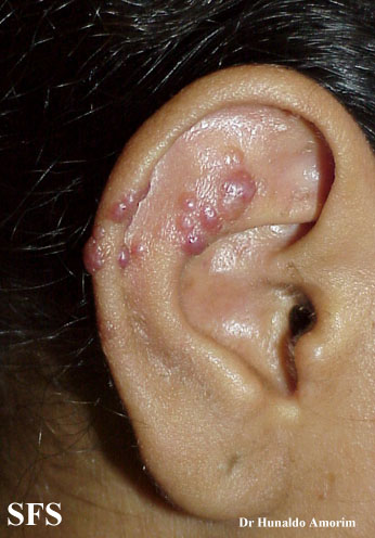
Epithelioid hemangioma. Adapted from Dermatology Atlas.<ref name="Dermatology Atlas">{{Cite
-
Epithelioid hemangioma. Adapted from Dermatology Atlas.<ref name="Dermatology Atlas">{{Cite
-
Epithelioid hemangioma. Adapted from Dermatology Atlas.<ref name="Dermatology Atlas">{{Cite
-
Epithelioid hemangioma. Adapted from Dermatology Atlas.<ref name="Dermatology Atlas">{{Cite
-
Epithelioid hemangioma. Adapted from Dermatology Atlas.<ref name="Dermatology Atlas">{{Cite
-
Epithelioid hemangioma. Adapted from Dermatology Atlas.<ref name="Dermatology Atlas">{{Cite
-
Epithelioid hemangioma. Adapted from Dermatology Atlas.<ref name="Dermatology Atlas">{{Cite
-
Epithelioid hemangioma. Adapted from Dermatology Atlas.<ref name="Dermatology Atlas">{{Cite
-
Epithelioid hemangioma. Adapted from Dermatology Atlas.<ref name="Dermatology Atlas">{{Cite
-
Epithelioid hemangioma. Adapted from Dermatology Atlas.<ref name="Dermatology Atlas">{{Cite


Laboratory Findings
- A eosinophilia in complete blood count is characteristic of angiolymphoid hyperplasia with eosinophilia.
- Eosinophils in urinalysis is also a feature.
Other Diagnostic Studies
- Angiolymphoid hyperplasia with eosinophilia may also be diagnosed using biopsy.
- Findings on biopsy include proliferation of small blood vessels with endothelial cells having a cobblestone appearance, a perivascular and interstitial infiltrate of lymphocytes and eosinophils.
Treatment
Medical Therapy
- The mainstay of therapy for angiolymphoid hyperplasia with eosinophilia is intralesional corticoid therapy and imiquimod.
- Imiquimod acts by induction of the production of interferon-α and induction or inhibition of certain cytokines, mainly interleukin-5.[5]
Surgery
- Surgery is the mainstay of therapy for angiolymphoid hyperplasia with eosinophilia.
- Mohs micrographic surgery with complete margin examination is performed.
- Other treatment options include radiotherapy, curettage, shave excision with electrodessication, cryotherapy, corticosteroids (topical, systemic or intralesional preparation), and laser therapy using continuous wave carbon dioxide and argon lasers.[6]
Prevention
- There are no primary preventive measures available for angiolymphoid hyperplasia with eosinophilia.
References
- ↑ 1.0 1.1 Wells GC, Whimster IW (1969). "Subcutaneous angiolymphoid hyperplasia with eosinophilia". Br J Dermatol. 81 (1): 1–14. PMID 5763634.
- ↑ Rapini, Ronald P.; Bolognia, Jean L.; Jorizzo, Joseph L. (2007). Dermatology: 2-Volume Set. St. Louis: Mosby. ISBN 1-4160-2999-0.
- ↑ 3.0 3.1 James, William; Berger, Timothy; Elston, Dirk (2005). Andrews' Diseases of the Skin: Clinical Dermatology. (10th ed.). Saunders. ISBN 0-7216-2921-0.
- ↑ Rapini, Ronald P.; Bolognia, Jean L.; Jorizzo, Joseph L. (2007). Dermatology: 2-Volume Set. St. Louis: Mosby. ISBN 1-4160-2999-0.
- ↑ Esteves, Paola; Barbalho, Marcella; Lima, Tiago; Quintella, Leonardo; Niemeyer-Corbellini, Jo�o Paulo; Ramos-e-Silva, Marcia (2015). "Angiolymphoid Hyperplasia with Eosinophilia: A Case Report". Case Reports in Dermatology. 7 (2): 113–116. doi:10.1159/000381843. ISSN 1662-6567. replacement character in
|first5=at position 3 (help) - ↑ Lembo S, Balato A, Cirillo T, Balato N (2011). "A Long-Term Follow-Up of Angiolymphoid Hyperplasia with Eosinophilia Treated by Corticosteroids: When a Traditional Therapy is Still Up-to-Date". Case Rep Dermatol. 3 (1): 64–7. doi:10.1159/000323182. PMC 3073756. PMID 21487464.