Alcoholic liver disease CT: Difference between revisions
Jump to navigation
Jump to search
No edit summary |
m (Bot: Removing from Primary care) |
||
| (8 intermediate revisions by 3 users not shown) | |||
| Line 1: | Line 1: | ||
__NOTOC__ | __NOTOC__ | ||
{{Alcoholic liver disease}} | {{Alcoholic liver disease}} | ||
{{CMG}}; {{AE}} {{MKA}} | |||
==Overview== | ==Overview== | ||
==CT== | |||
* | Abdominal [[CT scan]] may be helpful in the diagnosis of alcoholic liver disease. Findings on [[Computed tomography|CT scan]] suggestive of [[hepatic steatosis]] and [[cirrhosis]] may be seen. | ||
* | |||
* | ==CT scan== | ||
*Findings on [[Computed tomography|CT scan]] suggestive of alcoholic liver disease include: | |||
**Non-contrast [[CT scan]] for detecting [[hepatic steatosis]]:<ref name="pmid4058482">{{cite journal |vauthors=Soliman R, Saad MA, Refai M |title=Studies on histoplasmosis farciminosii (epizootic lymphangitis) in Egypt. III. Application of a skin test ('Histofarcin') in the diagnosis of epizootic lymphangitis in horses |journal=Mykosen |volume=28 |issue=9 |pages=457–61 |year=1985 |pmid=4058482 |doi= |url=}}</ref><ref name="pmid6934563">{{cite journal |vauthors=Piekarski J, Goldberg HI, Royal SA, Axel L, Moss AA |title=Difference between liver and spleen CT numbers in the normal adult: its usefulness in predicting the presence of diffuse liver disease |journal=Radiology |volume=137 |issue=3 |pages=727–9 |year=1980 |pmid=6934563 |doi=10.1148/radiology.137.3.6934563 |url=}}</ref> | |||
***[[Macroscopic]] [[fat]] in the [[liver]] | |||
***[[Liver]] to [[spleen]] [[attenuation]] ratio greater than ten [[hounsfield units]] indicates [[hepatic steatosis]] | |||
**[[Cirrhosis]]:<ref name="pmid7718279">{{cite journal |vauthors=Rofsky NM, Fleishaker H |title=CT and MRI of diffuse liver disease |journal=Semin. Ultrasound CT MR |volume=16 |issue=1 |pages=16–33 |year=1995 |pmid=7718279 |doi= |url=}}</ref> | |||
***[[Atrophy]] of the right lobe of the [[liver]] | |||
***[[Hypertrophy (medical)|Hypertrophy]] of the [[Caudate lobe of liver|caudate lobe of the liver]] | |||
***[[Hypertrophy (medical)|Hypertrophy]] of the lateral segment of the left [[Lobe (anatomy)|lobe]] | |||
***[[Parenchymal]] nodularity | |||
***[[Attenuation]] of [[hepatic]] [[vasculature]] | |||
***[[Splenomegaly]] | |||
***[[Venous]] collaterals | |||
***[[Ascites]] | |||
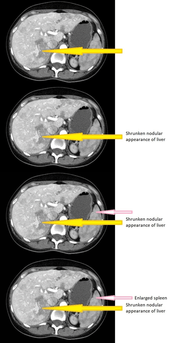
*CT of a [[Cirrhosis|cirrhotic]] patient shows a [[liver]] with a shrunken, [[Nodule (medicine)|nodular]] appearance. | |||
[[File:Output qzgZxt.gif|500px|center|thumb|Liver Cirrhosis <br> Source: Wikimedia commons <ref name="urlFile:Morbus-Osler-CT-Leber-ax-012.jpg - Wikimedia Commons">{{cite web |url=https://commons.wikimedia.org/wiki/File:Morbus-Osler-CT-Leber-ax-012.jpg |title=File:Morbus-Osler-CT-Leber-ax-012.jpg - Wikimedia Commons |format= |work= |accessdate=}}</ref>]] | |||
==References== | ==References== | ||
{{reflist|2}} | {{reflist|2}} | ||
| |||
{{WS}} | |||
{{WH}} | {{WH}} | ||
[[Category: | [[Category:Surgery]] | ||
[[Category:Gastroenterology]] | [[Category:Gastroenterology]] | ||
[[Category: | [[Category:Up-To-Date]] | ||
[[Category:Hepatology]] | [[Category:Hepatology]] | ||
[[Category:Medicine]] | |||
[[Category:Radiology]] | |||
Latest revision as of 20:19, 29 July 2020
|
Alcoholic liver disease Microchapters |
|
Diagnosis |
|---|
|
Treatment |
|
Case Studies |
|
Alcoholic liver disease CT On the Web |
|
American Roentgen Ray Society Images of Alcoholic liver disease CT |
|
Risk calculators and risk factors for Alcoholic liver disease CT |
Editor-In-Chief: C. Michael Gibson, M.S., M.D. [1]; Associate Editor(s)-in-Chief: M. Khurram Afzal, MD [2]
Overview
Abdominal CT scan may be helpful in the diagnosis of alcoholic liver disease. Findings on CT scan suggestive of hepatic steatosis and cirrhosis may be seen.
CT scan
- Findings on CT scan suggestive of alcoholic liver disease include:
- Non-contrast CT scan for detecting hepatic steatosis:[1][2]
- Macroscopic fat in the liver
- Liver to spleen attenuation ratio greater than ten hounsfield units indicates hepatic steatosis
- Cirrhosis:[3]
- Atrophy of the right lobe of the liver
- Hypertrophy of the caudate lobe of the liver
- Hypertrophy of the lateral segment of the left lobe
- Parenchymal nodularity
- Attenuation of hepatic vasculature
- Splenomegaly
- Venous collaterals
- Ascites
- Non-contrast CT scan for detecting hepatic steatosis:[1][2]
- CT of a cirrhotic patient shows a liver with a shrunken, nodular appearance.

Source: Wikimedia commons [4]
References
- ↑ Soliman R, Saad MA, Refai M (1985). "Studies on histoplasmosis farciminosii (epizootic lymphangitis) in Egypt. III. Application of a skin test ('Histofarcin') in the diagnosis of epizootic lymphangitis in horses". Mykosen. 28 (9): 457–61. PMID 4058482.
- ↑ Piekarski J, Goldberg HI, Royal SA, Axel L, Moss AA (1980). "Difference between liver and spleen CT numbers in the normal adult: its usefulness in predicting the presence of diffuse liver disease". Radiology. 137 (3): 727–9. doi:10.1148/radiology.137.3.6934563. PMID 6934563.
- ↑ Rofsky NM, Fleishaker H (1995). "CT and MRI of diffuse liver disease". Semin. Ultrasound CT MR. 16 (1): 16–33. PMID 7718279.
- ↑ "File:Morbus-Osler-CT-Leber-ax-012.jpg - Wikimedia Commons". External link in
|title=(help)