African trypanosomiasis laboratory findings: Difference between revisions
Aditya Ganti (talk | contribs) |
m (Bot: Removing from Primary care) |
||
| (19 intermediate revisions by 8 users not shown) | |||
| Line 1: | Line 1: | ||
__NOTOC__ | __NOTOC__ | ||
{{African trypanosomiasis}} | {{African trypanosomiasis}} | ||
{{CMG}}; {{AOEIC}} Pilar Almonacid | {{CMG}}; {{AOEIC}} Pilar Almonacid, {{JH}} | ||
==Overview== | ==Overview== | ||
The diagnosis of African trypanosomiasis rests | The diagnosis of [[African trypanosomiasis]] rests on demonstrating [[trypanosomes]] by [[microscopic examination]] of [[chancre]] fluid, [[lymph node]] [[Aspirate|aspirates]], [[blood|blood]], [[bone marrow|bone marrow]], and [[cerebrospinal fluid]] in the late stages of [[infection]]. | ||
==Laboratory Findings== | ==Laboratory Findings== | ||
The diagnosis of African trypanosomiasis rests | The diagnosis of [[African trypanosomiasis]] rests on demonstrating [[trypanosomes]] by [[microscopic examination]] of [[chancre]] fluid, [[lymph node]] aspirates, [[blood]], [[bone marrow]] or, in the late stages of [[infection]], in [[cerebrospinal fluid]]. | ||
===Blood smear=== | ===Blood smear=== | ||
*Acute | *Acute disease is often diagnosed by visual detection of the ''[[Trypanosoma brucei rhodesiense]] '' [[Parasites|parasite]] on [[peripheral blood smear]]. | ||
*Peripheral blood smears are usually stained with Giemsa stain for adequate visualization of the parasite. | *[[Peripheral blood smear|Peripheral blood smears]] are usually stained with [[Giemsa stain]] for adequate visualization of the [[Parasites|parasite]]. | ||
{| class="wikitable" | {| class="wikitable" | ||
!Microscopy | !Microscopy | ||
!Findings | !Findings | ||
|- | |- | ||
|[[Image:African trypanosomiasis.jpg|left|African trypanosomiasis]] | |[[Image:African trypanosomiasis.jpg|left|African trypanosomiasis]] | ||
| | | | ||
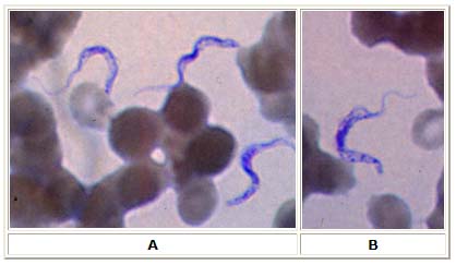
* Thin blood smear stained with Giemsa. | * Thin blood smear stained with [[Giemsa stain|Giemsa]]. | ||
* Typical trypomastigote stages (the only stages found in patients), with a posterior kinetoplast, a centrally located nucleus, an undulating membrane, and an anterior flagellum. | * Typical trypomastigote stages (the only stages found in patients), with a posterior [[kinetoplast]], a centrally located [[Cell nucleus|nucleus]], an undulating membrane, and an anterior [[flagellum]]. | ||
* The two | * The two ''[[Trypanosoma brucei]]'' [[species]] that cause human trypanosomiasis, ''[[Trypanosoma brucei gambiense]]'' and ''[[Trypanosoma brucei rhodesiense]]'', are indistinguishable [[Morphology|morphologically]]. | ||
* The | * The [[Trypanosome|trypanosome's]] length ranges from 14 to 33 µm. | ||
|- | |- | ||
|[[Image:African trypanosomiasis 5.jpg|left|African trypanosomiasis 5]] | |||
| | | | ||
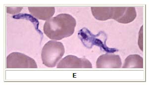
* Parasite exhibiting division (on right). | |||
|} | |} | ||
===Electrolyte and | ===Electrolyte and biomarker studies=== | ||
*Three | *[[Serology]] is not usually helpful in acute [[disease]]. | ||
*wb-CATT | *Detection of anti-trypanosomal [[Immunoglobulin G|IgG antibodies]] is helpful in detection of African trypanosomiasis [[Infection|infections]]. | ||
* | *Three [[Serological testing|serologica]]<nowiki/>l tests are available for detection of the [[Parasites|parasite]]: | ||
**Micro-CATT (uses dried blood) | |||
**wb-CATT (uses whole blood) | |||
**wb-LATEX (uses whole blood) | |||
*wb-CATT is the most efficient test for diagnosis, while wb-LATEX is a better exam for situations in which greater [[sensitivity]] is required.<ref>{{cite journal |author=Truc P, Lejon V, Magnus E, ''et al.'' |title=Evaluation of the micro-CATT, CATT/Trypanosoma brucei gambiense, and LATEX/T b gambiense methods for serodiagnosis and surveillance of human African trypanosomiasis in West and Central Africa |journal=Bull. World Health Organ. |volume=80 |issue=11 |pages=882–6 |year=2002 |pmid=12481210 |pmc=2567684 |doi= |url=}}</ref> | |||
*Detection of [[antibodies]] among [[infants]] may be difficult due to the presence of [[maternal]] [[antibodies]] early following birth. Accordingly, [[Serology|serologic]] testing for [[infants]] is only recommended at least 9 months after birth. | |||
==Gallery== | ==Gallery== | ||
| Line 86: | Line 60: | ||
==References== | ==References== | ||
{{reflist|2}} | {{reflist|2}} | ||
{{WH}} | |||
{{WS}} | |||
[[Category:Disease]] | [[Category:Disease]] | ||
[[Category:Up-To-Date]] | |||
[[Category:Dermatology]] | |||
[[Category:Neurology]] | [[Category:Neurology]] | ||
[[Category:Emergency medicine]] | |||
[[Category:Infectious disease]] | [[Category:Infectious disease]] | ||
Latest revision as of 20:19, 29 July 2020
|
African trypanosomiasis Microchapters |
|
Diagnosis |
|---|
|
Treatment |
|
Case Studies |
|
African trypanosomiasis laboratory findings On the Web |
|
American Roentgen Ray Society Images of African trypanosomiasis laboratory findings |
|
Risk calculators and risk factors for African trypanosomiasis laboratory findings |
Editor-In-Chief: C. Michael Gibson, M.S., M.D. [1]; Associate Editor(s)-In-Chief: Pilar Almonacid, Jesus Rosario Hernandez, M.D. [2]
Overview
The diagnosis of African trypanosomiasis rests on demonstrating trypanosomes by microscopic examination of chancre fluid, lymph node aspirates, blood, bone marrow, and cerebrospinal fluid in the late stages of infection.
Laboratory Findings
The diagnosis of African trypanosomiasis rests on demonstrating trypanosomes by microscopic examination of chancre fluid, lymph node aspirates, blood, bone marrow or, in the late stages of infection, in cerebrospinal fluid.
Blood smear
- Acute disease is often diagnosed by visual detection of the Trypanosoma brucei rhodesiense parasite on peripheral blood smear.
- Peripheral blood smears are usually stained with Giemsa stain for adequate visualization of the parasite.
| Microscopy | Findings |
|---|---|
 |
|
 |
|
Electrolyte and biomarker studies
- Serology is not usually helpful in acute disease.
- Detection of anti-trypanosomal IgG antibodies is helpful in detection of African trypanosomiasis infections.
- Three serological tests are available for detection of the parasite:
- Micro-CATT (uses dried blood)
- wb-CATT (uses whole blood)
- wb-LATEX (uses whole blood)
- wb-CATT is the most efficient test for diagnosis, while wb-LATEX is a better exam for situations in which greater sensitivity is required.[1]
- Detection of antibodies among infants may be difficult due to the presence of maternal antibodies early following birth. Accordingly, serologic testing for infants is only recommended at least 9 months after birth.
Gallery
-
African trypanosomiasis. Adapted from Public Health Image Library (PHIL). [2]
-
African trypanosomiasis. Adapted from Public Health Image Library (PHIL). [2]
-
African trypanosomiasis. Adapted from Public Health Image Library (PHIL). [2]
-
African trypanosomiasis. Adapted from Public Health Image Library (PHIL). [2]
-
African trypanosomiasis. Adapted from Public Health Image Library (PHIL). [2]
-
African trypanosomiasis. Adapted from Public Health Image Library (PHIL). [2]
-
African trypanosomiasis. Adapted from Public Health Image Library (PHIL). [2]
-
African trypanosomiasis. Adapted from Public Health Image Library (PHIL). [2]
References
- ↑ Truc P, Lejon V, Magnus E; et al. (2002). "Evaluation of the micro-CATT, CATT/Trypanosoma brucei gambiense, and LATEX/T b gambiense methods for serodiagnosis and surveillance of human African trypanosomiasis in West and Central Africa". Bull. World Health Organ. 80 (11): 882–6. PMC 2567684. PMID 12481210.
- ↑ 2.0 2.1 2.2 2.3 2.4 2.5 2.6 2.7 "Public Health Image Library (PHIL)".
![African trypanosomiasis. Adapted from Public Health Image Library (PHIL). [2]](/images/f/f9/African_trypanosomiasis01.jpg)
![African trypanosomiasis. Adapted from Public Health Image Library (PHIL). [2]](/images/1/1c/African_trypanosomiasis02.jpeg)
![African trypanosomiasis. Adapted from Public Health Image Library (PHIL). [2]](/images/5/5f/African_trypanosomiasis03.jpeg)
![African trypanosomiasis. Adapted from Public Health Image Library (PHIL). [2]](/images/3/33/African_trypanosomiasis05.jpeg)
![African trypanosomiasis. Adapted from Public Health Image Library (PHIL). [2]](/images/f/f8/African_trypanosomiasis06.jpeg)
![African trypanosomiasis. Adapted from Public Health Image Library (PHIL). [2]](/images/c/c9/African_trypanosomiasis07.jpeg)
![African trypanosomiasis. Adapted from Public Health Image Library (PHIL). [2]](/images/6/6d/African_trypanosomiasis08.jpeg)
![African trypanosomiasis. Adapted from Public Health Image Library (PHIL). [2]](/images/2/29/African_trypanosomiasis09.jpeg)