Acute coronary syndrome resident survival guide
Patient Presentation
Common presentation
- Substernal / precordial chest pressure / heaviness / pain
- Pain radiation to shoulder or arm / neck / jaw
- Nausea and/or vomiting
- Shortness of breath
- Diaphoresis
- Heartburn/ burning sensation in chest
- Dizziness
- Palpitations
- Near syncope / Syncope
Initial Evaluation and Orders
- Ensure patency of airway, check for adequate breathing and circulation.
- Vital signs (Pulse rate, blood pressure, respiratory rate, temperature, and oxygen saturation)
- 12-lead EKG (compare with old EKG if possible)
- Cardiac enzymes (three sets of troponin, CK, CK-MB at six hour intervals; first set may be normal, but order all three sets)
- Chest x-ray
- Oxygen (titrate for oxygen saturation levels >92%)
- IV access
- 325mg non-enteric coated aspirin by mouth (or per rectum if patient cannot take orally)
- If patient is not hypotensive and inferior myocardial infarction has been ruled out by EKG, give 0.4mg nitroglycerin sublingually up to three times, at 5 minute intervals, until chest pain improves.
- If pulmonary embolism is suspected D-dimers should be obtained.
| Important! |
|---|
| Follow up with all pending tests and lab results as soon as these become available. For information on evaluating the results go to the apppropriate section on this page. |
History and Symptoms
History of Present Illness:
- Chest pain history; ask about onset, duration, nature, intensity, location, progression, radiation (to arm, neck, jaw= acute coronary syndrome, or back=aortic dissection), aggravating (pleurtic and pericarditis chest pain worsens with respiration) and relieving factors (relieved by nitrates), constant or intermittent.
- Ask if pain is associated with (head to toe). Headache, confusion, fever, photophobia, vision changes, bleeding, nausea, vomiting, apetite, weight loss, shortness of breath, palpitations, cough, sputum, abdominal pain, bowel symptoms, urinary symptoms.
Physical Examination
- General
- Check for alertness, and orientation with time, place, and person
- Patient leaning forward can point towards pericarditis
- Cardiovascular:
- Pulse Rate (rate, rhythm. volume, quality, symmetry, all 4 limbs. Aortic dissection- Diminution or absence of pulses)
- Blood pressure (check for symmetry in all the limbs)
- Auscultate carotid artery (check for bruit)
- Jugular venous distension, check for hepatojugular reflex
- Inspection: Check for displacement of the apex
- Palpation: Confirm the findings of inspection (cardiac apex), musculo-skeletal tenderness, crepitus (esophageal rupture,subcutaneous emphysema), feel for any thrill (possible regurgitation), heave (right ventricular hypertrophy)
- Auscultation:
- Heart sounds (muffled in cardiac tamponade, Pericardial effusion), S3 and S4 (Heart failure)
- Murmur (commonly regurgitation murmur)
- Pericardial rub - (Pericarditis, commonly tricuspid area sounds like scratching), and gallop
Differential Diagnosis
EKG findings
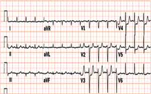
Electrocardiogram in Unstable angina / NSTEMI
The resting electrocardiogram in the patient with unstable angina / non ST elevation MI may show any of the following:
- No changes
- Non specific ST / T wave changes
- Flipped or inverted T waves
- ST Depression as shown below. ST depression carries the poorest prognosis. Greater magnitudes of downsloping ST depression are associated with a poorer prognosis.

Electrocardiogram in STEMI
The electrocardiographic definition of ST elevation MI requires the following: at least 1 mm (0.1 mV) of ST segment elevation in 2 or more anatomically contiguous leads.[1] While these criteria are sensitive, they are not specific as thromboctic coronary occlusion is not the most common cause of ST segment elevation in chest pain patients.[2]
Chest X ray Findings
- Aortic dissection: Suspect aortic dissection if findings include increased aortic diameter, widened mediastinum, and/or pleural effusion (hemothorax) in the absence of CHF
- Pulmonary embolism: May not appreciate any abnormalities on chest x ray.
- Tension pneumothorax: No pulmonary vessels are visible beyond the visceral pleural line (the collection of pleural gas is avascular). Tracheal deviation away from the collapsed area of lung will be seen.
2. Pulmonary Embolism
- Next study to do:
- Spiral CT scanning is now a standard modality to non-invasively diagnose PE.
- Initial studies reported sensitivities for diagnosing emboli to the segmental level (4th order branch) as high as 98%
3. Tension Pneumothorax
- Supportive signs and symptoms include"
- Sudden shortness of breath, cyanosis (turning blue) and pain felt in the chest and/or back are the main symptoms.
- In penetrating chest wounds, the sound of air flowing through the puncture hole may indicate pneumothorax, hence the term "sucking" chest wound.
- The flopping sound of the punctured lung is also occasionally heard.
- Spontaneous pneumothoraces are reported in young people with a tall stature. As men are generally taller than women, there is a preponderance among males.
- Pneumothorax can also occur as part of medical procedures, such as the insertion of a central venous catheter (an intravenous catheter) in the subclavian vein or jugular vein. While rare, it is considered a serious complication and needs immediate treatment. Other causes include mechanical ventilation, emphysema and rarely other lung diseases (pneumonia).
4. Esophageal Rupture
- Supportive signs and symptoms include:
- The classic Meckler's triad of symptoms includes vomiting, lower chest pain, and cervical subcutaneous emphysema following overindulgence in food or alcohol, but is observed in only half of the cases.
- The most common chest radiograph findings in spontaneous esophageal rupture (SER) are pleural effusion (91%) and pneumothorax (80%).
- The initial sign on a plain film may be pneumomediastinum or subcutaneous emphysema.
- Up to 12% of patients with SER may have a normal chest radiograph.
- Next study to do:
- Contrast-enhanced esophageal radiography is diagnostic in 75% to 85% of cases.