Acral lentiginous melanoma: Difference between revisions
m (Bot: Automated text replacement (-{{SIB}} + & -{{EH}} + & -{{EJ}} + & -{{Editor Help}} + & -{{Editor Join}} +)) |
No edit summary |
||
| Line 24: | Line 24: | ||
* [[desmoplasia]] | * [[desmoplasia]] | ||
* [[hyperplastic]] [[Epidermis (skin)|epidermis]] | * [[hyperplastic]] [[Epidermis (skin)|epidermis]] | ||
==Diagnosis== | |||
===Physical Examination=== | |||
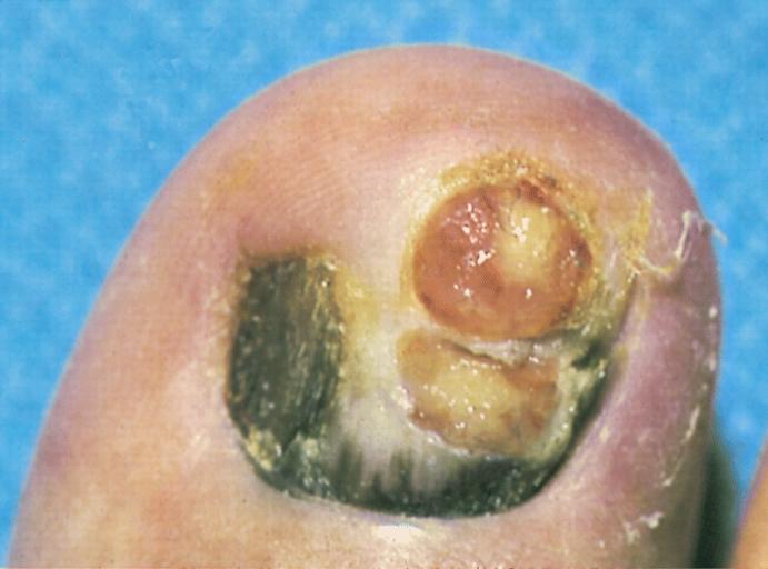
An example of a subungual melanoma is shown in the image below. | |||
[http://www.peir.net Image shown below is courtesy of Professor Peter Anderson DVM PhD and published with permission © PEIR, University of Alabama at Birmingham, Department of Pathology] | |||
<div align="left"> | |||
<gallery heights="175" widths="175"> | |||
image:Subungual melanoma.jpg|This is a subungual radial growth phase of acral-lentiginous type with two amelanotic vertical growth phase tumor nodules. Radial growth phase is just beginning to grow onto the cuticle of the nail, displaying a subtle example of "Hutchinson's sign." | |||
</gallery> | |||
</div> | |||
==External links== | ==External links== | ||
Revision as of 15:38, 15 August 2012
Template:DiseaseDisorder infobox
Overview
Acral lentiginous melanoma is a kind of skin melanoma. It is also known as subungual melanoma. It is seen on the palms, soles and under the nails. This is the most common form of melanoma in Asians and Blacks. The average age at diagnosis is between sixty and seventy years. It also occurs in Caucasians and in young people. This type of melanoma occurs on non hair baring surfaces of the body which may or may not be exposed to sunlight.
Typical symptoms include:
- longitudinal tan, black, or brown streak on a finger or toe nail (melanonychia striata)
- pigmentation of proximal nail fold
- areas of dark pigmentation on palms of hands or soles of feet
Any new area of pigmentation or an existing one that shows change should be checked by a dermatologist. If caught early this type of melanoma has a similar cure rate as the other types of superficial spreading melanoma.
The microscopic hallmarks are:
- atypical melanocytes in junctional nests
- dermal invasion
- desmoplasia
- hyperplastic epidermis
Diagnosis
Physical Examination
An example of a subungual melanoma is shown in the image below.
-
This is a subungual radial growth phase of acral-lentiginous type with two amelanotic vertical growth phase tumor nodules. Radial growth phase is just beginning to grow onto the cuticle of the nail, displaying a subtle example of "Hutchinson's sign."

External links