Acanthocytosis laboratory findings
|
Acanthocytosis Microchapters |
|
Diagnosis |
|---|
|
Treatment |
|
Case Studies |
|
Acanthocytosis laboratory findings On the Web |
|
American Roentgen Ray Society Images of Acanthocytosis laboratory findings |
|
Risk calculators and risk factors for Acanthocytosis laboratory findings |
Editor-In-Chief: C. Michael Gibson, M.S., M.D. [1]
Please help WikiDoc by adding more content here. It's easy! Click here to learn about editing.
Laboratory Findings
In human biology and medicine, the term refers to pathological red blood cells, which are coarse and irregularly crenelated resembling many-pointed stars. They are seen on blood films in, among others, lipid abnormalities, liver disease, chorea acanthocytosis, McLeod syndrome and several inherited neurological disorders, such as neuroacanthocytosis.
-
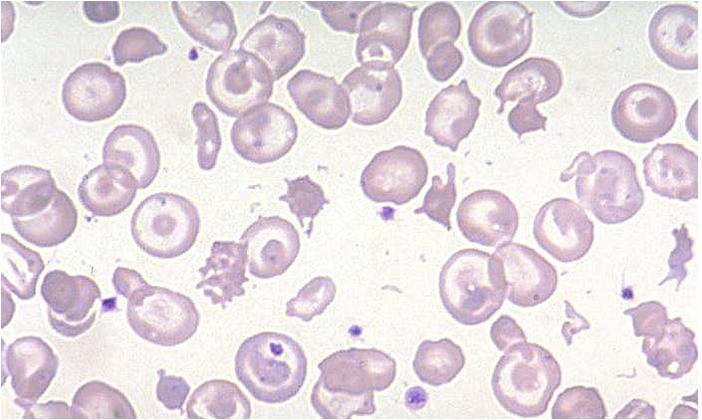
Acanthocytes
-
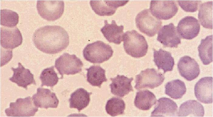
Acanthocytes

