WBR0586
| Author | [[PageAuthor::Rim Halaby, M.D. [1]]] |
|---|---|
| Exam Type | ExamType::USMLE Step 1 |
| Main Category | MainCategory::Pathophysiology |
| Sub Category | SubCategory::Head and Neck, SubCategory::Neurology |
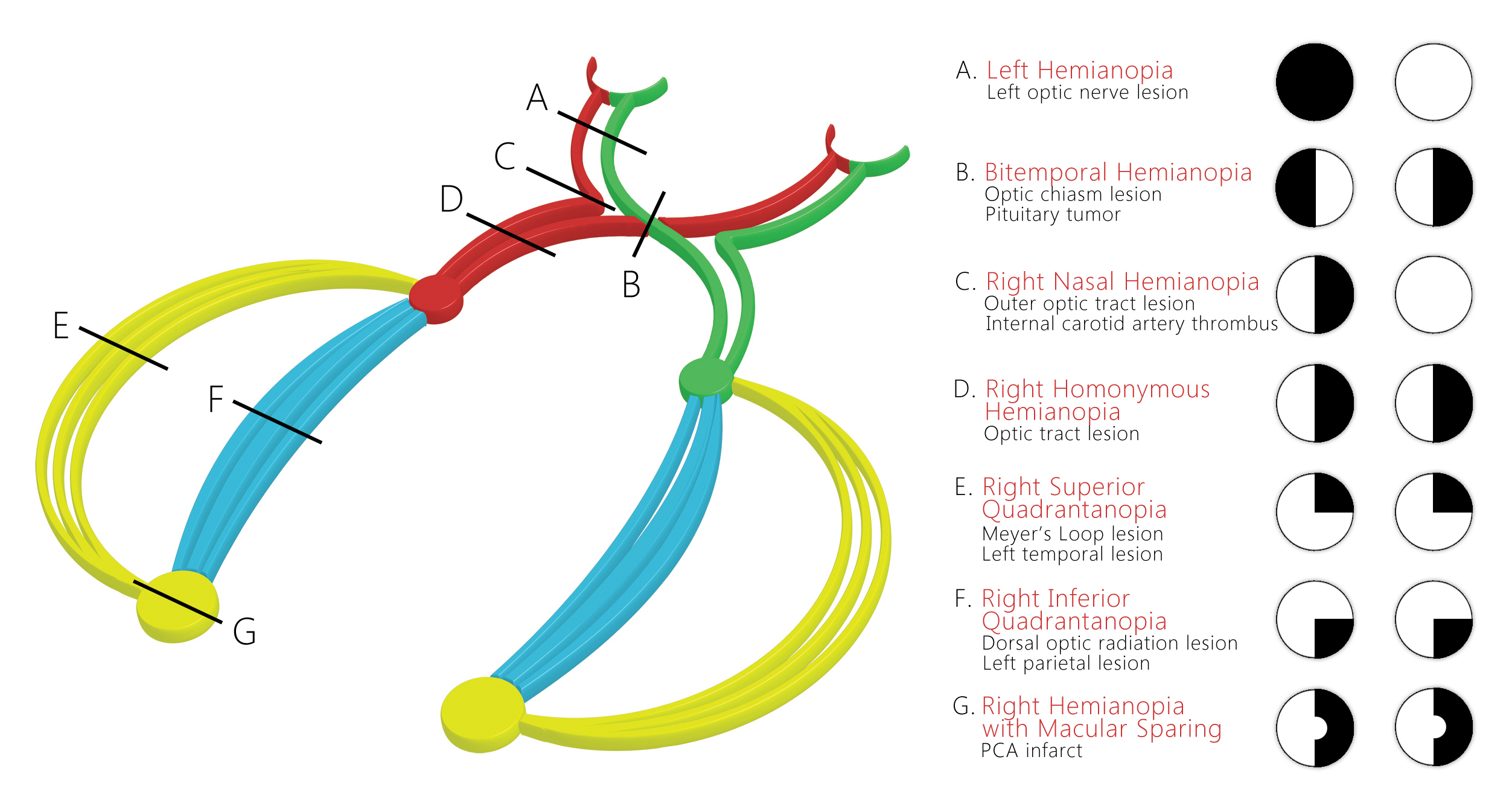
| Prompt | [[Prompt::A 45 year old woman with past medical history significant for resected breast cancer presents to the emergency department after suffering a tonic-clonic seizure lasting 3 minutes. She explains that she was walking her dog and then found herself on the ground with several people surrounding her, not knowing what had happened. The patient explains that she had been recovering well after her last chemotherapy cycle, and had no complaints except an annoying area of visual field disturbance she noticed recently. On physical exam, you notice the pattern of visual loss shown below. You order a brain MRI that shows a intracranial lesion with high suspicion for metastasis. Where is the most likely location of the lesion given the patient's symptoms? |
| Answer A | AnswerA:: |
| Answer A Explanation | [[AnswerAExp::Homonymous hemianopia involves loss of vision on one side. It usually occurs due to a lesion to the optic tracts or a PCA stroke although the latter usually has associated macular sparing. This lesion is not seen in patients with carotid artery aneurysms.]] |
| Answer B | [[AnswerB:: ]] ]]
|
| Answer B Explanation | [[AnswerBExp::Right upper quadrantopia is characterized by loss of vision in the right upper quadrant of the visual field. It usually occurs with left temporal lesions due to the interruption of the left Meyer's loop. This pattern is unusual with carotid artery aneurysms.]] |
| Answer C | [[AnswerC:: ]] ]]
|
| Answer C Explanation | [[AnswerCExp::Left nasal hemianopia usually occurs with lesions of the internal carotid artery (internal carotid thrombosis or anyrysms) at the origin of the ophthalmic artery. The lesion is usually located laterally to the optic chiasm interrupting part of the optic nerve as it becomes the optic tract. Our patient would best fit this pattern of visual field loss.]] |
| Answer D | [[AnswerD:: ]] ]]
|
| Answer D Explanation | AnswerDExp::This lesion portrays bitemporal hemianopia usually seen in large prolactinomas that compress the optic chiasm. It is unusual in cases with internal carotid artery lesions. |
| Answer E | [[AnswerE:: ]] ]]
|
| Answer E Explanation | AnswerEExp::Right lower quadrantopia is characterized by loss of vision in the right lower quadrant of the visual field. It usually occurs with left parietal lesions due to the interruption of the left dorsal optic radiations. |
| Right Answer | RightAnswer::C |
| Explanation | [[Explanation::
Jay WM. Visual field defects. Am Fam Physician. 1981;24(2):138-42. |
| Approved | Approved::No |
| Keyword | WBRKeyword::Upper quadrantopia, WBRKeyword::Temporal lesions, WBRKeyword::Visual field defects |
| Linked Question | Linked:: |
| Order in Linked Questions | LinkedOrder:: |