Enterobiasis laboratory findings
Template:Pinworm Editor-In-Chief: C. Michael Gibson, M.S., M.D. [1]
Overview
Diagnosis is often made clinically by observing the female worm (or many worms) in the peri-anal region, but can also be made using the "scotch-tape" test, in which the sticky side of a strip of cellophane tape is pressed against the peri-anal skin, then examined under a microscope for pinworm eggs.
Laboratory Findings
Scotch Tape Test
Sticky side of a strip of cellophane tape is pressed against the peri-anal skin, then examined under a microscope for pinworm eggs. The actual worms may be seen in the host's feces; however the eggs are invisible to the naked eye. The diagnostic characteristics of egg are: size 50-60 µm by 20-32 µm; typical elongated shape, with one convex side and one flattened side and colorless shell.[1] On histologic cross-section, alae or wings (running the length of the worm) are identifying features of the pinworm (see micrograph).[2]
-
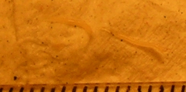
Two pinworms, captured on emergence from the anus. Markings are 1 mm apart.
-
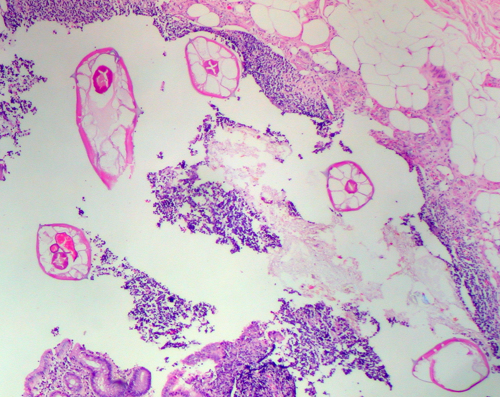
Pinworms are sometimes diagnosed incidentally by pathology. Micrograph of pinworms in the appendix. H&E stain.
-
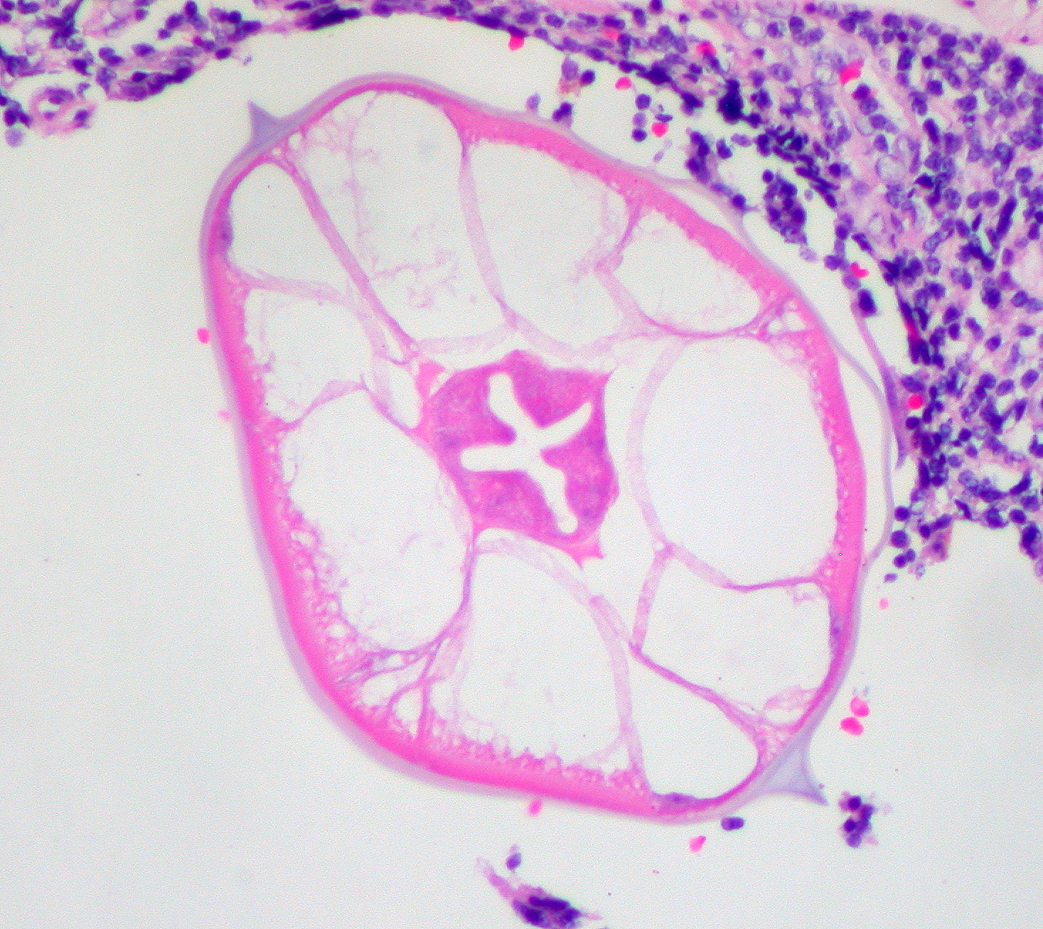
High magnification micrograph of a pinworm in cross-section in the appendix. H&E stain.
-
High magnification micrograph of a pinworm in cross-section in the appendix. H&E stain.
-
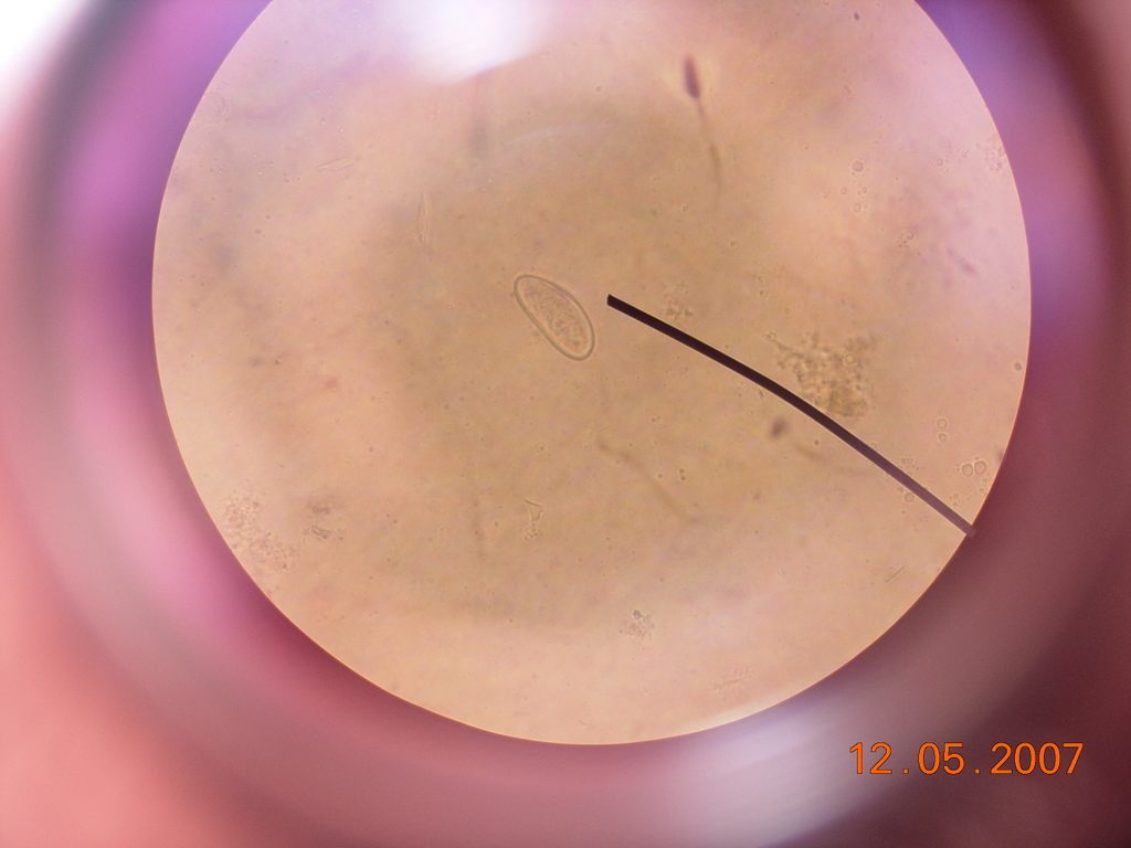
Enterobius vermicularis egg under a light microscope.
-
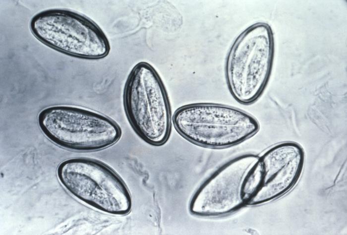
Pinworm eggs are easily seen under a microscope.
-
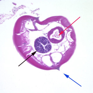
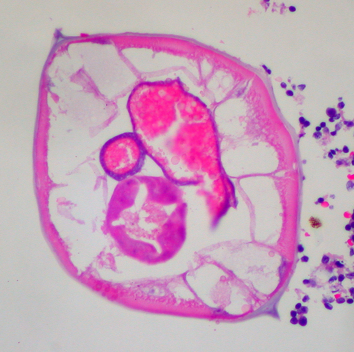
Pinworms are sometimes diagnosed incidentally by pathology. Micrograph of male pinworm in cross-section. Alae (blue arrow), intestine (red arrow) and testis (black arrow). H&E stain.
-
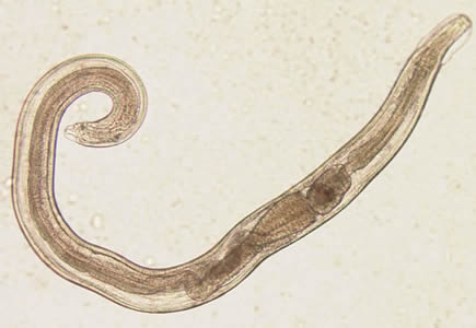
A pinworm (Enterobius vermicularis).

.jpg)
.jpg)
.jpg)


References
- ↑ "PARASITOLOGY - CHAPTER FOUR NEMATODES (Round Worms)". The Board of Trustees of the University of South Carolina. Retrieved 2007-10-18.
- ↑ Diagnostic Findings Enterobiasis. Centers for Disease Control and Prevention. URL:http://www.dpd.cdc.gov/dpdx/HTML/Enterobiasis.htm. Accessed on: August 6, 2008.