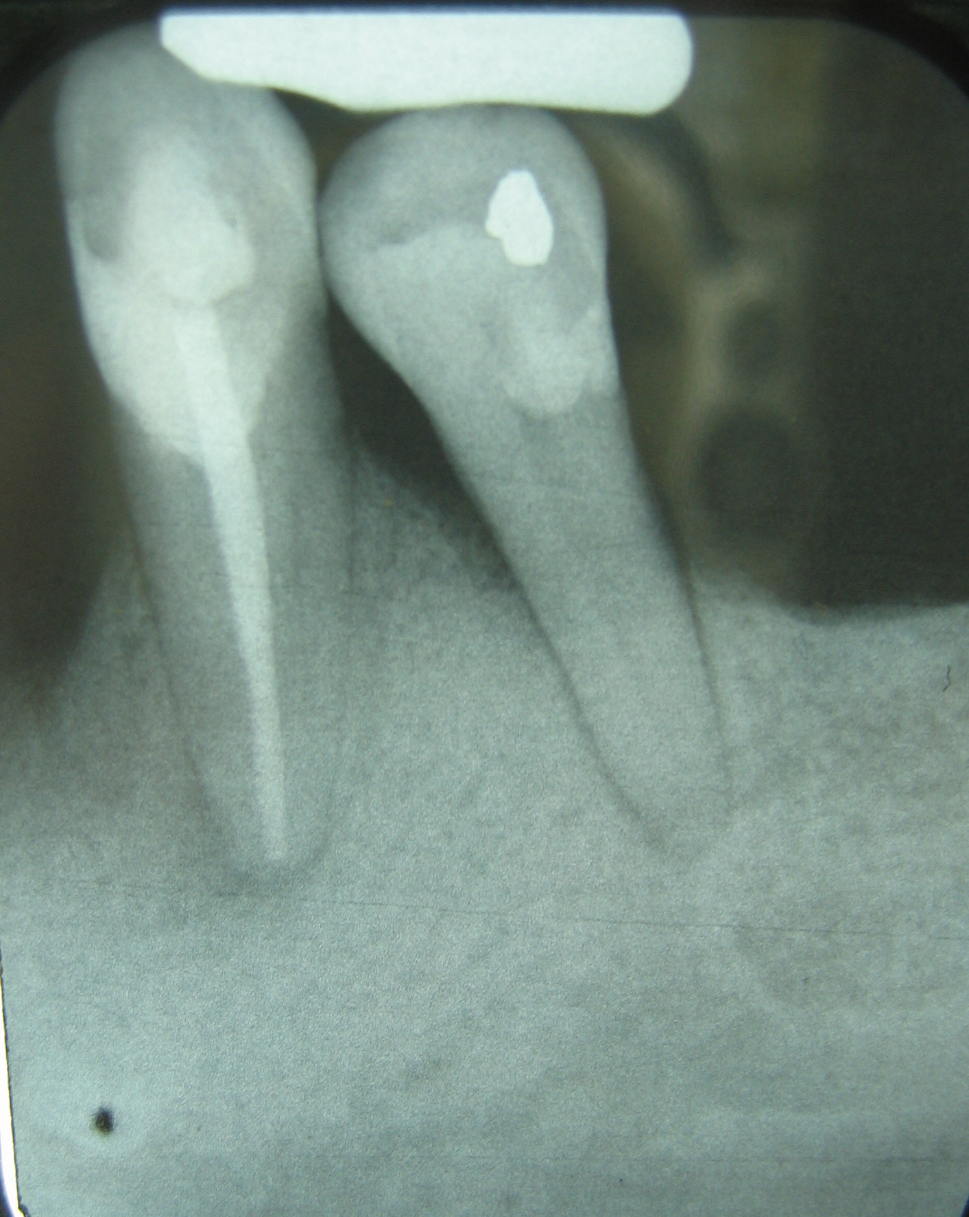
Periodontitis x ray
|
Periodontitis Microchapters |
|
Diagnosis |
|---|
|
Treatment |
|
Case Studies |
|
Periodontitis x ray On the Web |
|
American Roentgen Ray Society Images of Periodontitis x ray |
Editor-In-Chief: C. Michael Gibson, M.S., M.D. [1]
Please help WikiDoc by adding more content here. It's easy! Click here to learn about editing.
X Ray

This X-ray film displays two lone-standing mandibular teeth, #21 and #22, or the lower left first premolar and canine, exhibiting severe bone loss of 30-50%. Widening of the PDL surrounding the premolar is due to secondary occlusal trauma.