File:Pineal parenchymal tumor with intermediate differentiation microscopic 2.jpg
No higher resolution available.
Pineal_parenchymal_tumor_with_intermediate_differentiation_microscopic_2.jpg (800 × 593 pixels, file size: 176 KB, MIME type: image/jpeg)
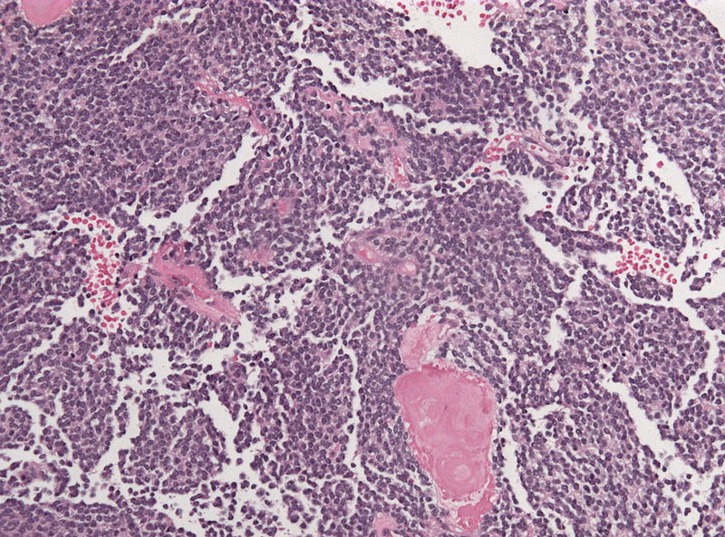
Histology specimen of a pineal parenchymal tumor of intermediate differentiation (HE stain; intermediate magnification) demonstrating several hyalinized vessels between the tumor cells in high density.
File history
Click on a date/time to view the file as it appeared at that time.
| Date/Time | Thumbnail | Dimensions | User | Comment | |
|---|---|---|---|---|---|
| current | 18:23, 23 November 2015 | | 800 × 593 (176 KB) | Sujit Routray (talk | contribs) | |
| 18:22, 23 November 2015 |  | 800 × 593 (176 KB) | Sujit Routray (talk | contribs) | Histology specimen of a pineal parenchymal tumor of intermediate differentiation (HE stain; intermediate magnification) demonstrating several hyalinized vessels between the tumor cells in high density. |
You cannot overwrite this file.
File usage
There are no pages that use this file.