File:Blastomycosis22.jpeg
No higher resolution available.
Blastomycosis22.jpeg (700 × 465 pixels, file size: 40 KB, MIME type: image/jpeg)
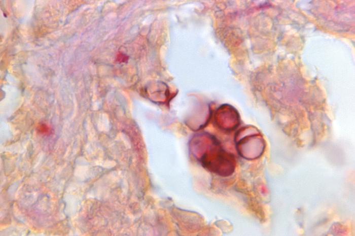
Magnified 1150X, this Gram-stained photomicrograph reveals the presence of a number of hyphae of the fungal organism, Exophiala castellanii. These fungi were harvested from a pus-laden lesion located on a patient’s buttock.
File history
Click on a date/time to view the file as it appeared at that time.
| Date/Time | Thumbnail | Dimensions | User | Comment | |
|---|---|---|---|---|---|
| current | 21:42, 24 November 2014 | | 700 × 465 (40 KB) | Jesus Hernandez (talk | contribs) | Magnified 1150X, this Gram-stained photomicrograph reveals the presence of a number of hyphae of the fungal organism, Exophiala castellanii. These fungi were harvested from a pus-laden lesion located on a patient’s buttock. |
You cannot overwrite this file.
File usage
The following file is a duplicate of this file (more details):
There are no pages that use this file.