Uncategorized files
Jump to navigation
Jump to search
Showing below up to 50 results in range #42,901 to #42,950.
-
 Neurofibroma (1).jpg 796 × 599; 219 KB
-
 Neurofibroma in the hip.png 500 × 500; 89 KB
-
 Neurofibroma oral 001.jpg 713 × 436; 273 KB
-
 Neurofibromatosis-type-1- 001.jpg 540 × 705; 47 KB
-
 Neurofibromatosis-type-1- 002.jpg 718 × 657; 63 KB
-
 Neurofibromatosis-type-1- 003.jpg 713 × 678; 74 KB
-
 Neurofibromatosis-type-1- 004.jpg 719 × 652; 60 KB
-
 Neurofibromatosis.jpg 1,200 × 1,600; 231 KB
-
 Neurofibromatosis2.jpg 512 × 369; 40 KB
-
 Neurofibromatosis21.jpg 465 × 381; 119 KB
-
 Neurofibromatosis30.jpg 469 × 378; 130 KB
-
 Neurofibromatosis33.jpg 469 × 377; 89 KB
-
 Neurofibromatosis case 001.jpg 673 × 450; 33 KB
-
 Neurofibromatosis case 002.jpg 668 × 450; 40 KB
-
 Neurofibromatosis case 003.jpg 674 × 450; 32 KB
-
 Neurofibromatosis case 004.jpg 625 × 450; 48 KB
-
 Neurofibromatosis case 005.jpg 603 × 450; 53 KB
-
 Neurofibromatosis case 006.jpg 598 × 450; 89 KB
-
 Neurofibromatosis case 007.jpg 609 × 450; 74 KB
-
 Neurofibromatosis case 008.jpg 599 × 450; 59 KB
-
 Neurofibromatosis plexiform neurofriboma 3.jpg 800 × 600; 25 KB
-
 Neurogenic-bladder-001.jpg 490 × 600; 32 KB
-
 Neuroglia.png ; 77 KB
-
 Neurolite image.jpg 720 × 354; 31 KB
-
 Neurolite ingredients and appearance.png 628 × 3,038; 283 KB
-
 Neurolite table4.png 634 × 813; 78 KB
-
 Neurolite table5.png 663 × 883; 92 KB
-
 Neurology.jpg 606 × 606; 100 KB
-
 Neuron-no labels.png 400 × 215; 29 KB
-
 Neuron colored.jpg 511 × 599; 15 KB
-
 Neurontin NDC 00710803.jpg 640 × 480; 55 KB
-
 Neurontin NDC 00710806.jpg 640 × 480; 106 KB
-
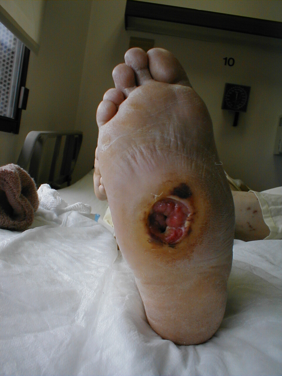 Neuropathic Ulcer in Patient with diabetic neuropathy.jpg 960 × 1,280; 320 KB
-
 Neuropathology case XII 01.jpg 1,500 × 1,112; 667 KB
-
 Neuropeptide Y.png 274 × 362; 4 KB
-
 Neurosarcoidosis-001.jpg 727 × 773; 75 KB
-
 Neurosarcoidosis-002.jpg 707 × 781; 59 KB
-
 Neurosarcoidosis-003.jpg 755 × 764; 63 KB
-
 Neurosarcoidosis-004.jpg 685 × 771; 67 KB
-
 Neurosarcoidosis-005.jpg 685 × 771; 69 KB
-
 Neurosarcoidosis-006.jpg 685 × 771; 58 KB
-
 Neurosarcoidosis-and-chiari-i-malformation.jpg 1,002 × 1,024; 68 KB
-
 Neurosarcoidosis.jpg 986 × 1,024; 66 KB
-
 Neurosarcoidosis MRI pre-post treatment arrows.gif 978 × 489; 182 KB
-
Neurovascular Bundle.svg 467 × 154; 5 KB
-
 Neurovascular compression of the trigeminal root.jpeg 506 × 1,280; 112 KB
-
 Neus1.jpg 876 × 1,034; 363 KB
-
 NeutronPort.gif 424 × 409; 58 KB
-
 Neutropenic enterocolitis.png 512 × 331; 221 KB
-
 Neutrophil, basophil and active lymphocyte 0001.jpg 505 × 331; 24 KB
.jpg)


