Uncategorized files
Jump to navigation
Jump to search
Showing below up to 50 results in range #40,691 to #40,740.
-
 Microcephaly.jpg 488 × 500; 22 KB
-
 Microcephaly 2.jpg 281 × 219; 8 KB
-
 Microcephaly 3.jpg 1,200 × 803; 317 KB
-
 Microcephaly Trisomy.JPG 1,013 × 789; 69 KB
-
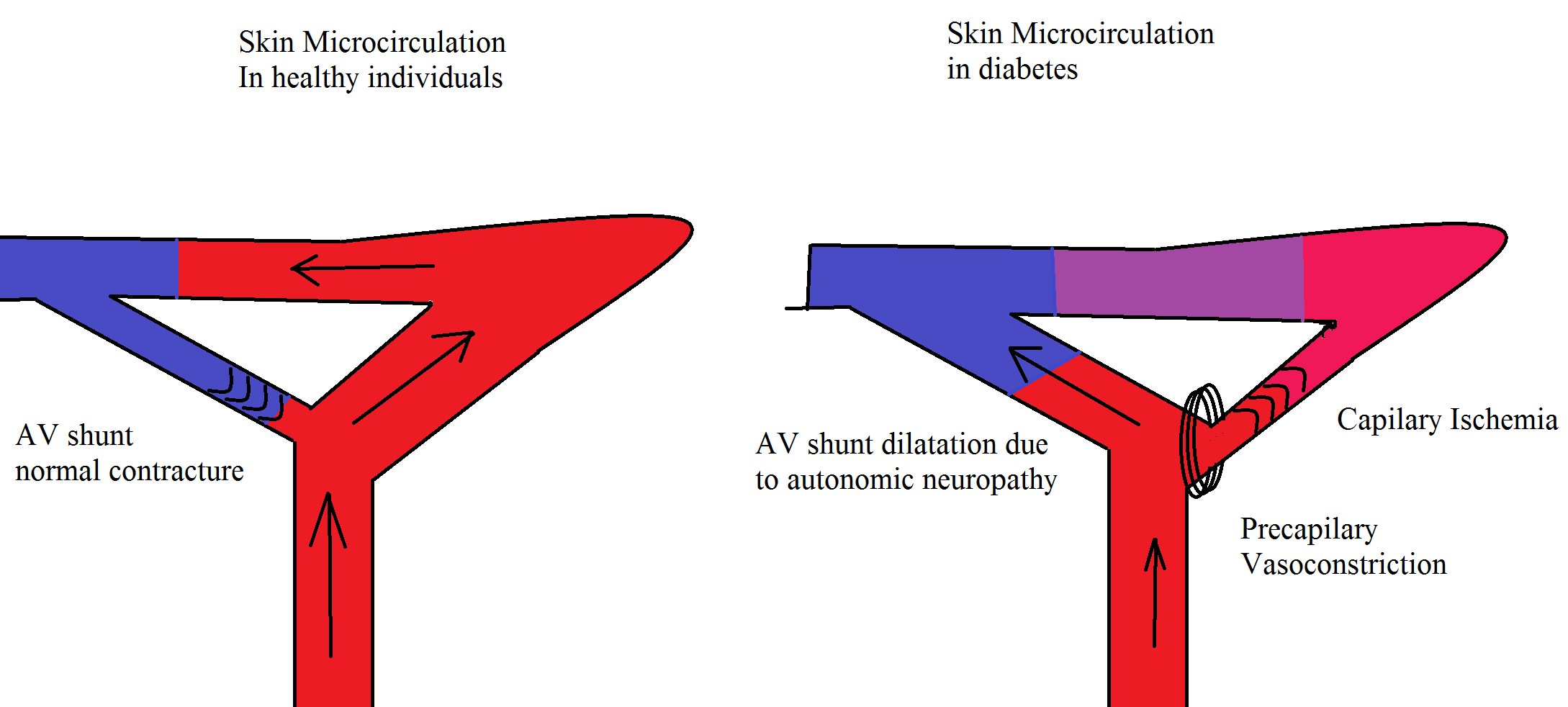 Microcirculation image.png 2,178 × 982; 96 KB
-
 Microcystic meningeoma.jpg 120 × 89; 6 KB
-
 Microfollicular stain.jpg 600 × 451; 222 KB
-
 Microgestin Fe NDC 525440631.jpg 1,024 × 768; 171 KB
-
 Micrograph of a cell nucleus.png 200 × 199; 33 KB
-
 Micronase NDC 00090171.jpg 640 × 480; 49 KB
-
 Micronized glyburide NDC 597623781.jpg 640 × 480; 70 KB
-
 Micronized glyburide NDC 597623782.jpg 640 × 480; 104 KB
-
 Micronized glyburide NDC 597623783.jpg 640 × 480; 89 KB
-
 Micropathology.jpg 600 × 226; 155 KB
-
 Micropathology osteoma.png 1,017 × 620; 1.07 MB
-
 Micropathologyneoplastic meningitis image 1.PNG 976 × 788; 458 KB
-
 Micropathologyneoplastic meningitis image 2.PNG 1,140 × 619; 408 KB
-
Microscope-optical path.svg 425 × 177; 458 KB
-
 Microscope And Digital Camera.JPG 3,072 × 2,304; 2.16 MB
-
 Microscope Zeiss 1879.jpg 251 × 520; 68 KB
-
 Microscope de HOOKE.png 458 × 507; 51 KB
-
Microscope diag.svg 673 × 343; 17 KB
-
 Microscope with stained slide.jpg 640 × 480; 51 KB
-
MicroscopesOverview.svg 905 × 651; 117 KB
-
 Microscopic Hematuria Clinical Presentaion and Risk factors.jpg 2,065 × 4,327; 1.2 MB
-

-
 Microscopic feature of Sclerosing adenosis.jpg 800 × 533; 180 KB
-
 Microscopic features of atrt 1.PNG 956 × 786; 858 KB
-
 Microscopic findings in GAE.jpg 566 × 400; 40 KB
-
 Microscopic hematuria.jpg 1,182 × 689; 213 KB
-
 Microscopic image of pineoblastoma 1.jpg 800 × 593; 155 KB
-
 Microscopic image of pineoblastoma 2.jpg 1,024 × 767; 353 KB
-
-
 Microscopic pathology.jpg 264 × 206; 36 KB
-
 Microscopic pathology2.jpg 254 × 200; 32 KB
-
 Microscopic pathology of phyllodes tumor.jpg 800 × 533; 179 KB
-
 Microscopic pathology of primary effusion lymphoma.jpg 1,200 × 894; 688 KB
-
 Microscopic takayasu.jpg 696 × 510; 43 KB
-
 Microscopy valley fever.jpg 300 × 205; 52 KB
-
 Microsporidiosis 01.jpg 457 × 600; 41 KB
-
 Microsporidiosis 01.png 457 × 600; 1.05 MB
-
 Microtia lvl3.jpg 240 × 180; 61 KB
-
 Microtiter plate.JPG 800 × 600; 79 KB
-
 Microtuble.jpg 300 × 286; 21 KB
-
 Microvilli.jpg 120 × 85; 2 KB
-
 Microvoltages.jpg 1,024 × 675; 271 KB
-
 Microvoltages02.jpg 1,024 × 675; 284 KB
-
 Microzosyn8.JPG 893 × 283; 64 KB
-
 Mid-S8-Dendritic Cells Dragging Conidia in Collagen.ogg.jpg 400 × 384; 21 KB
-
 Mid Diastolic Murmur.jpg 151 × 66; 3 KB


