Uncategorized files
Jump to navigation
Jump to search
Showing below up to 50 results in range #39,241 to #39,290.
-
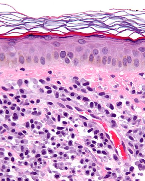 Mastocytosis Histology.jpg 480 × 600; 79 KB
-
 Mastoid Osteoma 13April05.jpg 480 × 360; 206 KB
-
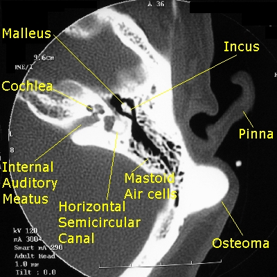 Mastoid Osteoma Labeled.jpg 400 × 400; 159 KB
-
 Mastoid Osteoma May 15 2005 480 SQ.jpg 480 × 480; 293 KB
-
 Mastoid Osteoma May SQ 480 15 2005 001 7 .jpg 480 × 480; 267 KB
-
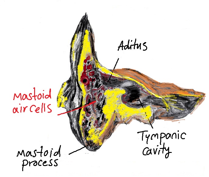 Mastoid air cells.jpg 686 × 580; 126 KB
-
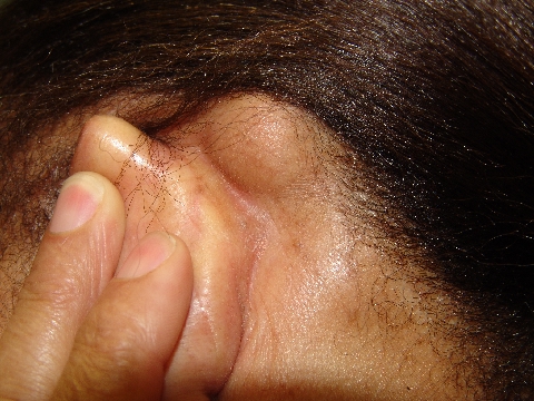
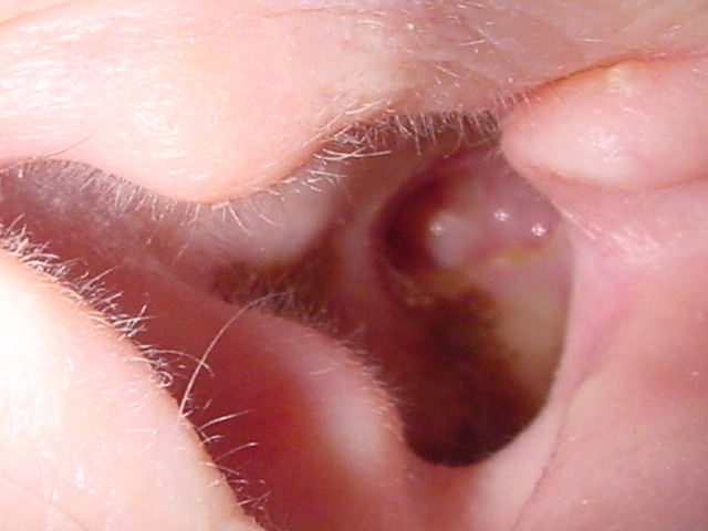
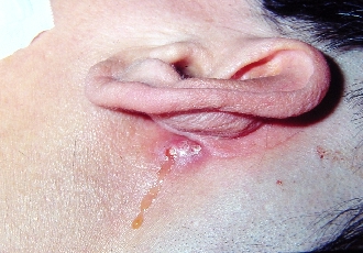 Mastoid cutaneous fistula trim.jpg 330 × 230; 92 KB
-
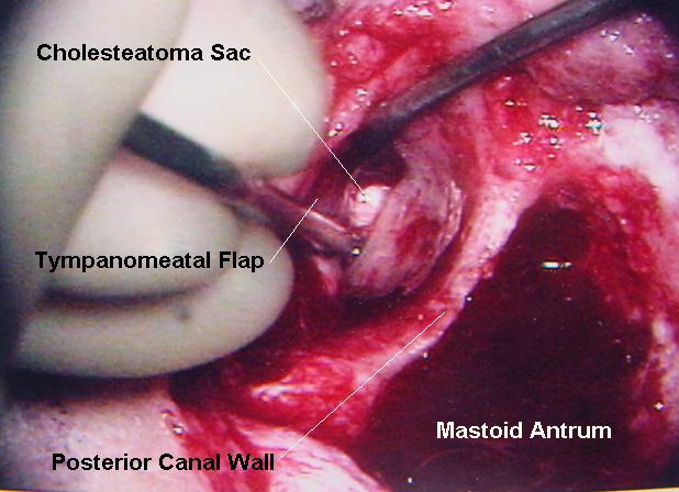
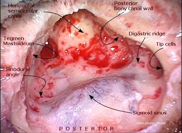 Mastoidectomy Arrowheads LABELED.jpg 626 × 459; 61 KB
-
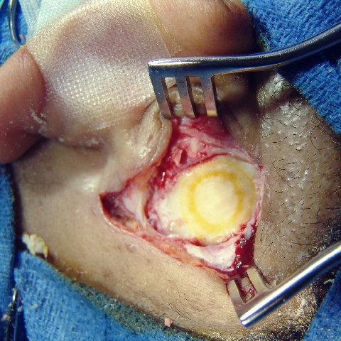
 Mastoidectomy Bowl.jpg 600 × 400; 178 KB
-
 Mastoidectomy Bowl 008.jpg 640 × 480; 62 KB
-
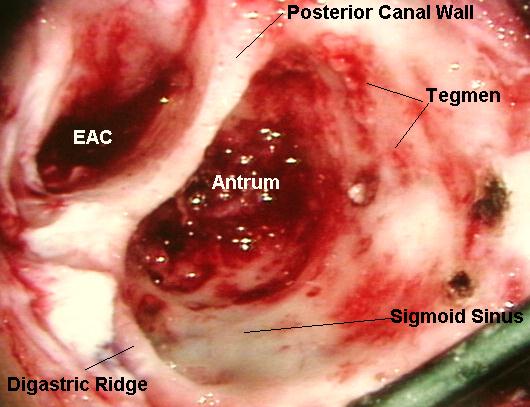
 Mastoidectomy labeled.jpg 618 × 448; 56 KB
-
 Mastoidectomy labeled1.jpg 530 × 407; 51 KB
-
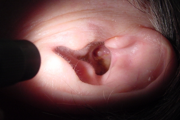
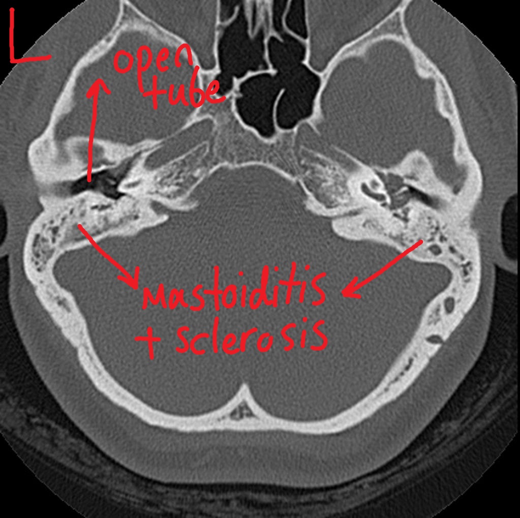 Mastoiditis.jpg 1,023 × 1,019; 206 KB
-
 Mastoiditisphe.jpg 350 × 418; 107 KB
-
 Mastomys.gif 175 × 99; 15 KB
-
 Mastomys natalensis.jpg 334 × 249; 58 KB
-
 Mastomys natalensis mammary.jpg 262 × 244; 50 KB
-
 Material safety data sheet.JPG 1,487 × 1,902; 394 KB
-
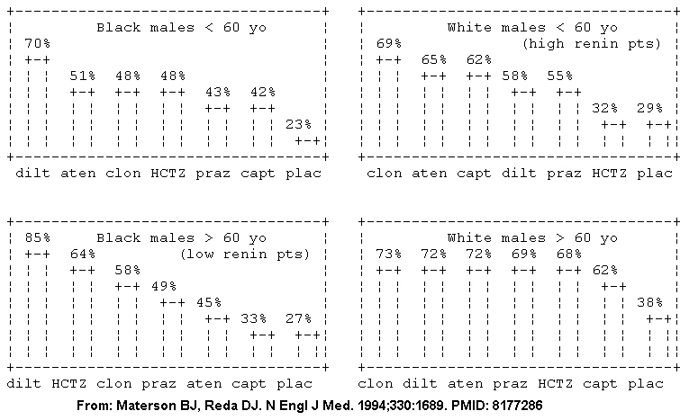 Materson et al. NEJM 1994. PMID 8177286.jpg 765 × 467; 152 KB
-
 Matrix Isolated.JPG 623 × 453; 45 KB
-
 MattP(1).png 3,600 × 3,600; 10.59 MB
-
 MattP.png 3,600 × 3,600; 10.59 MB
-
 Matt Reynold.jpg 55 × 75; 16 KB
-
 Matulane NDC 544820053.jpg 640 × 480; 82 KB
-
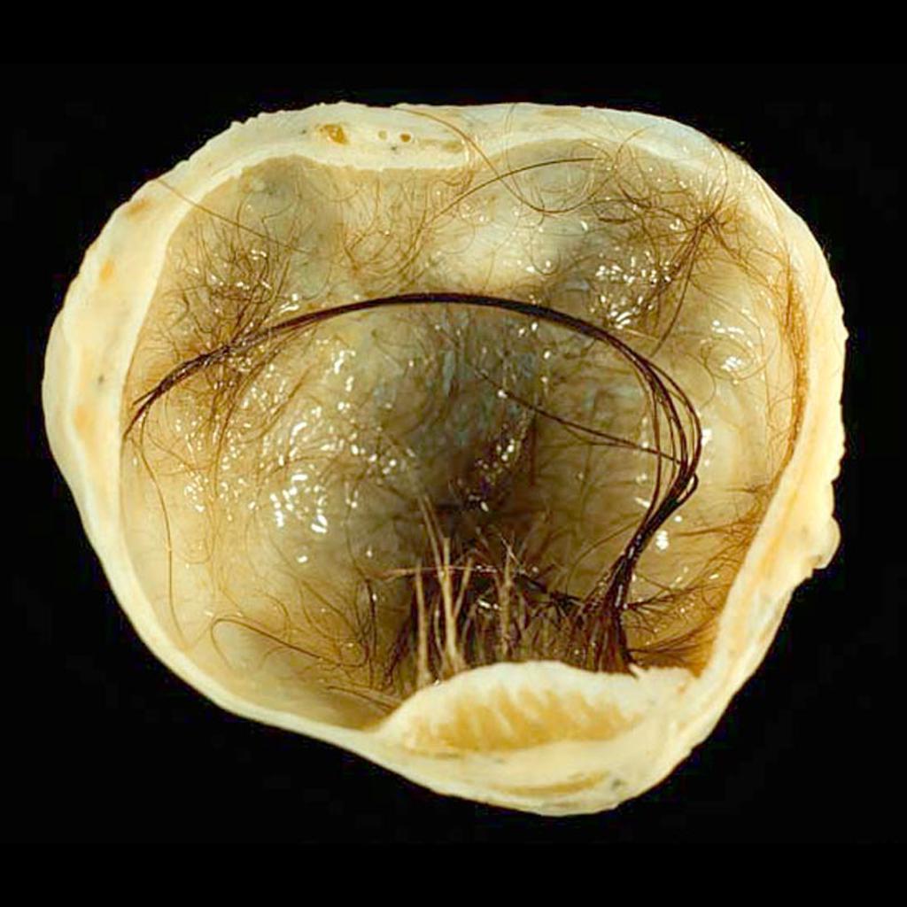
 Mature-cystic-ovarian-teratoma-6 (1).jpg 829 × 1,024; 116 KB
-
 Mature-cystic-teratoma-of-the-ovary.jpg 1,024 × 1,024; 110 KB
-
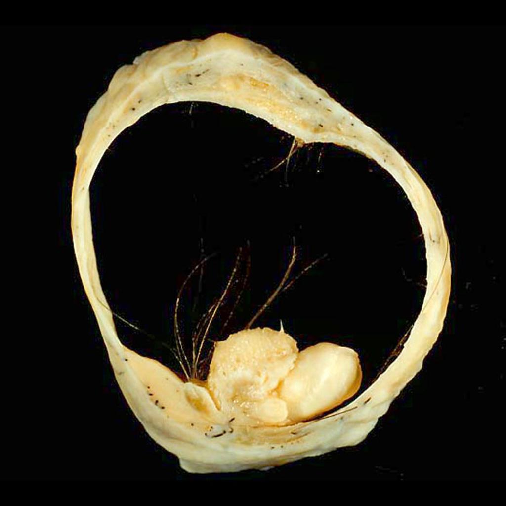 Mature-cystic-teratoma-of-the-ovary (2).jpg 1,024 × 1,024; 73 KB
-
 Mature Cystic Teratoma of the Ovary Bone Tissue (4047143950).jpg 1,510 × 1,441; 1.18 MB
-
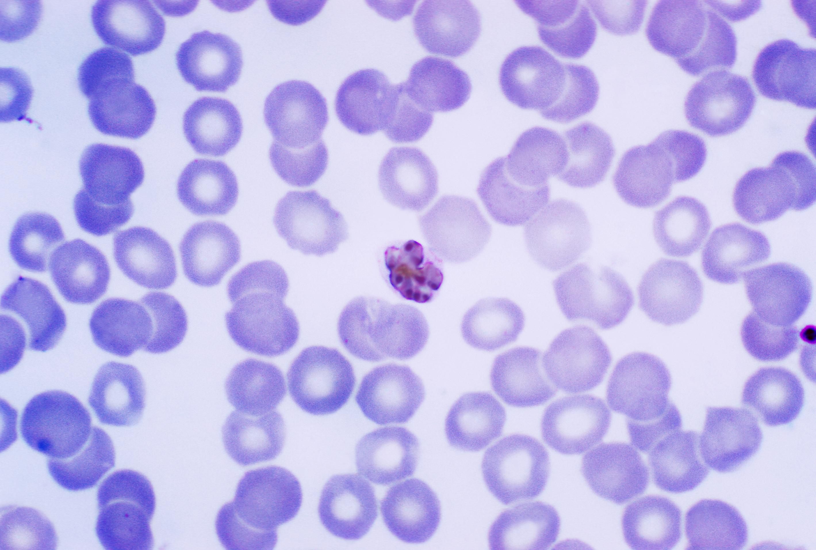 Mature Plasmodium malariae schizont PHIL 2715 lores.jpg 2,838 × 1,914; 522 KB
-
 Mature cystic teratoma 001.jpg 600 × 501; 56 KB
-
 Mature cystic teratoma 002.jpg 600 × 501; 54 KB
-
 Mature cystic teratoma 003.jpg 600 × 501; 53 KB
-
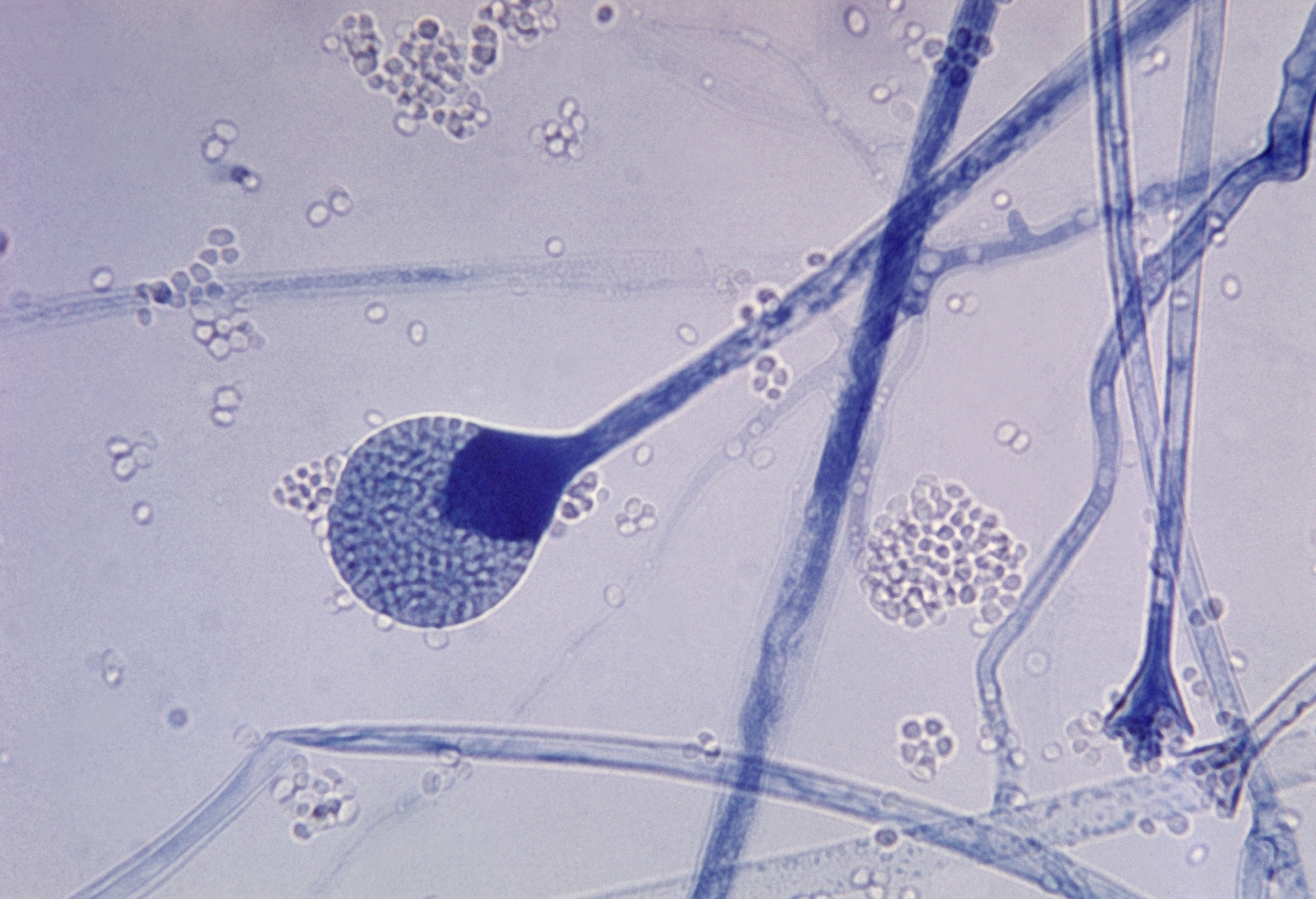 Mature sporangium of a Mucor sp. fungus.jpg 2,797 × 1,911; 3.63 MB
-
 Mature sporangium of a Mucor sp. fungus PHIL 3960 lores.jpg 200 × 137; 6 KB
-
 Matzim LA NDC 525440691.jpg 1,024 × 781; 148 KB
-
 Matzim LA NDC 525440694.jpg 1,024 × 781; 149 KB
-
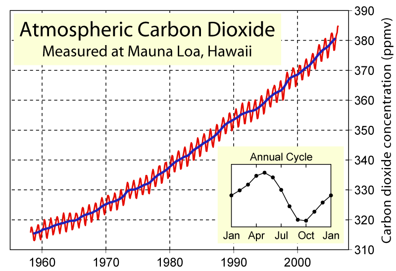 Mauna Loa Carbon Dioxide.png 800 × 547; 34 KB
-
 Mauriciocohen.jpg 400 × 400; 23 KB
-
 Mavacoxib.png 512 × 411; 14 KB
-
 Mavik NDC 00742278.jpg 640 × 480; 89 KB
-
 Mavik NDC 00742279.jpg 640 × 480; 88 KB
-
 Mavik NDC 00742280.jpg 640 × 480; 119 KB
-
 MaxiMounds.jpg 350 × 264; 15 KB
-
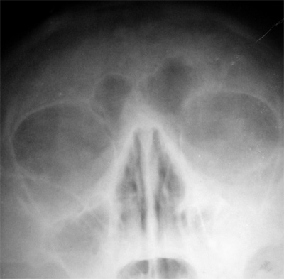 Maxilar sinusites.jpg 400 × 393; 37 KB
-
 Maxillary artery.png 552 × 647; 180 KB
-
 Maxillary central incisors01-01-06.jpg 168 × 158; 5 KB
-

-
 Maxillary first molars01-01-06.png 168 × 158; 49 KB
-
 Maxillary second premolars01-01-06.png 168 × 158; 49 KB
-
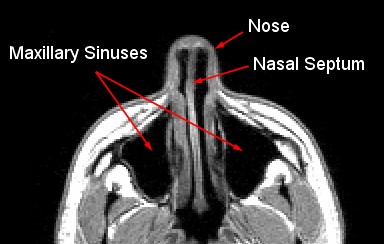 Maxillary sinus MRI.jpg 384 × 244; 25 KB


.png)


.jpg)

.jpg)
.jpg)


