Unused files
Jump to navigation
Jump to search
The following files exist but are not embedded in any page. Please note that other websites may link to a file with a direct URL, and so may still be listed here despite being in active use.
Showing below up to 50 results in range #3,731 to #3,780.
-
 Aortic disscetionDB1.jpg 850 × 930; 101 KB
-
 Aortic disscetion DB2.jpg 850 × 930; 96 KB
-
 Aortic disscetionDB3.jpg 850 × 930; 100 KB
-
 Situs inversus.jpg 700 × 876; 36 KB
-
 Takayasu arteritis.JPG 1,064 × 915; 59 KB
-
 Double aortic arch frontal.JPG 824 × 829; 50 KB
-
 Jeannine Parvati Baker circa 1990.jpg 400 × 279; 23 KB
-
 Churg strauss arteritis.jpg 403 × 270; 28 KB
-
 Churg strauss cxr.jpg 240 × 240; 31 KB
-
 Wide-mediastinum-ao-dissection-3-mos-prior.jpg 251 × 224; 7 KB
-
 Wide-mediastinum-ao-dissection-on-adm.jpg 251 × 224; 7 KB
-
 Wikidocsearchresults 614.xls ; 17 KB
-
 Scleroderma-001.jpg 512 × 512; 87 KB
-
 Scleroderma-002.jpg 512 × 512; 84 KB
-
 Scleroderma-003.jpg 512 × 512; 83 KB
-
 Scleroderma-004.jpg 512 × 512; 61 KB
-
 Scleroderma-005.jpg 512 × 512; 66 KB
-
 Scleroderma-006.jpg 512 × 512; 65 KB
-
 Scleroderma-007.jpg 512 × 512; 64 KB
-
 Scleroderma-008.jpg 512 × 512; 63 KB
-
 Scleroderma-301.jpg 438 × 738; 34 KB
-
 Scleroderma-302.jpg 425 × 737; 28 KB
-
 Scleroderma-303.jpg 582 × 688; 37 KB
-
 Sickle cell disease H shaped vertebral bodies 001.jpg 600 × 611; 64 KB
-
 Sickle cell disease H shaped vertebral bodies 002.jpg 600 × 829; 76 KB
-
 Sickle cell disease H shaped vertebral bodies 003.jpg 600 × 937; 133 KB
-
 Germ-cell-tumor-002.jpg 600 × 407; 59 KB
-
 Pulmonary-angiosarcoma-001.jpg 600 × 600; 63 KB
-
 Pulmonary-angiosarcoma-002.jpg 600 × 600; 66 KB
-
 Pulmonary-angiosarcoma-003.jpg 600 × 600; 61 KB
-
 Pulmonary-angiosarcoma-004.jpg 600 × 600; 61 KB
-
 Metastatic melanoma1.JPG 1,396 × 1,153; 150 KB
-
 Metastatic melanoma2.JPG 1,155 × 1,054; 115 KB
-
 Anomalous Origin Coronary Artery4.jpg 768 × 512; 43 KB
-
 Buprenorphine structure.jpg 608 × 403; 27 KB
-
 Elektronenmikroskop.jpg 504 × 472; 83 KB
-
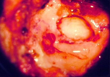
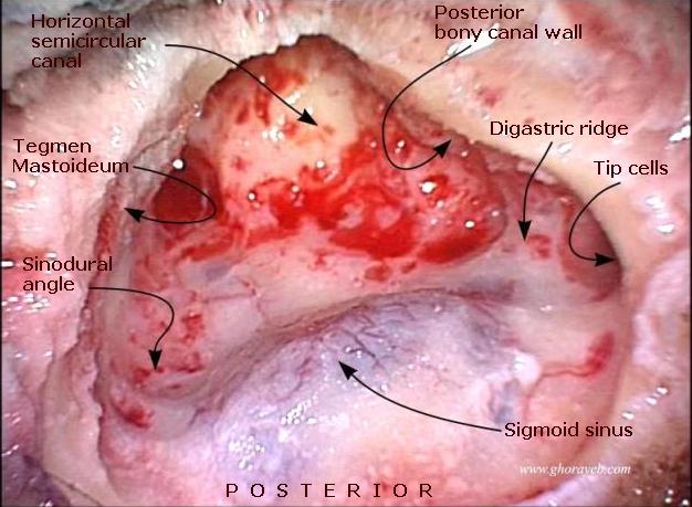 Mastoidectomy Arrowheads LABELED.jpg 626 × 459; 61 KB
-
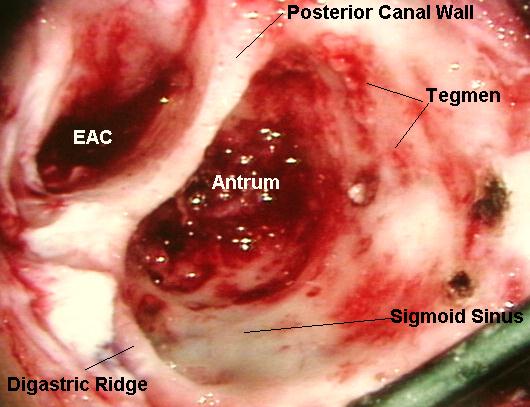 Mastoidectomy labeled1.jpg 530 × 407; 51 KB
-
 Coupled assay.svg 498 × 200; 14 KB
-
 Mastoidectomy labeled.jpg 618 × 448; 56 KB
-
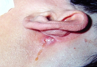 Mastoid cutaneous fistula trim.jpg 330 × 230; 92 KB
-
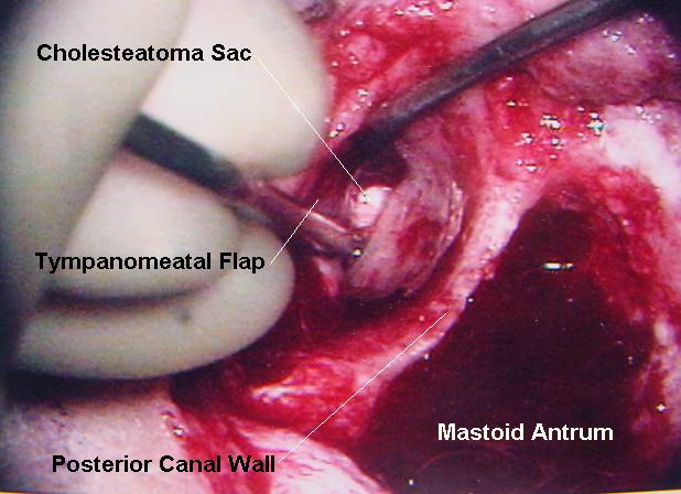
 Cholesteatoma sac eroding mastoid.jpg 375 × 265; 102 KB
-
Finasteride-3D-balls.png ; 175 KB
-
Finasteride.svg 1,577 × 1,064; 16 KB
-
 Sue'sBrain.jpg 640 × 480; 98 KB
-
Ecoli colonies.png ; 427 KB
-
 Hematoma.JPG 858 × 859; 52 KB
-
 Trypanosoma sp. PHIL 613 lores.jpg 190 × 135; 6 KB
-
 Bodydone.GIF 300 × 502; 8 KB
-
 Trichomoniasis 01.png 494 × 599; 40 KB


