Unused files
Jump to navigation
Jump to search
The following files exist but are not embedded in any page. Please note that other websites may link to a file with a direct URL, and so may still be listed here despite being in active use.
Showing below up to 50 results in range #26,091 to #26,140.
-
 SymdekTable8.png 591 × 404; 83 KB
-
 SymdekTable9.png 688 × 803; 150 KB
-
 SymdekTable10.png 601 × 787; 150 KB
-
 SymdekTable11.png 615 × 761; 158 KB
-
 SymdekTable12.png 508 × 1,277; 257 KB
-
 SymdekFigure2.png 780 × 438; 92 KB
-
 SymdekMedGuide.png 508 × 3,596; 761 KB
-
 Pulmonary embolism-1540.pdf ; 4 KB
-
 Blau syndrome BS.jpg 1,152 × 864; 210 KB
-
 Blau syndrome (BS).jpg 1,152 × 864; 210 KB
-
 Skin model.png 1,280 × 720; 620 KB
-
 ThyroidnodularSatturwar10.jpg 1,511 × 1,350; 615 KB
-
 Extramammary Paget's Disease.png 545 × 390; 224 KB
-
 1024px-SkinTumors-P9270829.jpg 1,024 × 768; 275 KB
-
 Skin dermatofibroma.jpg 112 × 83; 1 KB
-
 Dr. Sara Mohsin.jpg 200 × 297; 92 KB
-
 PMC3283526 1748-717X-6-182-4.png 512 × 500; 232 KB
-
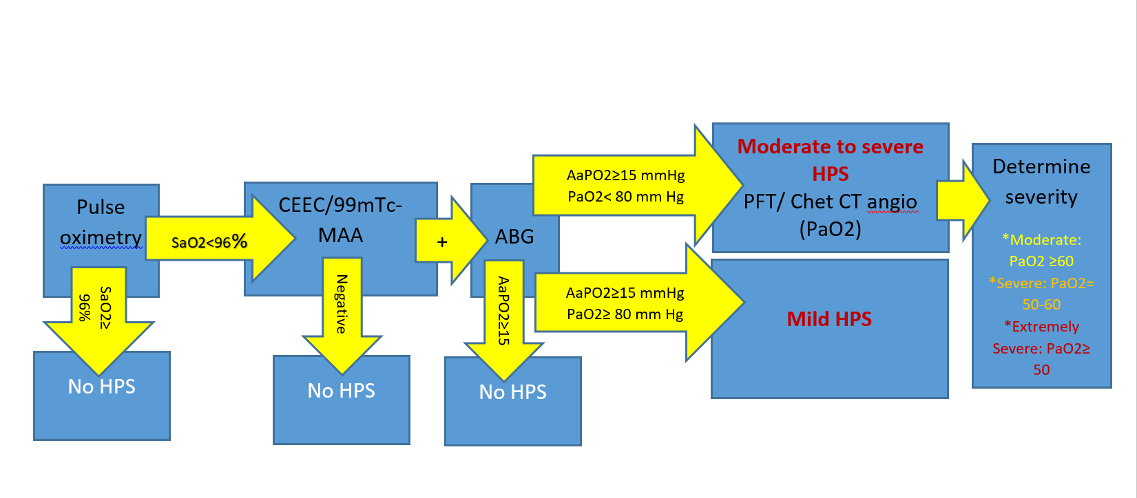 HPS algorithm.png 1,651 × 724; 70 KB
-
 Atypical-teratoid-rhabdoid-tumour-atrt v2.jpg 1,024 × 1,024; 197 KB
-
 Rhabdoid tumor v2.jpg 725 × 512; 409 KB
-
 PMC5124667 CRIPU2016-1035601.003.png 512 × 419; 501 KB
-
 Study tools.jpg 290 × 76; 7 KB
-
 Anaplastic-astrocytoma-2.jpg 928 × 1,024; 89 KB
-
 Glioblastoma-nos-butterfly-morphology.jpg 819 × 1,024; 106 KB
-
 Amyloidosis cutis dyschromica.png 1,495 × 1,127; 3.65 MB
-
RCT vs Observational Studies.pdf ; 1.45 MB
-
 3ch-CMR-CA.mp4 ; 411 KB
-
 3ch LGE.jpg 512 × 512; 126 KB
-
 Cardiac-amyloidosis-3 (1).jpg 1,656 × 1,234; 199 KB
-
 Cardiac-amyloidosis-Nuclear-anterior.jpg 512 × 1,024; 134 KB
-
 Cardiac-amyloidosis-Nuclear-posterior.jpg 512 × 1,024; 157 KB
-
 MRI FLAIR sequence Wernicke Encephalopathy.jpg 800 × 837; 62 KB
-
 D435dff9c313eed96624ff2361b64d big gallery.jpeg 630 × 429; 30 KB
-
 LBBB+RADandTransplant.png 765 × 862; 214 KB
-
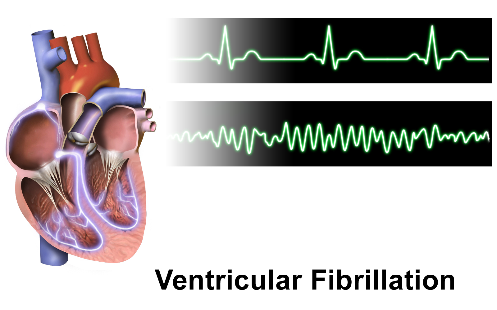 Ventricular Fibrillation.png 1,600 × 1,000; 684 KB
-
 Aortic-intramural-haematoma-1.jpg 792 × 792; 112 KB
-
 Aortic-intramural-haematoma-1 (1).jpg 512 × 512; 52 KB
-
 CDC coronavirus confirmed cases.jpg 1,193 × 593; 47 KB
-
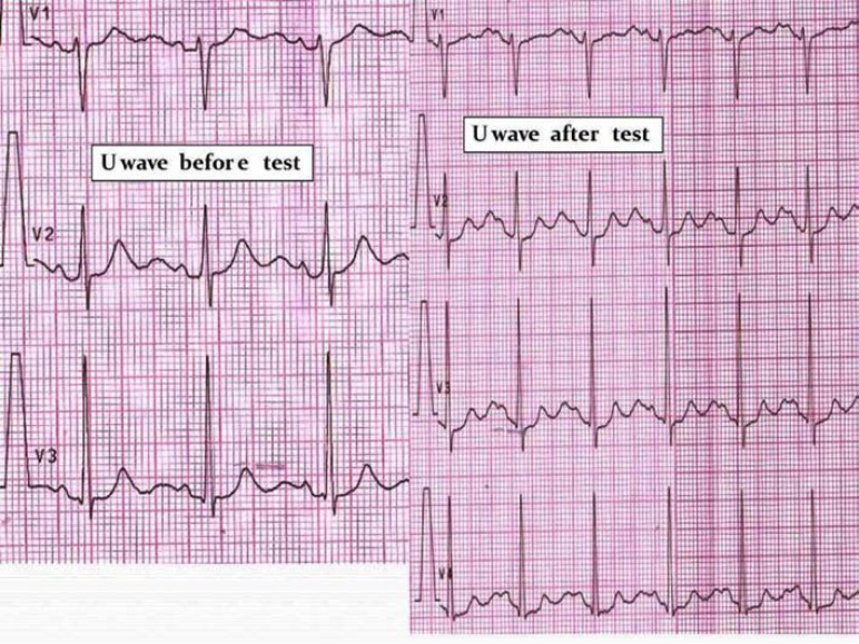 U-wave amplitude after “adrenaline test” in an ATS1 patient.jpg 772 × 579; 179 KB
-
 Slide14gggg.jpg 960 × 720; 106 KB
-
 597px-2019-nCoV-CDC-23312 without background.png 597 × 599; 508 KB
-
 Transesophageal echocardiogram of a patent ductus arteriosus.jpg 654 × 250; 39 KB
-
 Right heart catheterization.jpg 562 × 322; 141 KB
-
 Normal ECG 2.png 1,320 × 782; 55 KB
-
 Normal ECG axis 11111.png 300 × 300; 14 KB
-
 Bapaye.jpg 800 × 1,067; 1.04 MB
-
 Bapaye-eye-hospital-logo-nashik.jpg 1,000 × 268; 213 KB
-
 DvmTK9sVYAEyest.jpg 4,028 × 1,674; 539 KB
-
 Multiple sclerosis.png 800 × 667; 494 KB
-
 Cardiac-event-recorder.jpg 2,500 × 2,048; 551 KB


.jpg)


.jpg)


.jpg)


