Unused files
Jump to navigation
Jump to search
The following files exist but are not embedded in any page. Please note that other websites may link to a file with a direct URL, and so may still be listed here despite being in active use.
Showing below up to 50 results in range #25,961 to #26,010.
-
 Splenic infiltrate.png 512 × 322; 421 KB
-
 Wrights stain.png 512 × 366; 490 KB
-
 Bone marrow infiltrate.png 512 × 452; 650 KB
-
 Splenic lymphoid infiltrate.png 512 × 331; 452 KB
-
 Bm aspirate.png 512 × 170; 173 KB
-
 Interstitial pneumonitis after rutximab.png 512 × 231; 107 KB
-
 CT bing neel syndrome.png 472 × 417; 169 KB
-
 CT wm.png 512 × 395; 126 KB
-
 Ct contrast before n after treatment.png 512 × 434; 224 KB
-
 CT spleenomegaly.png 512 × 265; 143 KB
-
 Mri diffuse meningeal enhancment.png 512 × 579; 215 KB
-
 Penile ulcer. wm.png 512 × 476; 496 KB
-
 Hyperpigmented nodules with hemorrhagic crusting.png 512 × 613; 534 KB
-
 Ulcers tingling.png 512 × 382; 465 KB
-
 Dilation and tortosity of retinal veins.png 501 × 501; 420 KB
-
 Fundoscopy.png 512 × 387; 294 KB
-
 Fluorescein angiography.png 512 × 236; 116 KB
-
 GM. kidneys.png 512 × 382; 532 KB
-
 EM. kidneys.png 512 × 478; 354 KB
-
 Kidneys. EM.png 512 × 451; 509 KB
-
 Kidneys.immunoflorescence.png 512 × 403; 395 KB
-
 Renal biopsy.immunofloresence.png 512 × 254; 118 KB
-
 AngiosarcomaGross.JPG 800 × 638; 110 KB
-
 AMP.jpg 954 × 2,522; 688 KB
-
 Mucoepidermoid carcinoma.jpg 717 × 538; 67 KB
-
 Thyroid pathology.jpg 256 × 256; 8 KB
-
 Mri gif.gif 512 × 512; 439 KB
-
 Adenocarcinoma with predominant lepidic growth pattern.jpg 150 × 150; 29 KB
-
 DRMARS.jpg 813 × 666; 141 KB
-
 Ultrasound choriocarcinoma.jpg 442 × 338; 16 KB
-
 Mature-cystic-ovarian-teratoma-6 (1).jpg 829 × 1,024; 116 KB
-
 Desmoid surgery.png 512 × 244; 92 KB
-
 Desmoid abd ct.png 512 × 341; 138 KB
-
 Juvenile granulosa cell tumour.jpeg 450 × 300; 57 KB
-
 450px-Juvenile granulosa cell tumour - very high mag.jpg 450 × 300; 57 KB
-
 ARVD Pathophysiology.png 654 × 1,186; 193 KB
-
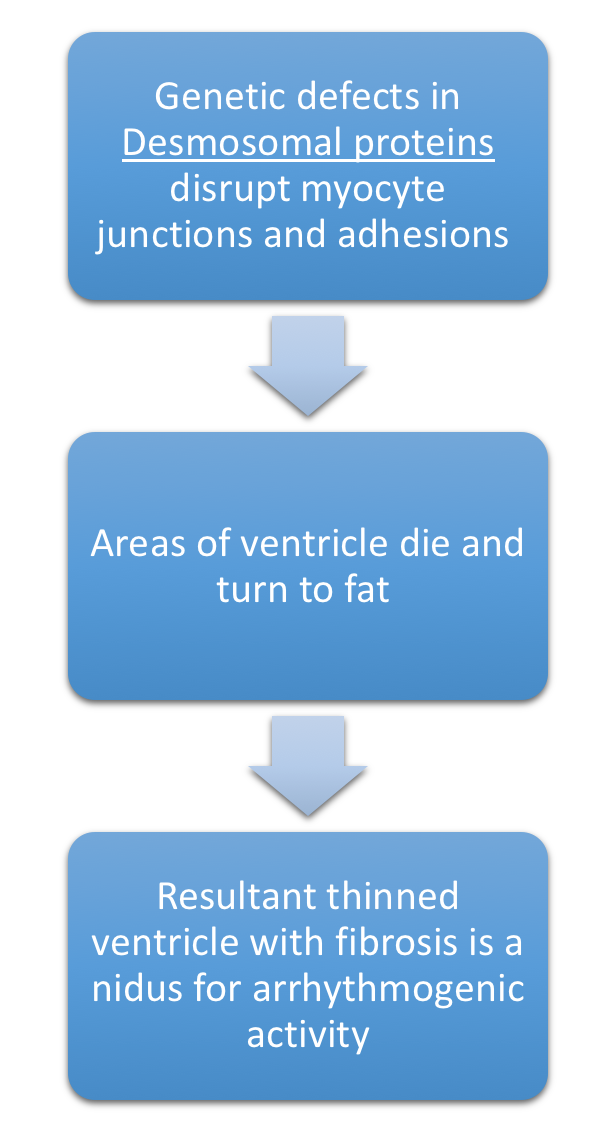 ARVC Pathophysiology.png 610 × 1,142; 193 KB
-
 Bladder neurofibroma.jpeg 500 × 303; 44 KB
-
 Hydronephrosis gif.gif 500 × 303; 147 KB
-
 Ct neurofb gif.gif 500 × 303; 134 KB
-
 Mri bladder mass gif.gif 500 × 292; 176 KB
-
 Sagittal mri.jpeg 500 × 454; 37 KB
-
 Axial mri gif.gif 500 × 292; 176 KB
-
 Mri gd gif.gif 500 × 331; 138 KB
-
 Enhanced gd gif.gif 500 × 305; 131 KB
-
 Gross neurofb.jpeg 500 × 443; 87 KB
-
 Nf histo.jpeg 500 × 390; 97 KB
-
 Neurofb histo.jpeg 500 × 393; 102 KB
-
 GB neurofb.png 512 × 339; 475 KB
-
 GB nb.jpg 576 × 383; 105 KB


.jpg)


