Unused files
Jump to navigation
Jump to search
The following files exist but are not embedded in any page. Please note that other websites may link to a file with a direct URL, and so may still be listed here despite being in active use.
Showing below up to 50 results in range #25,921 to #25,970.
-
 Hypopharyngeal-squamous-cell-carcinoma CT.jpg 1,024 × 1,024; 85 KB
-
 Polycythemia vera, blood smear (2).jpg 734 × 512; 247 KB
-
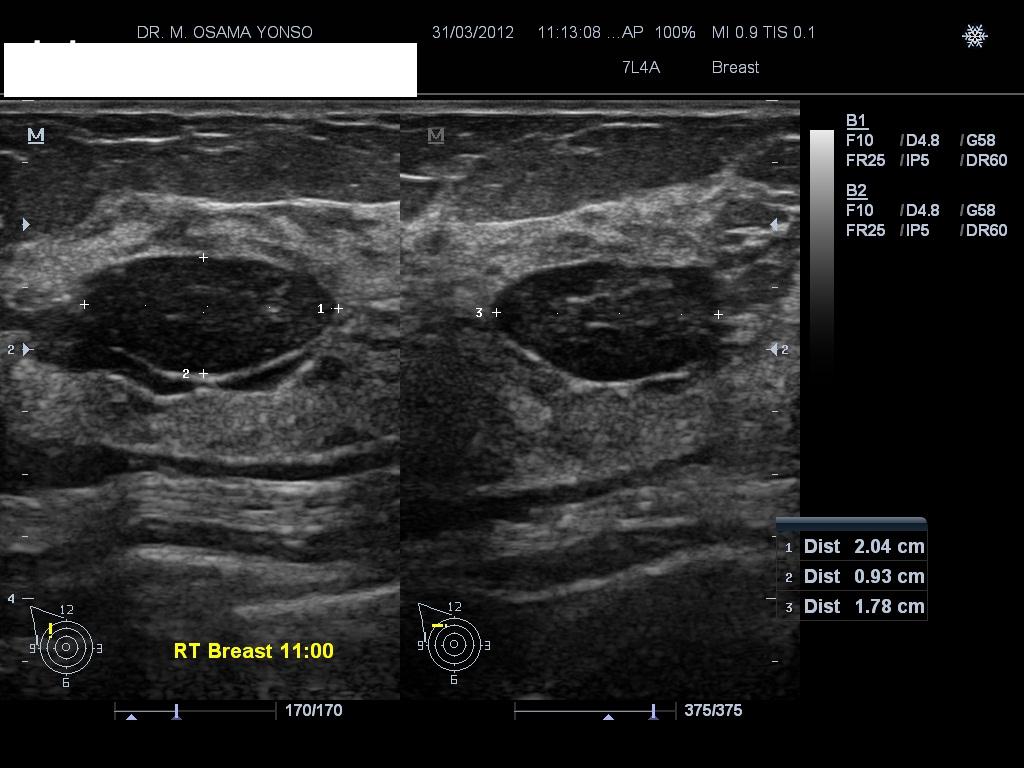 Ultrasound showing fib 1.jpg 1,024 × 768; 99 KB
-
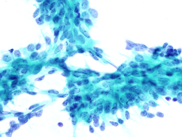 Histology fibro.jpg 700 × 527; 70 KB
-
 Thymoma.1.jpeg 442 × 442; 50 KB
-
 Webp.net-gifmaker thymoma.gif 192 × 288; 16 KB
-
 Retro peritoneal abscess Complicated appendicitis.JPG 986 × 921; 121 KB
-
 Lipomatosis gif.gif 1,280 × 720; 365 KB
-
 Thymoma gif 2.gif 384 × 288; 16 KB
-
 Gross pathology fibroadenoma.jpg 918 × 925; 346 KB
-
 Gonadoblastoma.jpg 4,272 × 2,848; 5.84 MB
-
 1280px-Gonadoblastoma - high mag.jpg 1,280 × 853; 454 KB
-
 Home logo.png 225 × 225; 1 KB
-
 Juhani Asem circle.png 300 × 300; 130 KB
-
 Memar Montazerin Sahar circle.png 300 × 300; 123 KB
-
 Venkatesan Swathi circle.png 300 × 300; 93 KB
-
 Venkatesan Swathi circle(2).png 300 × 300; 92 KB
-
 BRRS.jpg 600 × 417; 62 KB
-
 Lipoma.jpg 600 × 634; 50 KB
-
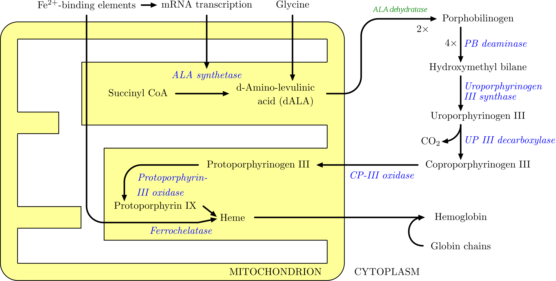 Heme.png 1,920 × 969; 161 KB
-
 Epithelial Ovarian carcinoma.jpg 256 × 256; 10 KB
-
 Craniopharyngioma-t1sagkm-006.jpg 512 × 513; 58 KB
-
 Necrotizing-sialometaplasia.png 773 × 556; 426 KB
-
 Nasopharyngeal angiofibroma.jpeg 4,272 × 2,848; 5.41 MB
-
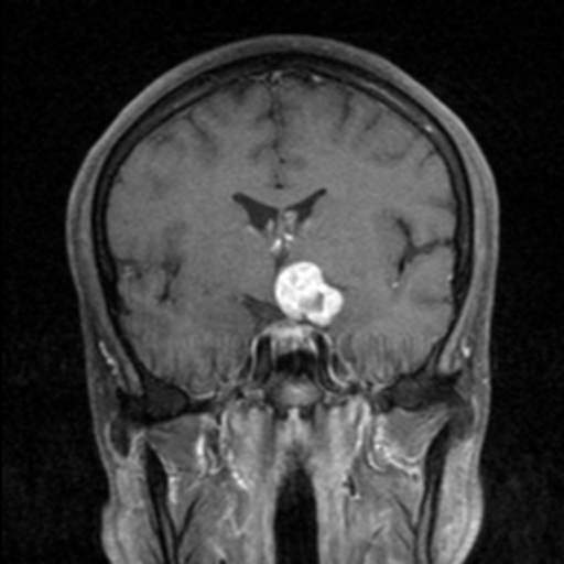 Craniopharyngioma-t1corkm-001.jpg 512 × 512; 21 KB
-
 Craniopharyngioma-t1corkm-003.jpg 512 × 512; 21 KB
-
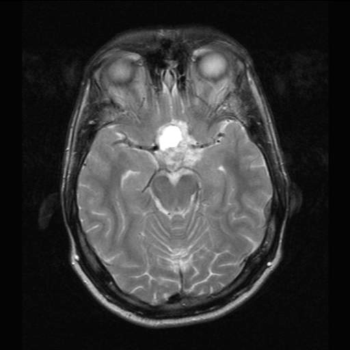 Craniopharyngioma-t2ax-003.jpg 512 × 512; 20 KB
-
 Thymoma GIF.gif 384 × 288; 21 KB
-
 Mediastinal Lipoma.gif 384 × 288; 48 KB
-
 BCC Nodular type.jpeg 256 × 388; 26 KB
-
 256px-BCC Nodular type.jpg 256 × 388; 26 KB
-
 BCC Nodular type peripheral palisading.jpeg 600 × 452; 204 KB
-
 Basal cell carcinoma histopathology (1).jpg 600 × 452; 204 KB
-
 CraniopharyngiomaCT.jpg 1,200 × 1,342; 177 KB
-
 TB gif.gif 384 × 288; 65 KB
-
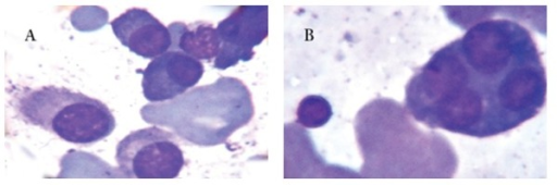
 Atypical B cell.png 512 × 357; 202 KB
-
 Neurenteric Cyst.gif 384 × 288; 165 KB
-
 WM.png 512 × 315; 297 KB
-
 Rouleaux formation.png 512 × 163; 190 KB
-
 CD20+ wm.png 512 × 277; 265 KB
-
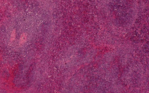 Splenic infiltrate.png 512 × 322; 421 KB
-
 Wrights stain.png 512 × 366; 490 KB
-
 Bone marrow infiltrate.png 512 × 452; 650 KB
-
 Splenic lymphoid infiltrate.png 512 × 331; 452 KB
-
 Bm aspirate.png 512 × 170; 173 KB
-
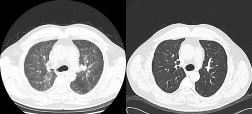 Interstitial pneumonitis after rutximab.png 512 × 231; 107 KB
-
 CT bing neel syndrome.png 472 × 417; 169 KB
-
 CT wm.png 512 × 395; 126 KB
-
 Ct contrast before n after treatment.png 512 × 434; 224 KB
-
 CT spleenomegaly.png 512 × 265; 143 KB

.jpg)


.png)


.jpg)


