Unused files
Jump to navigation
Jump to search
The following files exist but are not embedded in any page. Please note that other websites may link to a file with a direct URL, and so may still be listed here despite being in active use.
Showing below up to 50 results in range #25,901 to #25,950.
-
 Slide2.PNG 960 × 720; 356 KB
-
 Colorectal cancer staging.png 563 × 504; 90 KB
-
 Appendiceal-adenocarcinoma-complicated-by-retroperitoneal-abscess (2).jpg 1,024 × 748; 183 KB
-
 Merkel cell cancer.jpg 320 × 235; 97 KB
-
 Chondrosarcoma ct.jpg 1,024 × 1,024; 153 KB
-
 Chondrosarcoma MRI.jpg 1,024 × 1,024; 106 KB
-
 Tibial plateau fracture intra op 1.jpeg 513 × 514; 55 KB
-
 Intra-op tibial plateau.jpeg 1,171 × 744; 177 KB
-
 Tbial plateau intra-op2.png 732 × 850; 1.72 MB
-
 Intra-op tibial plateau 2.jpeg 743 × 774; 72 KB
-
 Intra-op tibial plateau 3.png 1,205 × 873; 2.92 MB
-
 Gross pathology of rhabdomyosarcoma.jpeg 1,486 × 1,074; 310 KB
-
 IMG 4360.jpg 184 × 302; 30 KB
-
 All.jpg 1,024 × 768; 105 KB
-
 51261392 2194265964026264 868264017259397120 n.jpg 960 × 960; 83 KB
-
 Acute-pancreatitis-and-walled-off-necrosis.jpg 1,024 × 747; 69 KB
-
 51097708 344211846175715 6986947914031431680 n.jpg 1,080 × 1,340; 139 KB
-
 51075927 322309311724764 1632750230099197952 n.jpg 720 × 960; 123 KB
-
 Allus5.gif 1,024 × 768; 1.13 MB
-
 Allxray.jpg 1,024 × 1,024; 180 KB
-
 Hypopharyngeal-squamous-cell-carcinoma CT.jpg 1,024 × 1,024; 85 KB
-
 Polycythemia vera, blood smear (2).jpg 734 × 512; 247 KB
-
 Ultrasound showing fib 1.jpg 1,024 × 768; 99 KB
-
 Histology fibro.jpg 700 × 527; 70 KB
-
 Thymoma.1.jpeg 442 × 442; 50 KB
-
 Webp.net-gifmaker thymoma.gif 192 × 288; 16 KB
-
 Retro peritoneal abscess Complicated appendicitis.JPG 986 × 921; 121 KB
-
 Lipomatosis gif.gif 1,280 × 720; 365 KB
-
 Thymoma gif 2.gif 384 × 288; 16 KB
-
 Gross pathology fibroadenoma.jpg 918 × 925; 346 KB
-
 Gonadoblastoma.jpg 4,272 × 2,848; 5.84 MB
-
 1280px-Gonadoblastoma - high mag.jpg 1,280 × 853; 454 KB
-
 Home logo.png 225 × 225; 1 KB
-
 Juhani Asem circle.png 300 × 300; 130 KB
-
 Memar Montazerin Sahar circle.png 300 × 300; 123 KB
-
 Venkatesan Swathi circle.png 300 × 300; 93 KB
-
 Venkatesan Swathi circle(2).png 300 × 300; 92 KB
-
 BRRS.jpg 600 × 417; 62 KB
-
 Lipoma.jpg 600 × 634; 50 KB
-
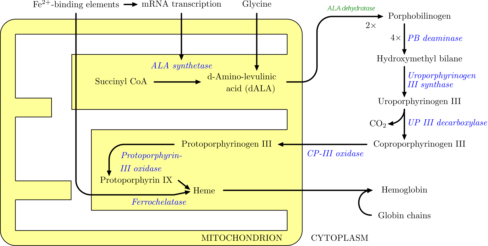 Heme.png 1,920 × 969; 161 KB
-
 Epithelial Ovarian carcinoma.jpg 256 × 256; 10 KB
-
 Craniopharyngioma-t1sagkm-006.jpg 512 × 513; 58 KB
-
 Necrotizing-sialometaplasia.png 773 × 556; 426 KB
-
 Nasopharyngeal angiofibroma.jpeg 4,272 × 2,848; 5.41 MB
-
 Craniopharyngioma-t1corkm-001.jpg 512 × 512; 21 KB
-
 Craniopharyngioma-t1corkm-003.jpg 512 × 512; 21 KB
-
 Craniopharyngioma-t2ax-003.jpg 512 × 512; 20 KB
-
 Thymoma GIF.gif 384 × 288; 21 KB
-
 Mediastinal Lipoma.gif 384 × 288; 48 KB
-
 BCC Nodular type.jpeg 256 × 388; 26 KB


.jpg)


.jpg)


.png)


