Unused files
Jump to navigation
Jump to search
The following files exist but are not embedded in any page. Please note that other websites may link to a file with a direct URL, and so may still be listed here despite being in active use.
Showing below up to 50 results in range #25,821 to #25,870.
-
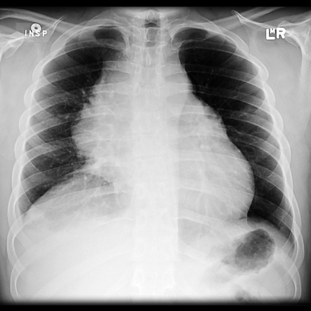
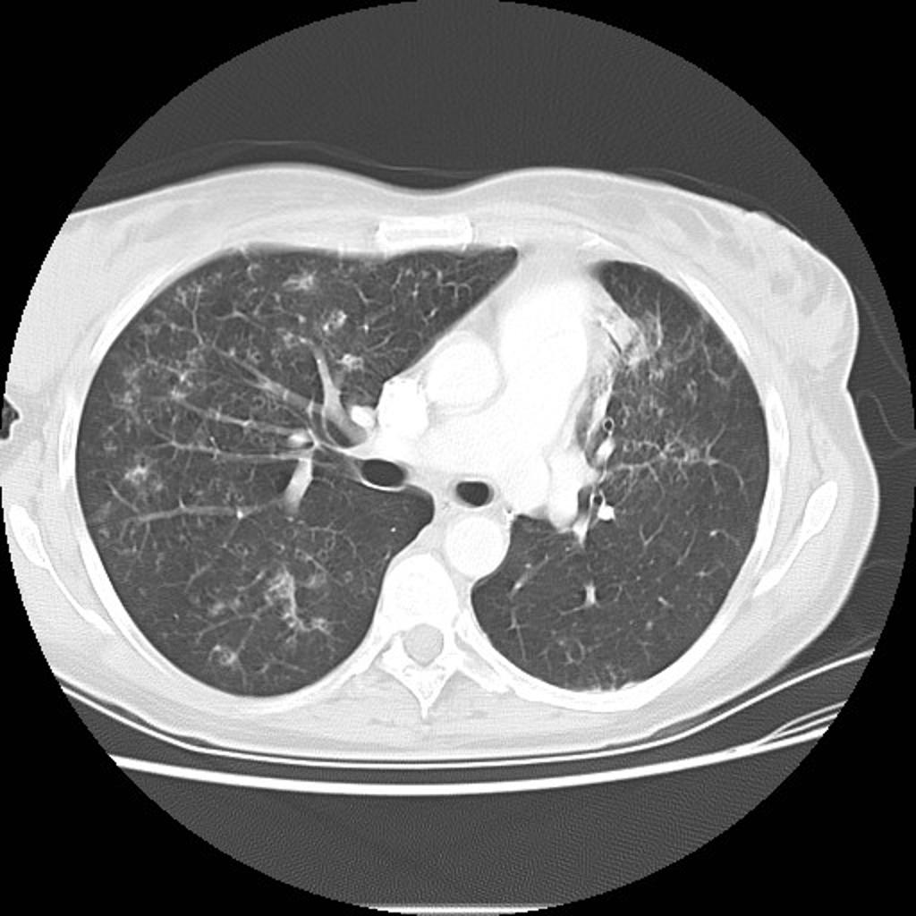 Multifocal-adenocarcinoma-of-lung-08.jpg 1,024 × 1,024; 102 KB
-
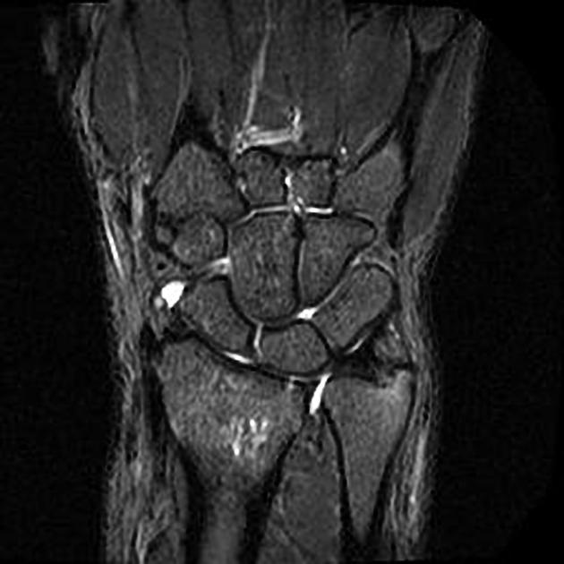 Ulnar fracture.jpg 630 × 630; 37 KB
-
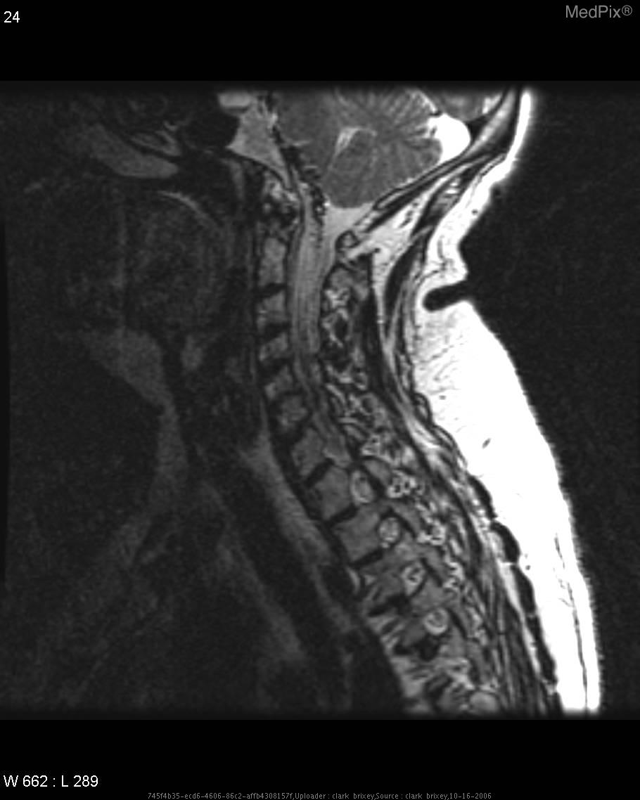
 Dural AVF, cervical spine 1.jpg 910 × 1,138; 69 KB
-
 Dural AVF, cervical spine 2.jpg 910 × 1,138; 69 KB
-
 Dural AVF, cervical spine 3.jpg 910 × 1,138; 69 KB
-
 Incidence, ulnar.gif 1,098 × 764; 36 KB
-
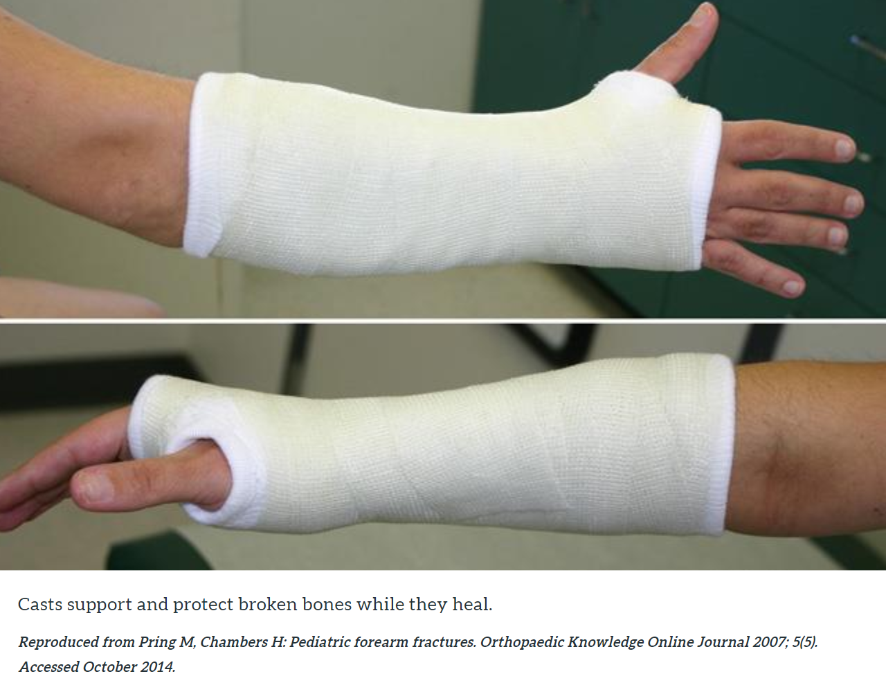 Ulnar Fracture Casting.png 1,280 × 986; 1.12 MB
-
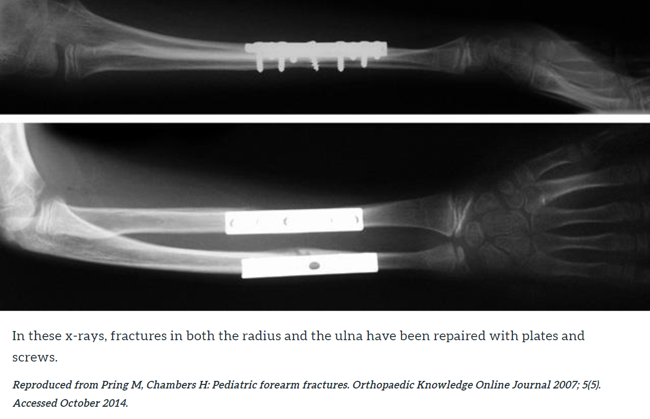 Ulnar bone surgical treatment intervention.png 1,276 × 824; 407 KB
-
 Ulna fracture intramedullary nailing.png 1,868 × 1,042; 906 KB
-
-
 Archdischild-2004-September-89-9-809-F1.large.jpg 1,280 × 847; 112 KB
-
 Pneumothorax-10.jpg 1,024 × 946; 84 KB
-
 Small-bowel-obstruction-12.jpg 839 × 1,024; 133 KB
-
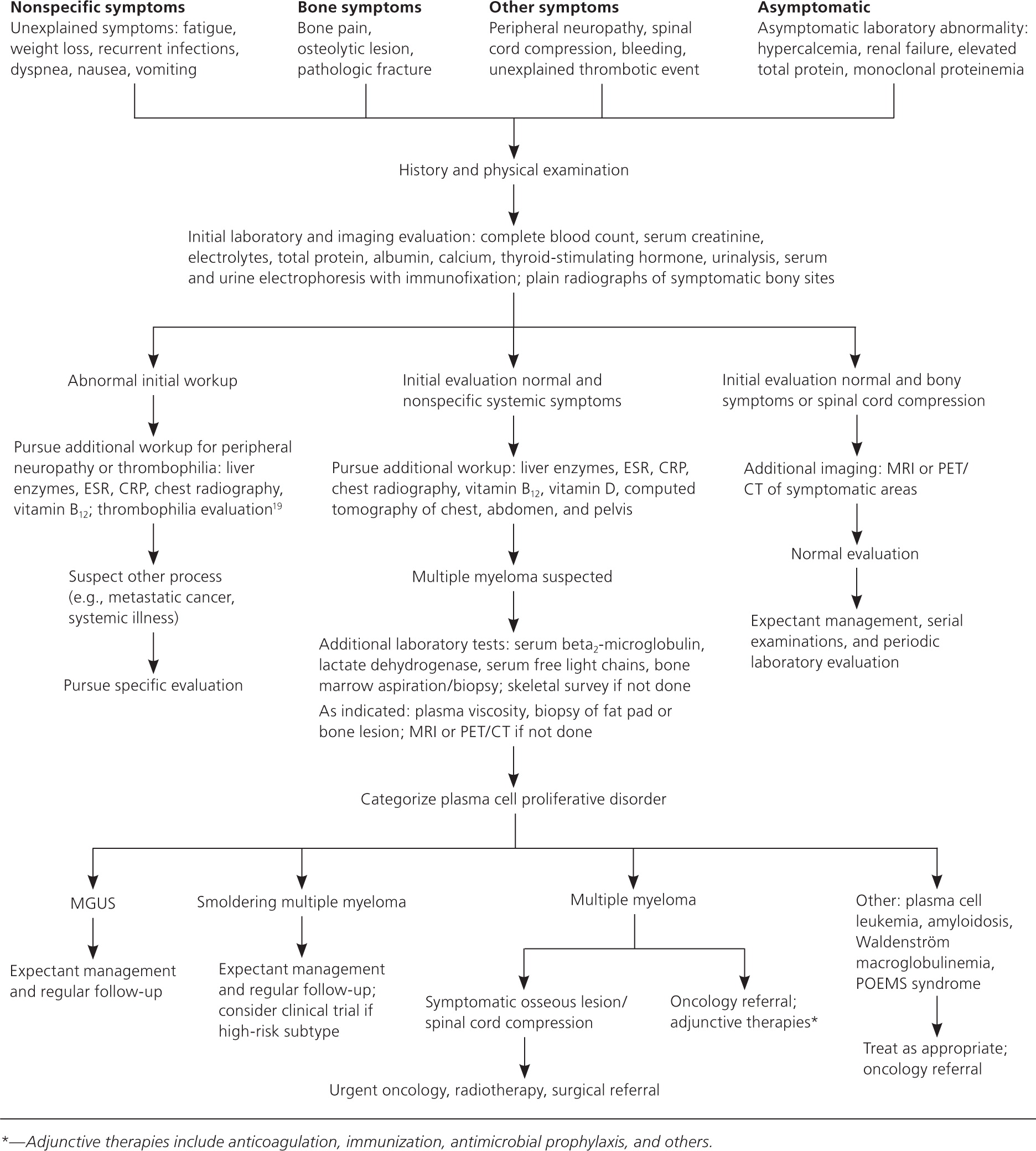 Evaluation of multiple myeloma.jpg 1,872 × 2,080; 562 KB
-
 Lytic lesion in right forearm.jpg 500 × 1,388; 47 KB
-
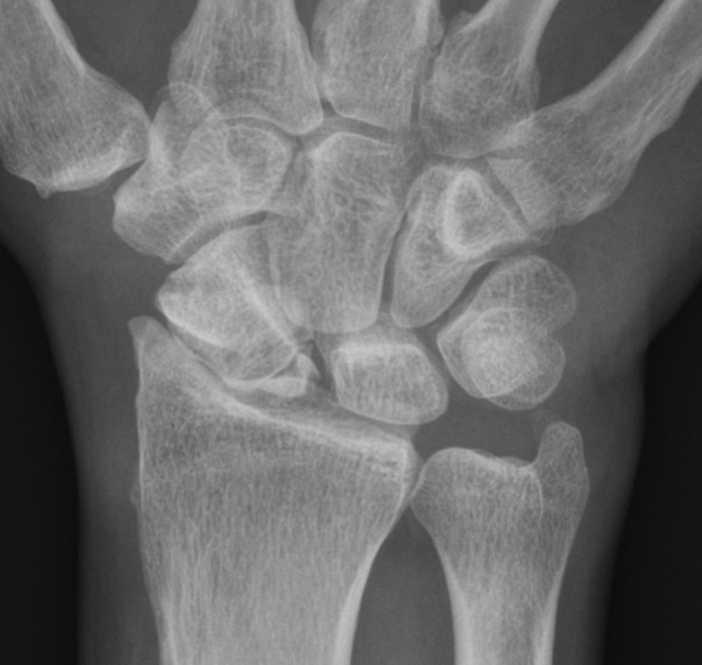 Scaph.jpg 1,024 × 970; 69 KB
-
 Ulnar.jpg 1,019 × 1,024; 102 KB
-
 PTCL NOS.jpg 256 × 256; 20 KB
-
 Hussain Syed Muhammad Dildar circle.png 300 × 300; 94 KB
-
 Action potential.svg 0 × 0; 13 KB
-
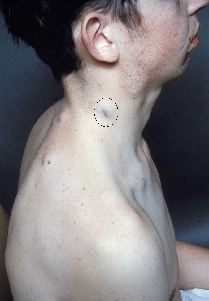 BRNS.jpg 675 × 970; 72 KB
-
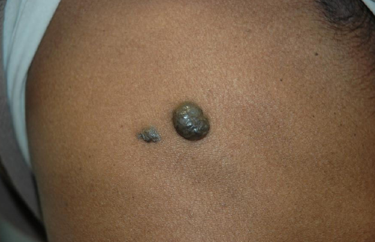 BRBNS.png 755 × 488; 555 KB
-
 Annular Needle Driving Part 1.mp4 ; 28.93 MB
-
 Yannam Chandrakala circle.png 307 × 300; 149 KB
-
 Rhabdomyoma -- very high mag.jpg 120 × 80; 6 KB
-
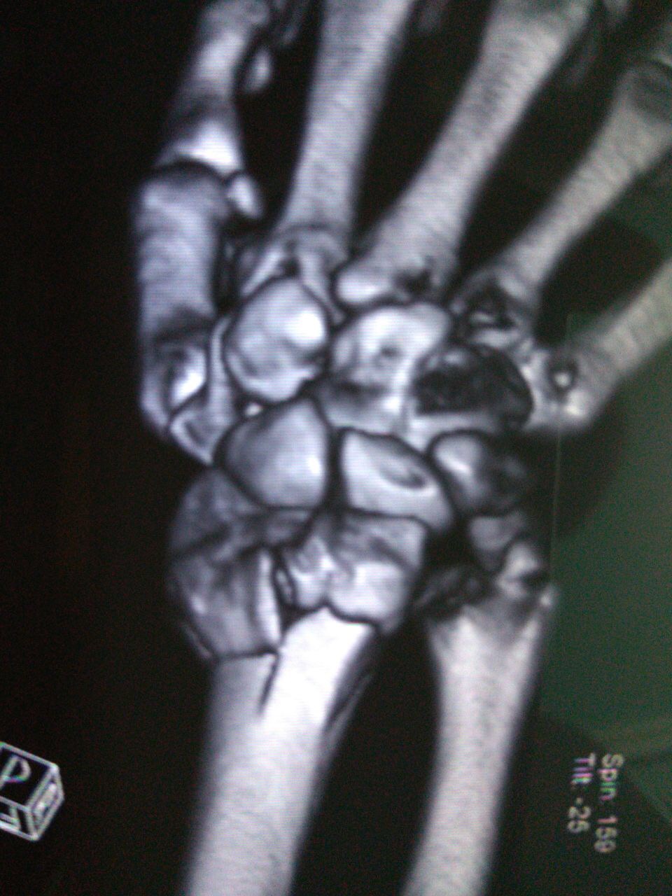 CT scan Wrist dorsal view.JPG 960 × 1,280; 233 KB
-
 CT scan wrist ventral view.JPG 960 × 1,280; 221 KB
-
 CT scan Lateral View with intraarticular step.JPG 820 × 843; 98 KB
-
 CT scan DER VENTRAL.JPG 960 × 1,280; 221 KB
-
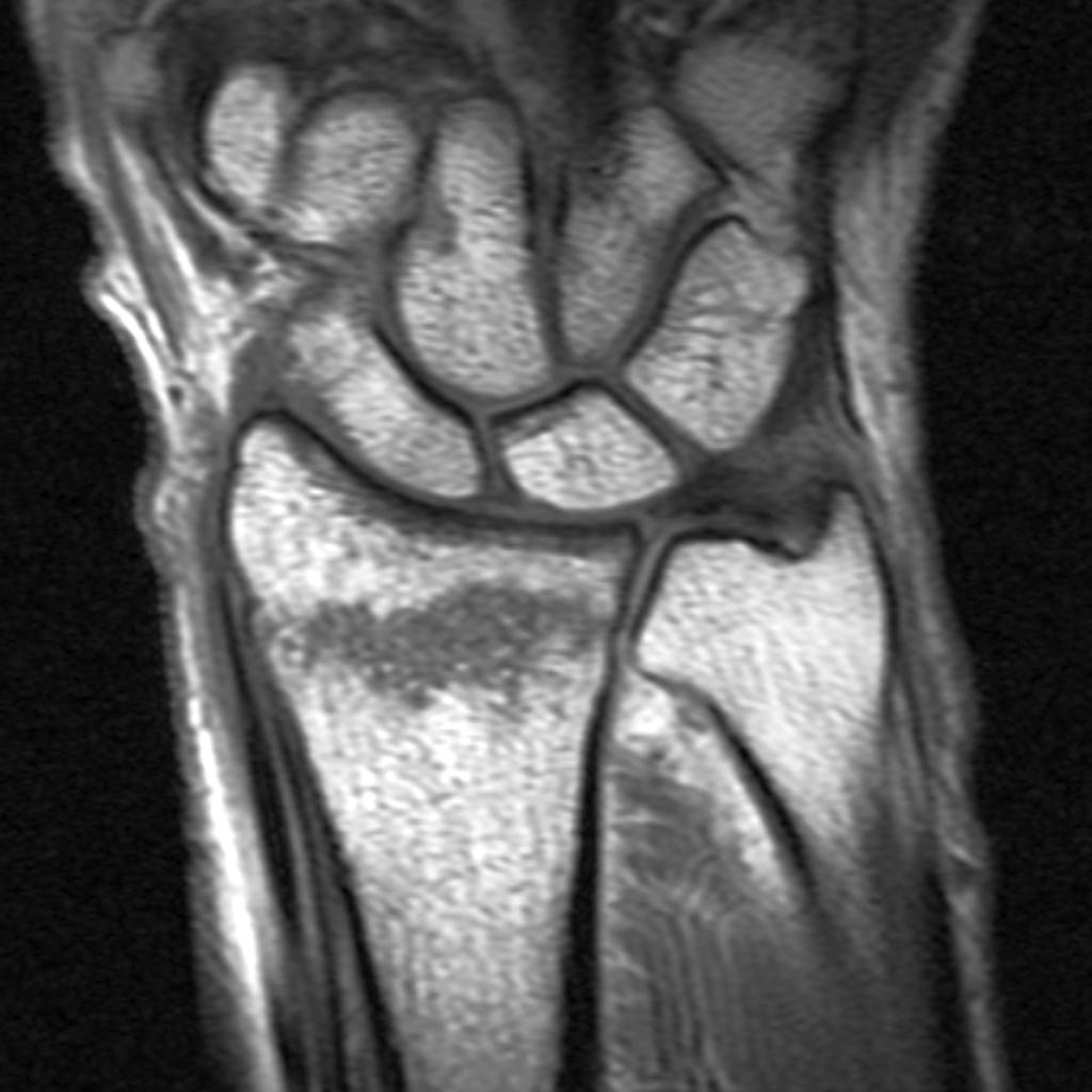 MRI- non-displaced-distal-radial-fracture-.jpg 1,024 × 1,024; 82 KB
-
 WIN 20181212 14 44 03 Pro.jpg 479 × 621; 68 KB
-
 Nima.jpg 48 × 47; 24 KB
-
 Xray Wrist AP and Lat view.JPG 1,280 × 960; 78 KB
-
 Nejmra021562 t1.jpeg 300 × 254; 26 KB
-
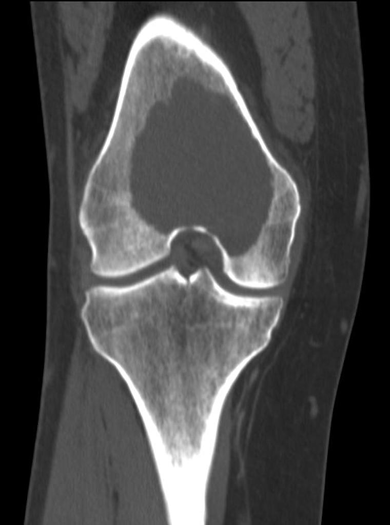 CT giant-cell-tumour-femur .jpg 760 × 1,024; 42 KB
-

-
 Badria Munir .jpg 569 × 744; 114 KB
-
 Primary mediastinal large B-cell lymphoma .gif 1,280 × 720; 193 KB
-
 Primary mediastinal-lymphoma-large-b-cell-1.jpg 1,024 × 1,024; 88 KB
-
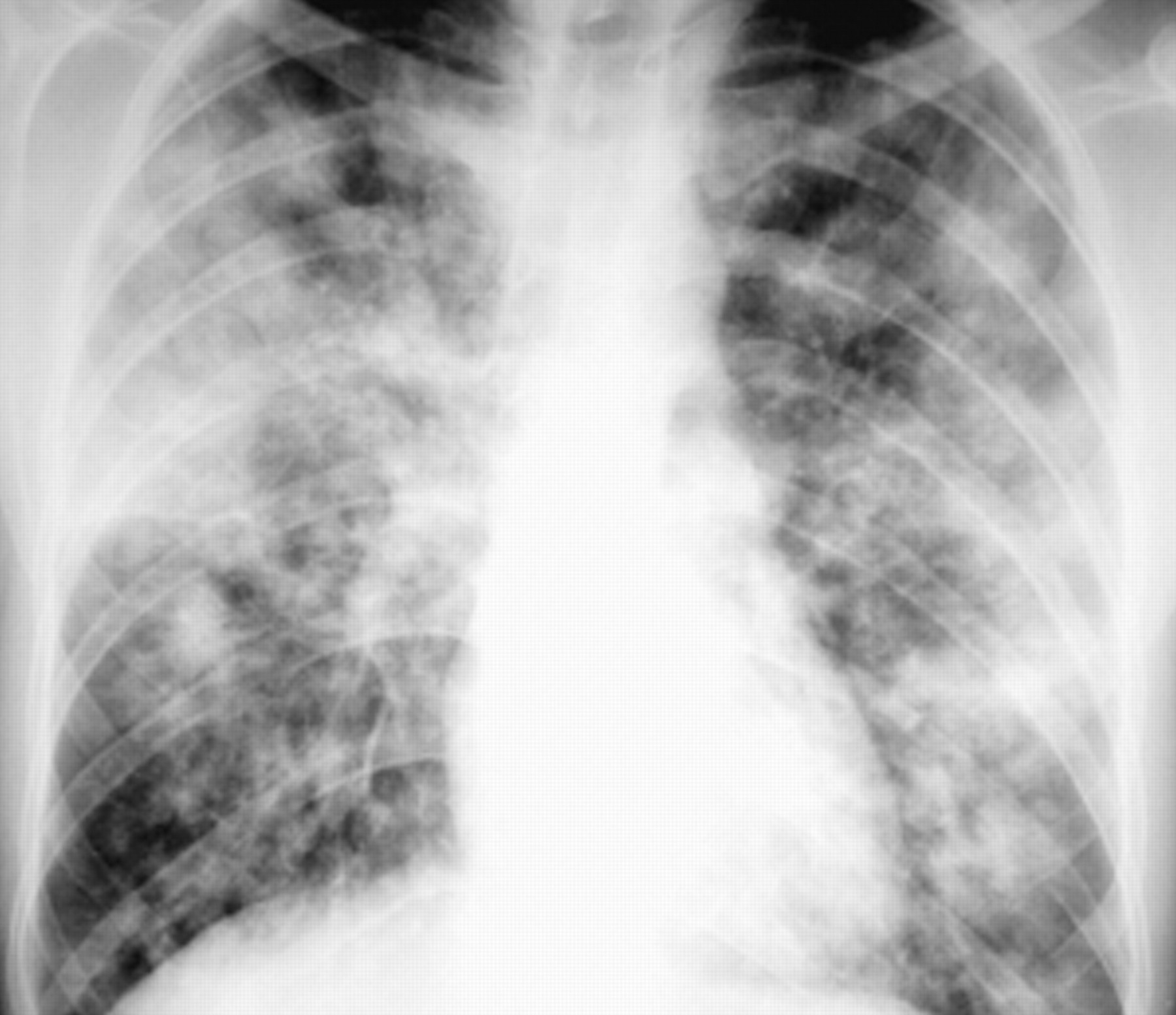 Bronchopneumonia caused by aspergillus.jpg 1,800 × 1,554; 267 KB
-
 Microscopic pathology of phyllodes tumor.jpg 800 × 533; 179 KB
-
 Sclerosing adenosis.jpg 800 × 533; 180 KB
-
 Pilocytic-astrocytoma.jpg 979 × 1,024; 52 KB
-
 Pilocytic-astrocytomaa.jpg 979 × 1,024; 52 KB
-
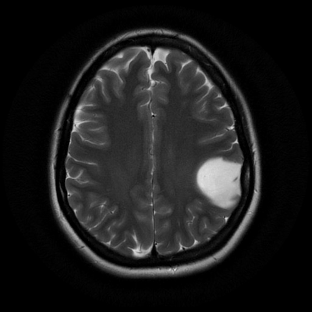 Diffuse-astrocytoma-nos-4.jpg 1,024 × 1,024; 87 KB
-
 Breast-hamartoma.jpg 941 × 1,024; 87 KB
-
 Breast-abscess-8.jpg 1,024 × 699; 86 KB
-
 Output OMCBD0.gif 672 × 378; 125 KB
-
 Breast hamartoma.gif 960 × 720; 253 KB
-
 Ali Syed Musadiq circle.png 300 × 300; 76 KB


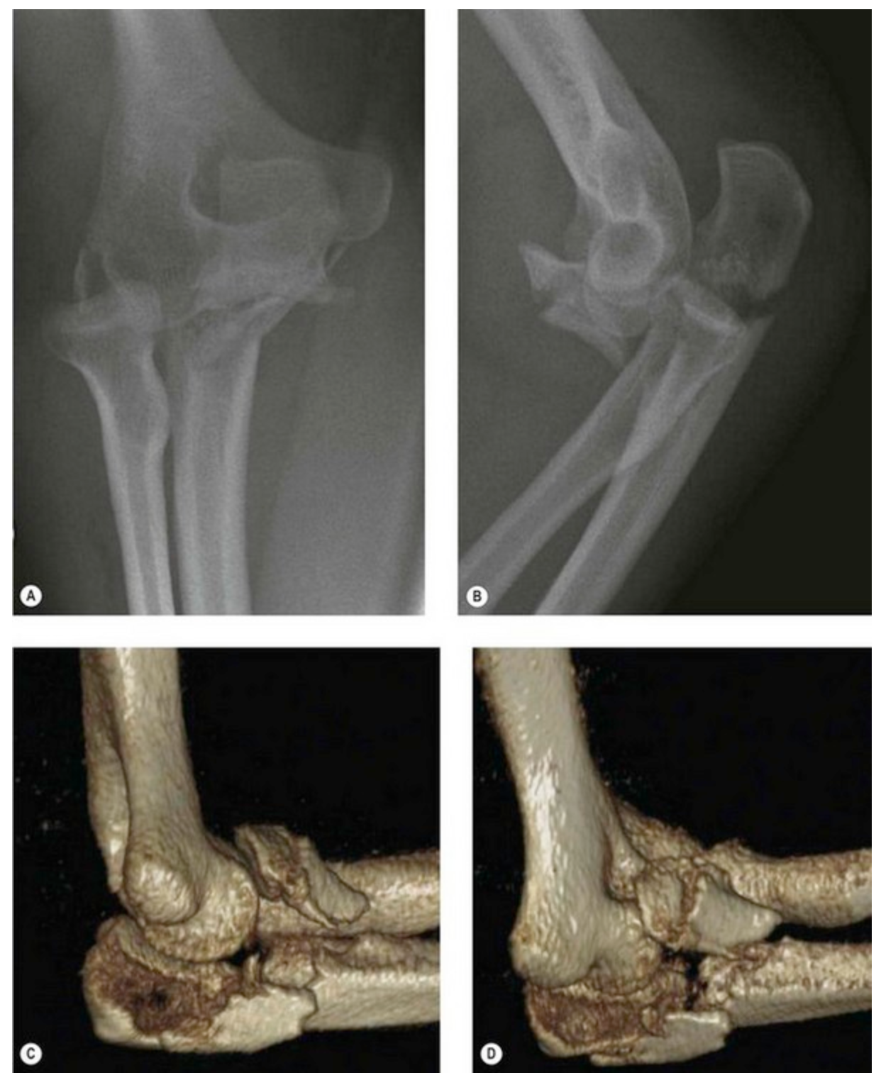
_The_radiographs_do_not_show_the_exact_anatomy_of_the_proximal_ulna_fracture._(C,_D)_CT_3-D_reconstruction_of_the_same_patient_clearly_identifies_the_fracture_morphology..png)