Unused files
Jump to navigation
Jump to search
The following files exist but are not embedded in any page. Please note that other websites may link to a file with a direct URL, and so may still be listed here despite being in active use.
Showing below up to 50 results in range #25,481 to #25,530.
-
 NET Pancreatic.jpg 4,272 × 2,848; 4.95 MB
-
 Gastrinoma.jpg 729 × 512; 339 KB
-
 400px-Gastric neuroendocrine tumour - high mag.jpg 400 × 600; 104 KB
-
 800px-Gastric neuroendocrine tumour - low mag.jpg 800 × 533; 129 KB
-
 400px-Gastric neuroendocrine tumour - intermed mag.jpg 400 × 600; 106 KB
-
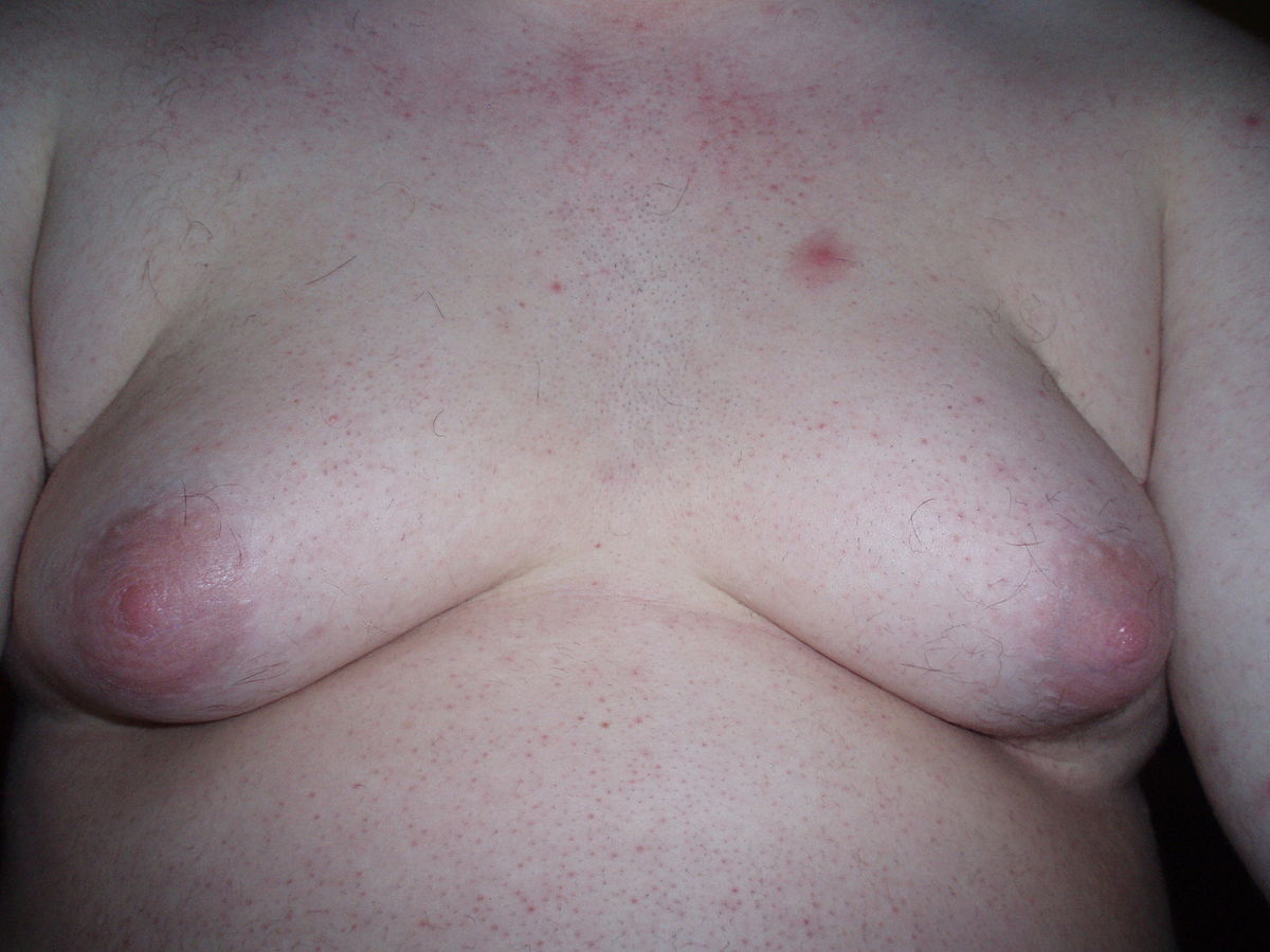 Gynecomastia 1.jpg 1,200 × 900; 113 KB
-
 Ekg brugada pattern.jpg 250 × 201; 10 KB
-
 Case courtesy of Dr Henry Knipe, Radiopaedia.org, rID 45995.jpg 1,024 × 804; 117 KB
-
 Gynecomastia 2 (1).jpg 800 × 533; 468 KB
-
 Furqan Muhammad circle.png 300 × 300; 86 KB
-
 Ghaffarpasand Eiman circle.png 300 × 300; 117 KB
-
 Jafarizade Mehrian circle.png 300 × 300; 110 KB
-
 Kothagadi Aravind Reddy circle.png 300 × 300; 79 KB
-
 DSC 0593.JPG 6,000 × 4,000; 6.17 MB
-
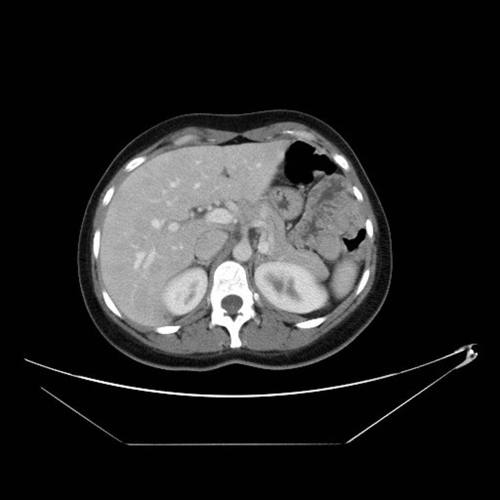
 Struma ovarii Chest CT.jpg 600 × 530; 45 KB
-
 Struma ovarii Chest Xray rmassive pleural effusion.jpg 551 × 229; 29 KB
-
 Struma ovarii Microscopic appearance of the resected tumor.jpg 600 × 450; 138 KB
-
 Struma ovarii Pelvic CT scan.jpg 600 × 267; 31 KB
-
 Struma ovarii Axial CT images.jpg 610 × 254; 83 KB
-
 Struma ovarii Transabdomen grayscale US.jpg 719 × 576; 99 KB
-
 Struma ovarii Transvaginal US.jpg 771 × 254; 88 KB
-
 Struma ovarii Transvaginal US 2.jpg 756 × 338; 117 KB
-
 Struma ovarii H&E.jpg 764 × 576; 267 KB
-
 Struma ovarii - Scintigraphy.jpg 396 × 467; 16 KB
-
 Struma ovarii - Scintigraphy 2.jpg 609 × 607; 52 KB
-
 Struma ovarii - Scintigraphy 3.jpg 307 × 849; 39 KB
-
 Struma ovarii - MR 2.jpg 609 × 553; 51 KB
-
 Struma ovarii - ultrasound.jpg 307 × 849; 39 KB
-
 Struma ovarii - ultrasound 2.jpg 609 × 875; 111 KB
-
 Struma ovarii - MR 3.jpg 609 × 303; 41 KB
-
 Struma ovarii - MR.jpg 609 × 607; 52 KB
-
 Pituitary-apoplexy-marked.jpg 992 × 1,024; 125 KB
-
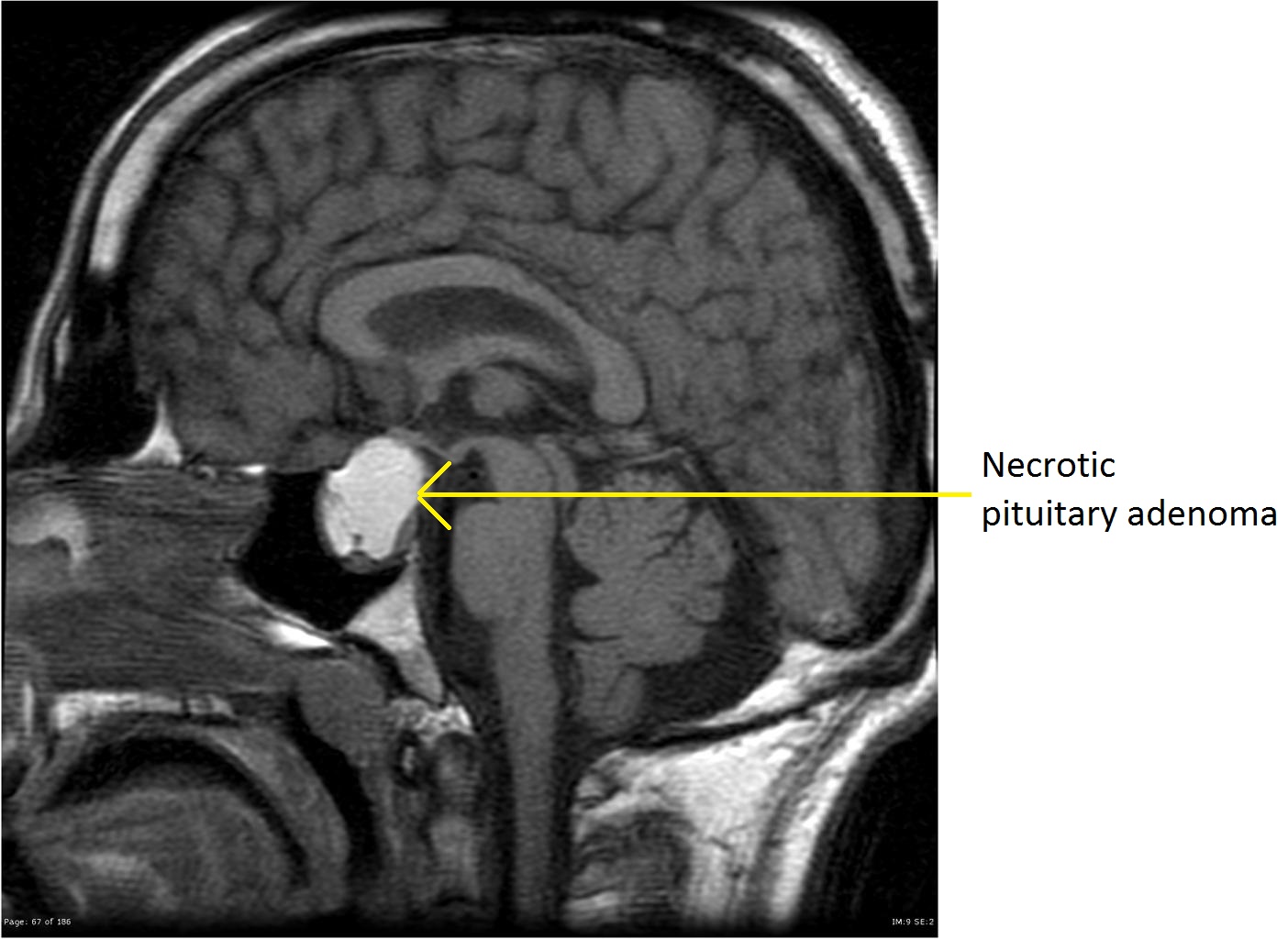 Pituitary-macroadenoma-necrotic-1marked.jpg 1,404 × 1,024; 185 KB
-
 Symptoms of menopause (vector).svg 0 × 0; 1.94 MB
-
 Wikidoc image.jpeg 160 × 207; 7 KB
-
 Pulmonary cryptococcosis (2) Alcian blue-PAS.jpg 240 × 181; 23 KB
-
 Ectopic-posterior-pituitary-1.jpg 1,024 × 1,024; 154 KB
-
 Ectopic-posterior-pituitary.jpg 1,024 × 1,024; 154 KB
-
 Pituitary-apoplexy-1.jpg 1,024 × 1,024; 146 KB
-
 Osteoporosis.gif 1,024 × 1,005; 3.97 MB
-
 Compressionfracture.gif 1,024 × 1,005; 6.89 MB
-
 Vertebra.jpg 1,024 × 1,024; 238 KB
-
 Osteoporosi.gif 512 × 672; 668 KB
-
 Empty-sella-4.jpg 896 × 1,024; 67 KB
-
 Path-pit-adenom ETX-PIT-3A-FIG-03.jpg 754 × 571; 194 KB
-
 Osteoporosis Locations.png 1,200 × 1,200; 1.44 MB
-
 Morbus Fabry MRT Osteoporosis 01.jpg 1,200 × 735; 147 KB
-
 Insulinoma-men-type-1.jpg 1,024 × 1,024; 49 KB
-
 Metastatic-insulinoma.jpg 1,024 × 1,024; 57 KB
-
 Insulinoma-4.jpg 1,024 × 1,024; 218 KB


.jpg)


.svg)

_Alcian_blue-PAS.jpg)


