Unused files
Jump to navigation
Jump to search
The following files exist but are not embedded in any page. Please note that other websites may link to a file with a direct URL, and so may still be listed here despite being in active use.
Showing below up to 50 results in range #25,451 to #25,500.
-
 A015828ba66db9da16b5a57d0cb2c6 jumbo.jpeg 842 × 1,024; 92 KB
-
 56dce39ec7860a50a4bf060db455b7 jumbo.jpeg 1,024 × 810; 402 KB
-
 Faa79e035d4c88b1029b3f6cd6e222 jumbo.jpeg 1,024 × 986; 57 KB
-
 2739a8754252754d33b8ee2d413894 big gallery.jpg 630 × 489; 46 KB
-
 9763697dab2e8388d7a54b513a3e42 jumbo.jpg 1,023 × 766; 52 KB
-
 9531e159d25c6beef968d6c1caf46c jumbo.jpg 1,024 × 910; 59 KB
-
 Ec9e2a6a4c1d377bdee5f0ccbbf180 big gallery.jpg 551 × 630; 38 KB
-
 D1797f6a1a111e62b0f9ba00fc93bb big gallery.jpg 630 × 630; 38 KB
-
 C13ada80fec4008d775777547a65ef big gallery.jpg 630 × 492; 31 KB
-
 SIADH causes.jpg 300 × 168; 12 KB
-
 CT showing intrasellar mas.jpg 718 × 581; 141 KB
-
 CT showing intrasellar mas2.jpg 718 × 581; 141 KB
-
 Dka.jpg 819 × 460; 77 KB
-
 ZES - GASTRINOMA TRIANGLE 2.jpg 1,653 × 1,479; 207 KB
-
 Vertebral-insufficiency-fractures-in-severe-osteoporosis.jpg 1,024 × 1,024; 96 KB
-
 Vertebral-insufficiency-fractures-in-severe-osteoporosis(4).jpg 1,024 × 1,024; 132 KB
-
 Scar endo....jpg 1,024 × 825; 157 KB
-
 Us.jpg 1,024 × 1,024; 168 KB
-
 Gynecomastia-etiolog MR-14-fig1.jpg 800 × 563; 195 KB
-
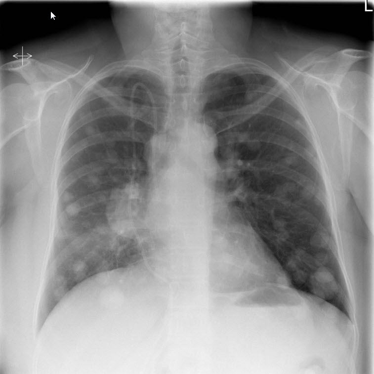 Lung cancer-2.jpg 757 × 757; 48 KB
-
 Octreo.jpg 630 × 511; 39 KB
-
 Shsh.png 1,203 × 531; 812 KB
-
 O0201af1.jpg 198 × 195; 23 KB
-
 Ct lung cancer.jpg 442 × 442; 27 KB
-
 MRI of brain abscess.jpg 630 × 630; 27 KB
-
 Mri subarachnoid hemorrhage.jpg 630 × 630; 44 KB
-
 Vertebra-plana.jpg 1,024 × 1,005; 82 KB
-
 Kazmi.Jpg 742 × 897; 119 KB
-
 Output 4prhSH.gif 963 × 1,024; 1.18 MB
-
 NET Duodenum.jpg 1,521 × 1,348; 967 KB
-
 NET Pancreatic.jpg 4,272 × 2,848; 4.95 MB
-
 Gastrinoma.jpg 729 × 512; 339 KB
-
 400px-Gastric neuroendocrine tumour - high mag.jpg 400 × 600; 104 KB
-
 800px-Gastric neuroendocrine tumour - low mag.jpg 800 × 533; 129 KB
-
 400px-Gastric neuroendocrine tumour - intermed mag.jpg 400 × 600; 106 KB
-
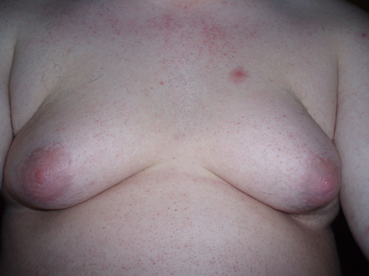 Gynecomastia 1.jpg 1,200 × 900; 113 KB
-
 Ekg brugada pattern.jpg 250 × 201; 10 KB
-
 Case courtesy of Dr Henry Knipe, Radiopaedia.org, rID 45995.jpg 1,024 × 804; 117 KB
-
 Gynecomastia 2 (1).jpg 800 × 533; 468 KB
-
 Furqan Muhammad circle.png 300 × 300; 86 KB
-
 Ghaffarpasand Eiman circle.png 300 × 300; 117 KB
-
 Jafarizade Mehrian circle.png 300 × 300; 110 KB
-
 Kothagadi Aravind Reddy circle.png 300 × 300; 79 KB
-
 DSC 0593.JPG 6,000 × 4,000; 6.17 MB
-
 Struma ovarii Chest CT.jpg 600 × 530; 45 KB
-
 Struma ovarii Chest Xray rmassive pleural effusion.jpg 551 × 229; 29 KB
-
 Struma ovarii Microscopic appearance of the resected tumor.jpg 600 × 450; 138 KB
-
 Struma ovarii Pelvic CT scan.jpg 600 × 267; 31 KB
-
 Struma ovarii Axial CT images.jpg 610 × 254; 83 KB
-
 Struma ovarii Transabdomen grayscale US.jpg 719 × 576; 99 KB


.jpg)


.jpg)


