Unused files
Jump to navigation
Jump to search
The following files exist but are not embedded in any page. Please note that other websites may link to a file with a direct URL, and so may still be listed here despite being in active use.
Showing below up to 50 results in range #24,941 to #24,990.
-
 Primary cutaneous follicle centre lymphoma image 06.jpg 1,200 × 1,594; 873 KB
-
 Primary cutaneous follicle centre lymphoma image 07.jpg 1,200 × 904; 383 KB
-
 Primary cutaneous follicle centre lymphoma image 08.jpg 1,200 × 593; 194 KB
-
 Primary cutaneous follicle centre lymphoma image 09.jpg 1,200 × 900; 189 KB
-
 Primary cutaneous follicle centre lymphoma image 10.jpg 1,200 × 900; 179 KB
-
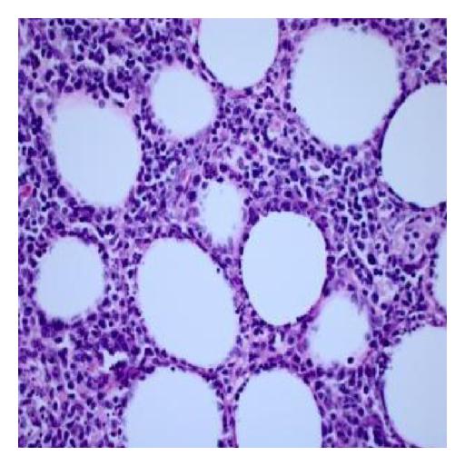
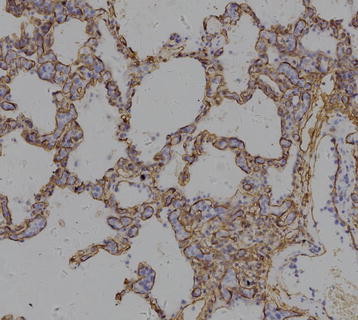
 Squamous lung cancer mircopathology.jpeg 1,599 × 1,066; 424 KB
-
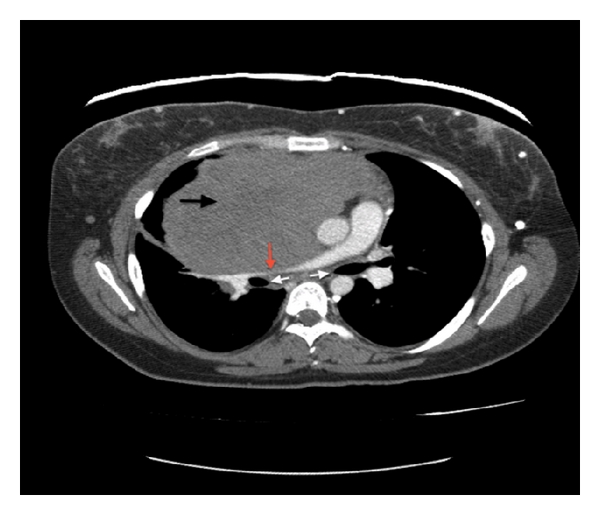 Primary mediastinal large B-cell lymphoma CT scan 1 .jpg 600 × 515; 92 KB
-
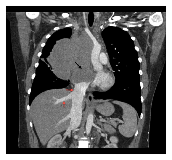 Primary mediatinsl large B-cell lymphoma coronal CT scan.jpg 600 × 560; 130 KB
-
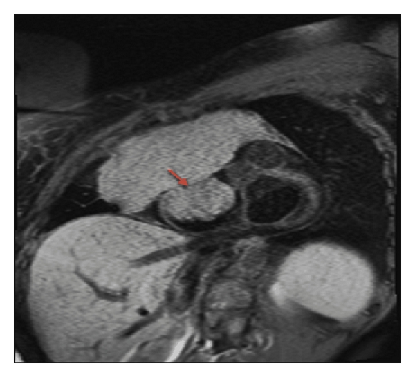 Primary medistinal large B-cell lymphoma MRI .jpg 600 × 545; 116 KB
-
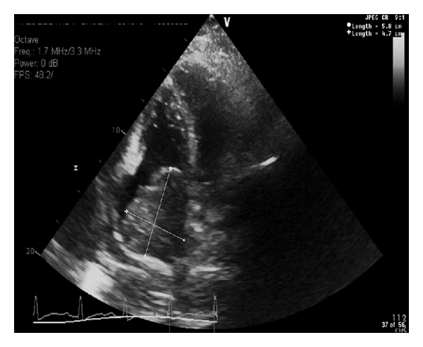 Primary mediastinal large B-cell lymphoma echo.jpg 600 × 492; 97 KB
-
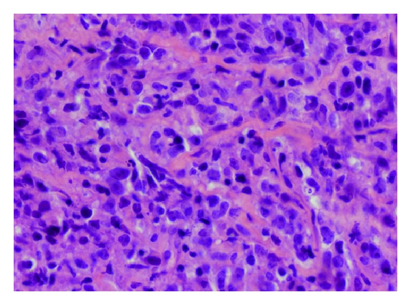 Primary mediastinal large B-cell lymphoma pathology .jpg 600 × 446; 202 KB
-
 Primary medistinal large b-cell lymphoma pathology 1 .jpg 600 × 449; 245 KB
-
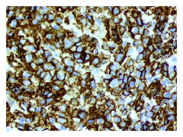
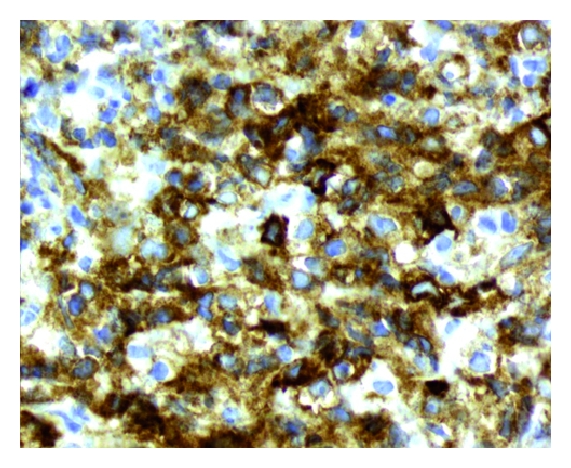 Primary mediastinal large B-cell lymphoma pathology 2.jpg 571 × 468; 223 KB
-
 German anti-smoking ad.jpeg 442 × 360; 36 KB
-
 Large cell carcinoma of the lung .jpg 701 × 512; 324 KB
-
 Large cell carcinoma of the lung.jpg 711 × 512; 308 KB
-
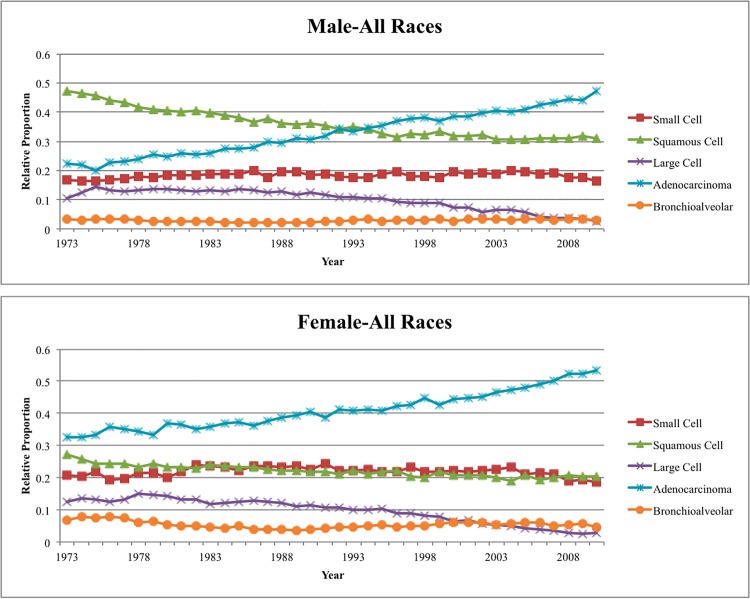 Gender nonsmallcell Graph.jpeg 750 × 599; 108 KB
-
 Subcutaneous panniculitis-like T-cell lymphoma biopsy 2.jpg 512 × 513; 42 KB
-
 Subcutaneous panniculitis-like T-cell lymphoma biopsy 4.jpg 512 × 512; 36 KB
-
 Subcutaneous panniculitis-like T-cell lymphoma biopsy 5.jpg 512 × 512; 44 KB
-
 Subcutaneous panniculitis-like T-cell lymphoma biopsy 6.jpg 512 × 512; 35 KB
-
 Subcutaneous panniculitis-like T-cell lymphoma biopsy 7.jpg 512 × 512; 31 KB
-
 Intravascular large B-cell lymphoma CT .jpg 600 × 363; 102 KB
-
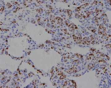
 Large-cell-neuroendocrine-carcinoma-of-the-lung.jpg 1,024 × 826; 329 KB
-
 Intravascular large b-cell lymphoma patology image 1 .jpg 600 × 460; 219 KB
-
 Intravascular large b-cell lymphoma pathophysiology image 2.jpg 600 × 463; 233 KB
-
 Intravascular large b-cell lymphoma image 3.jpg 600 × 463; 244 KB
-
 Intravascular large B-cell lymphoma pathophysiology image 5.jpg 358 × 288; 37 KB
-
 Intravascular large B-cell lymphoma pathology image 6.jpg 358 × 307; 30 KB
-
 Intravascular large B-cell lymphoma pathology image 7 .jpg 358 × 343; 42 KB
-
 Intravascular large B-cell lymphoma pathology image 8.jpg 358 × 320; 45 KB
-
 Intravascular large B-cell lymphoma pathology image 9.jpg 358 × 286; 41 KB
-
 Mri myelofibrosis.jpg 630 × 565; 41 KB
-
 Ct image of myelofibrosis 1.jpg 1,024 × 658; 48 KB
-
 Parotitis002.jpeg 587 × 630; 25 KB
-
 Probable-peritumoral-haemorrhage-in-metastatic-melanoma-ct-halo-sign.jpg 1,008 × 1,024; 61 KB
-
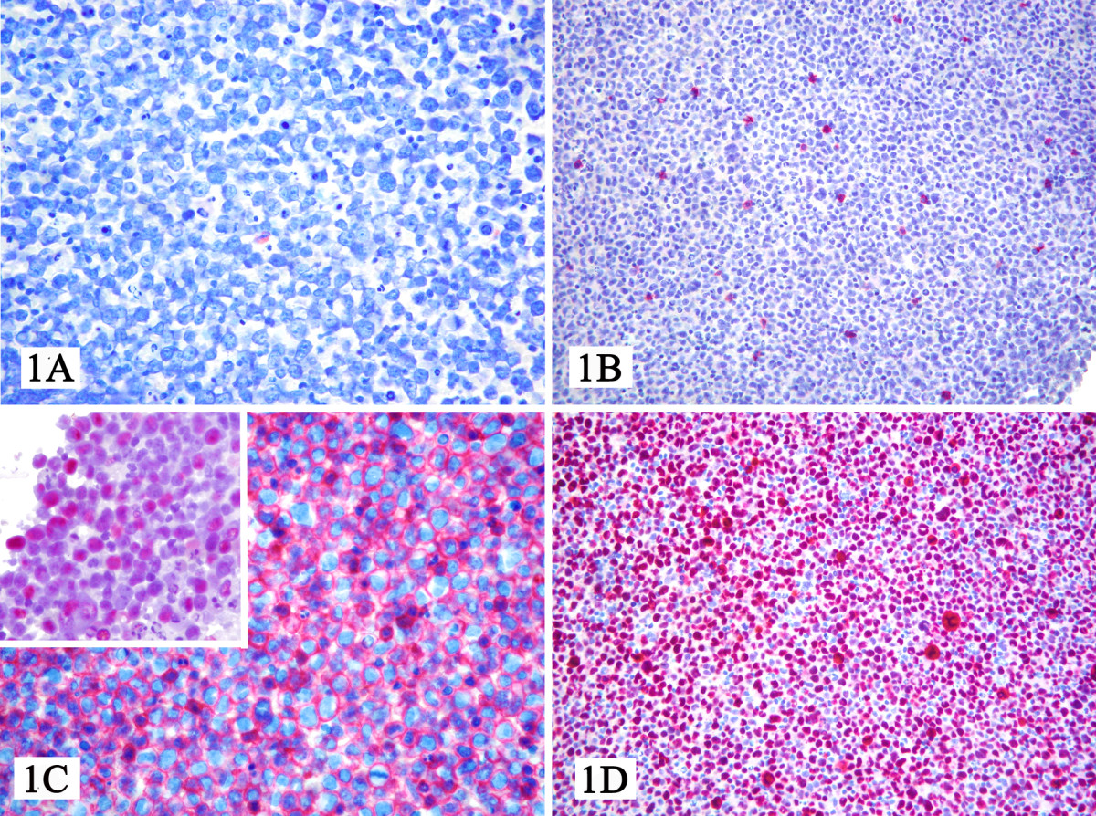 Microscopic pathology of primary effusion lymphoma.jpg 1,200 × 894; 688 KB
-
 Terry's nails.jpg 2,448 × 3,264; 1.59 MB
-
 Dot sign livermass.png 1,004 × 1,004; 753 KB
-
 FNH livermass.png 1,296 × 1,130; 1.16 MB
-
 Pyogenic-liver-abscess-2.jpg 1,024 × 876; 97 KB
-
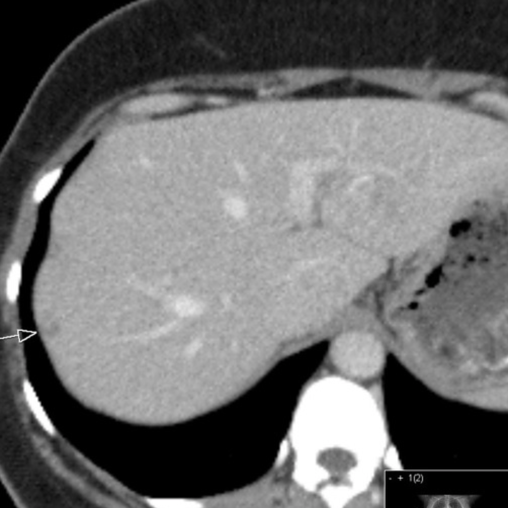
 Hepatoblastoma-1.jpg 1,024 × 1,024; 46 KB
-
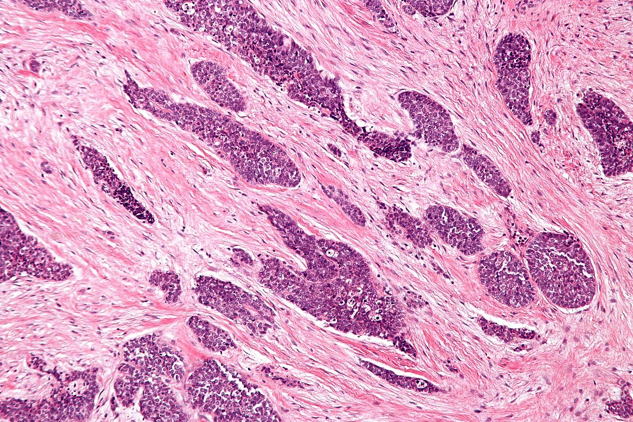 Desmoplastic small round cell tumour - intermed mag.jpg 1,280 × 853; 424 KB
-

-
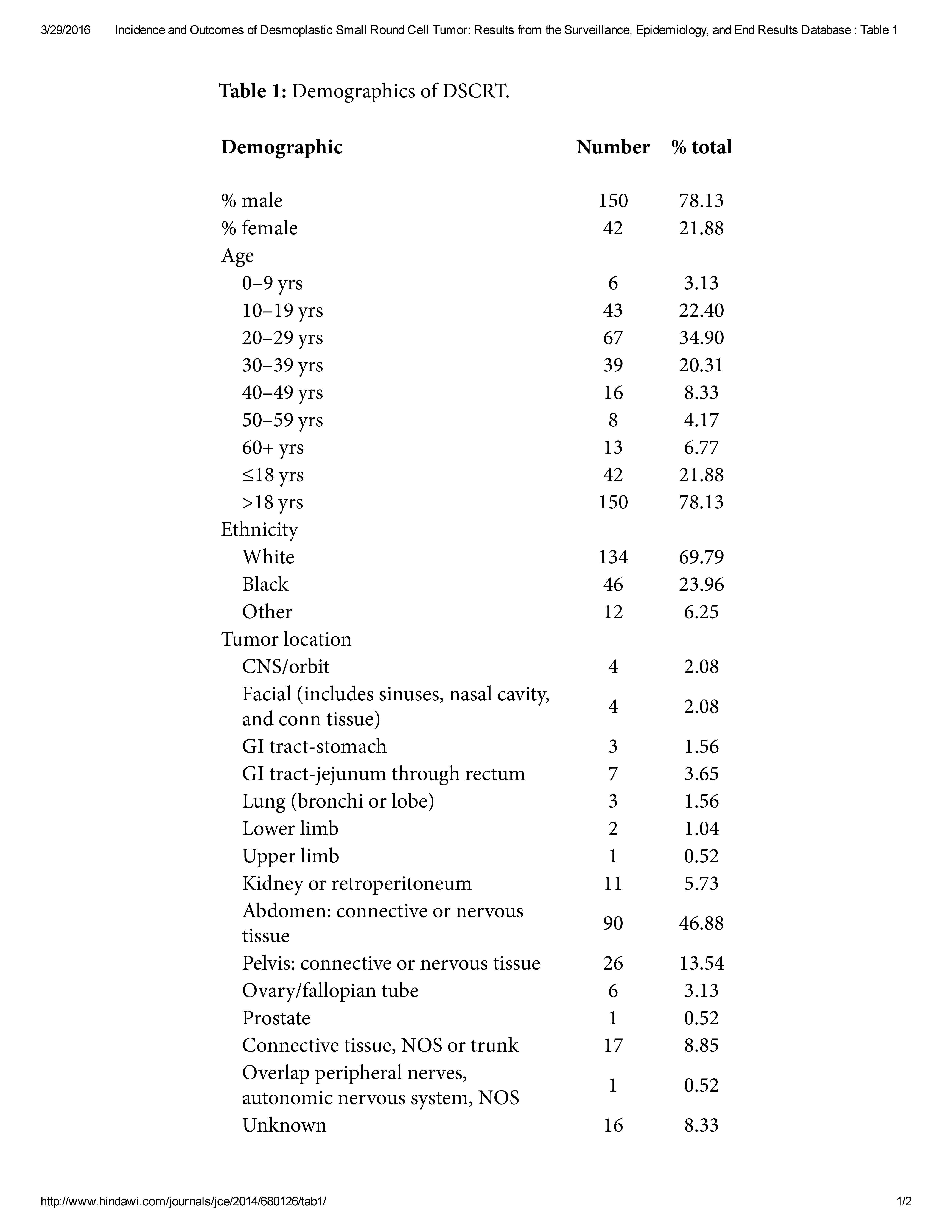 DSRCT.jpg 5,100 × 6,600; 1.12 MB
-
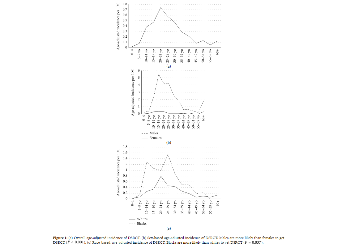 Gender and race DSRCT.PNG 1,106 × 790; 74 KB
-
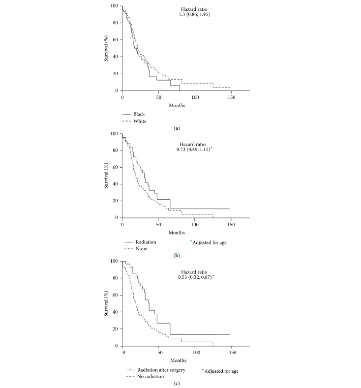 Prognosis DSRCT.PNG 734 × 789; 40 KB
-
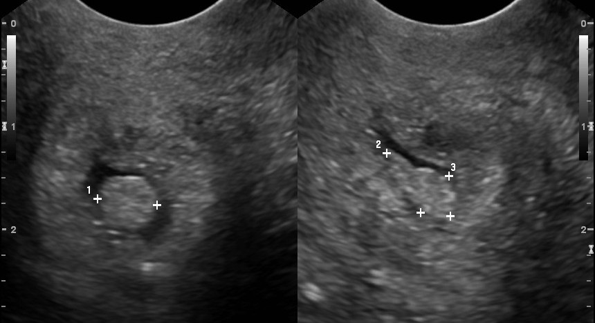 Cervical polyp.jpg 860 × 467; 69 KB
-
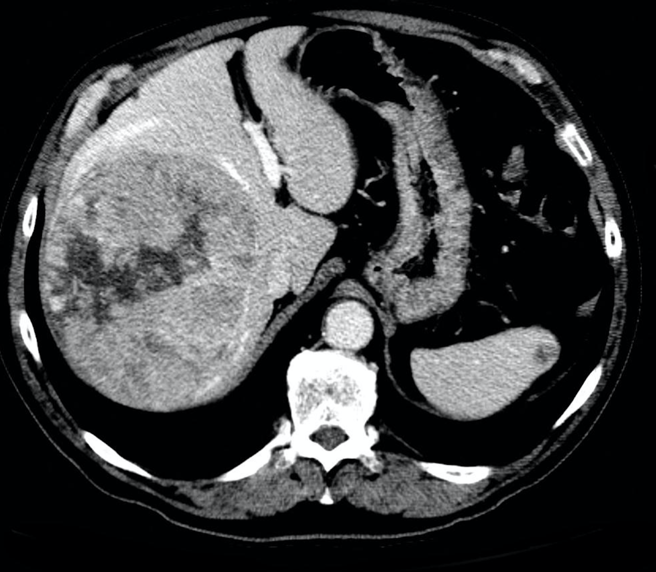
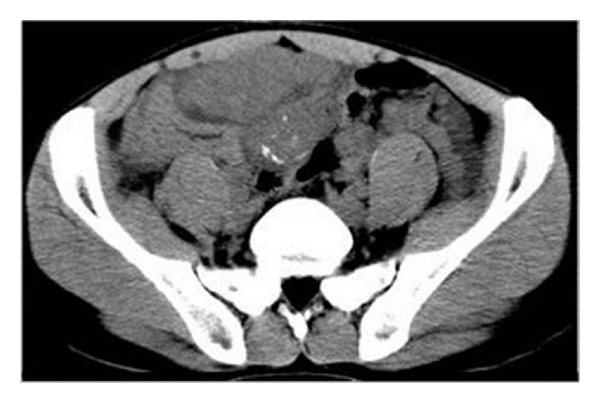 Ct image dsrct.jpg 600 × 403; 92 KB
-
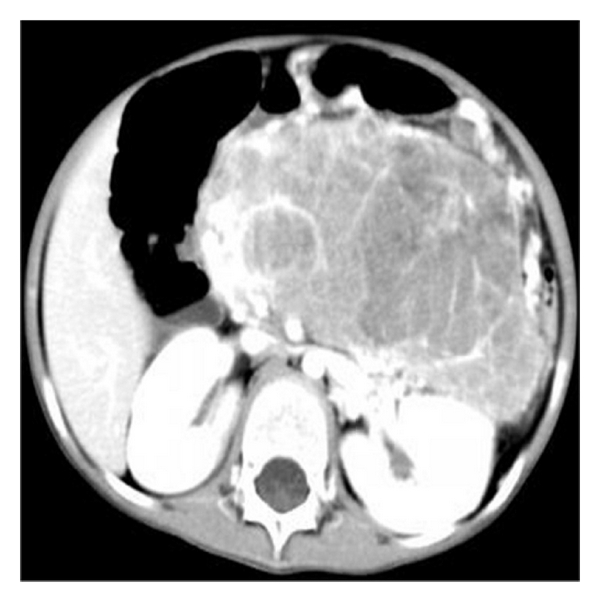 Ct image 2 dsrct.jpg 600 × 602; 101 KB


