Unused files
Jump to navigation
Jump to search
The following files exist but are not embedded in any page. Please note that other websites may link to a file with a direct URL, and so may still be listed here despite being in active use.
Showing below up to 50 results in range #24,871 to #24,920.
-
 120px-Skin Vulva PagetDisease GATA 15BR---.jpg 120 × 86; 7 KB
-
 120px-Skin Vulva PagetDisease MP 15BR---.jpg 120 × 86; 7 KB
-
 120px-Skin Vulva PagetDisease p63 15BR---.jpg 120 × 86; 6 KB
-
 Tick-borne encephalitis pathogenisis.jpg 535 × 314; 47 KB
-
 Tick-borne encephalitis epi.jpg 572 × 293; 52 KB
-
 Lyme Map Epi.jpg 500 × 386; 95 KB
-
 Germinoma1.jpg 800 × 593; 76 KB
-
 G2.jpg 800 × 593; 214 KB
-
 G3.jpg 800 × 593; 108 KB
-
 G4.jpg 800 × 593; 136 KB
-
 Epithelioid sarcoma - smarcb1 - high mag.jpg 800 × 533; 157 KB
-
 Pathological fracture.jpeg 600 × 720; 86 KB
-
 Geographic skull multiple m.jpeg 128 × 176; 5 KB
-
 Sagital T1 pineal germinoma.jpg 604 × 630; 44 KB
-
 Sagital T1 C1+.jpg 630 × 630; 52 KB
-
 VZV Encephalitis CT.jpeg 358 × 442; 29 KB
-
 Extranodal NK-T cell lymphoma.jpg 600 × 461; 59 KB
-
 Extranodal NK-T cell lymphoma image 1.jpg 600 × 601; 23 KB
-
 Extranodal NK-T cell lymphoma PET-CT .jpg 600 × 601; 28 KB
-
 Submental dermoid cyst.jpg 600 × 416; 151 KB
-
 Submental dermoid cyst2.jpg 600 × 481; 182 KB
-
 Dermoid cyst HPE1.jpg 600 × 423; 231 KB
-
 Dermoid cyst HPE2.jpg 600 × 632; 328 KB
-
 Submental dermoid cyst CT1.jpg 600 × 511; 137 KB
-
 Submental dermoid cystCT2.jpg 600 × 564; 189 KB
-
 Submental dermoid cyst US1.jpg 600 × 645; 177 KB
-
 Submental dermoid cyst US2.gif 100 × 70; 5 KB
-
 Scintiscan1.jpg 600 × 452; 178 KB
-
 Scintiscan2.jpg 600 × 592; 252 KB
-
 Spinal dermoid cyst MRI1.jpg 600 × 1,213; 178 KB
-
 Spinal dermoid cyst MRI2.gif 100 × 181; 27 KB
-
 Mucoepidermoid-carcinoma-parotid-1.jpg 1,023 × 957; 72 KB
-
 Extranodal Nk-T cell lymphoma image 2.jpg 600 × 461; 53 KB
-
 Ovarian-dermoid-2.jpg 1,024 × 834; 92 KB
-
 Race non small cell lung cancer.png 1,458 × 750; 118 KB
-
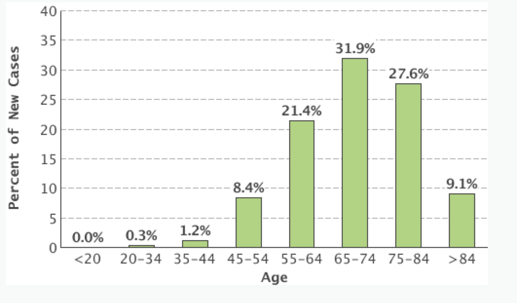 Age adjusted incidence non small cell lung cancer.png 1,062 × 622; 76 KB
-
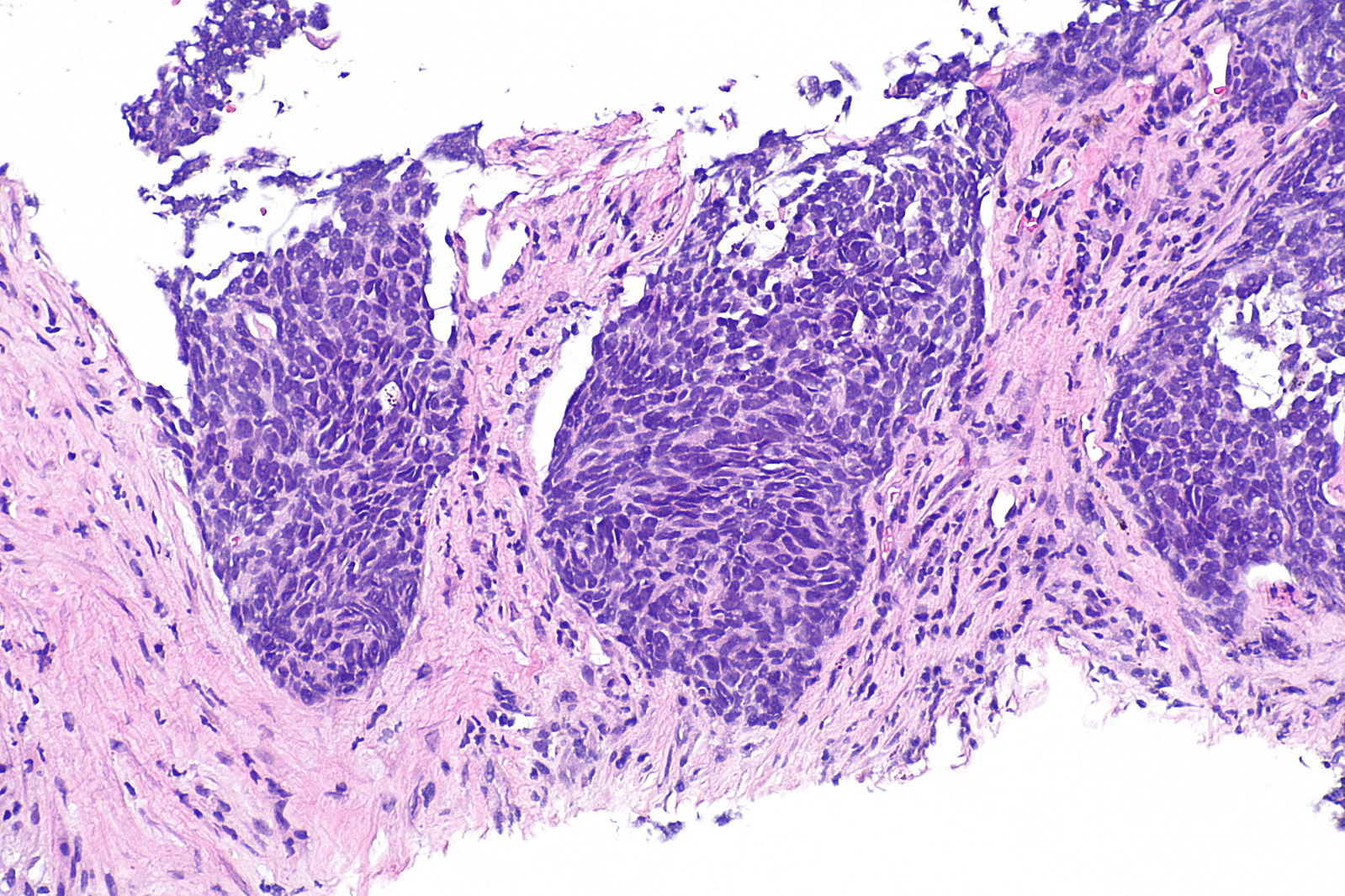 Nonsmallcell micropathology1.jpeg 1,599 × 1,066; 466 KB
-
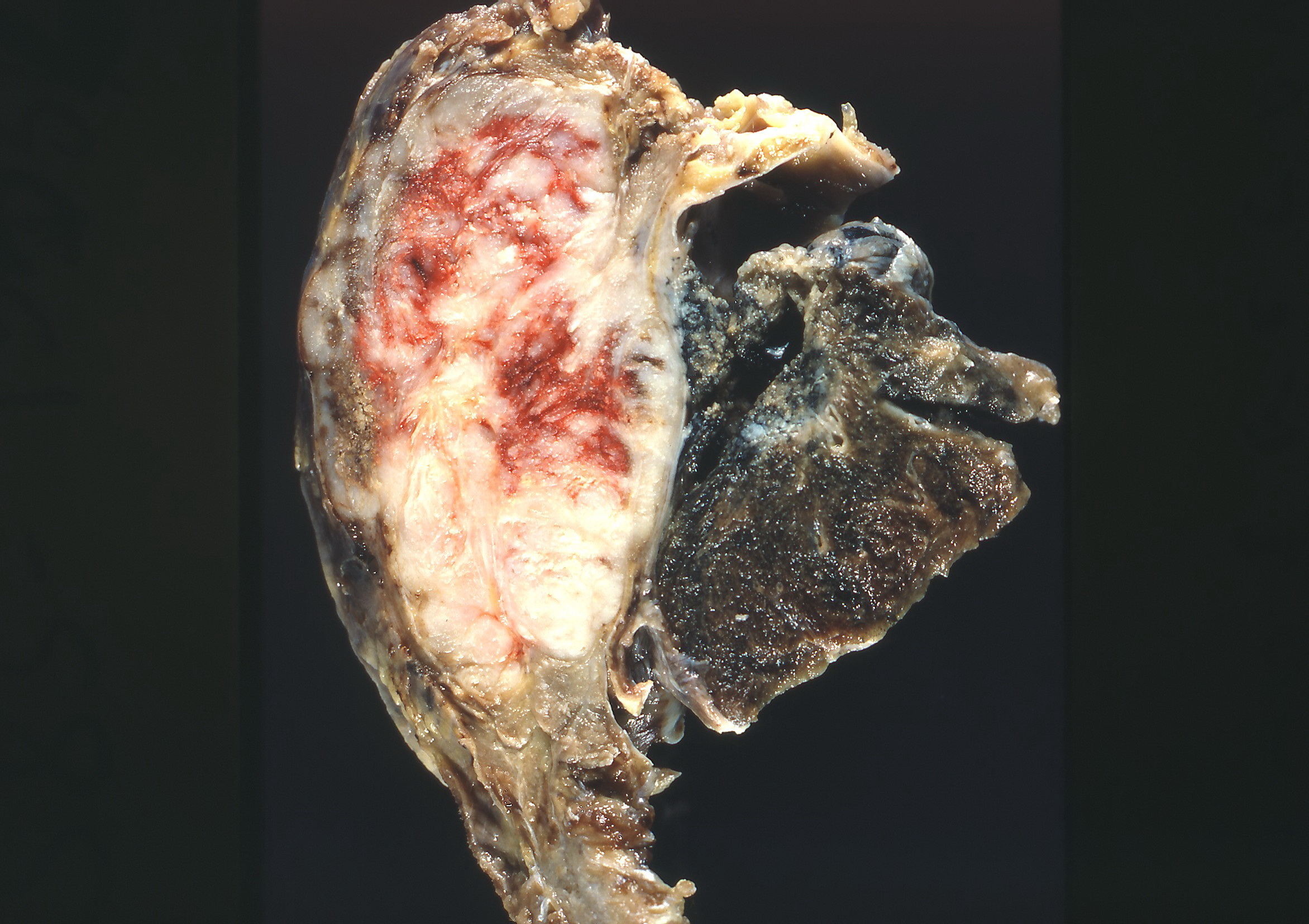 Pheripheal adenocarcinoma chest wall.jpg 2,344 × 1,656; 809 KB
-
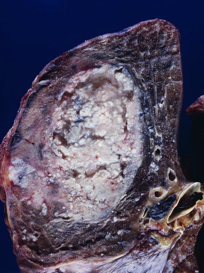 Gross pathology squamous cell.jpg 673 × 898; 196 KB
-
 Cavitation squamouscell.jpg 1,376 × 2,160; 993 KB
-
 Hepatosplenic T cell lymphoma peripheral blood smear.jpg 600 × 512; 90 KB
-
 Golden-s-sign-1.jpg 1,024 × 1,024; 86 KB
-
 Golden-s-sign.jpg 1,024 × 1,024; 85 KB
-
 Pleural-effusion-unilateral-malignant.jpg 1,024 × 1,024; 86 KB
-
 960px-Ovarian fibroma - intermed mag.jpg 960 × 640; 194 KB
-
 960px-Oral fibroma -- low mag.jpg 960 × 640; 199 KB
-
 960px-Ovarian fibroma - high mag.jpg 960 × 640; 170 KB
-
 426px-Oral fibroma -- intermed mag.jpg 426 × 639; 110 KB
-
 120px-Bone ChondromyxoidFibroma HP2 PA (2).jpg 120 × 90; 4 KB
-
 120px-Bone ChondromyxoidFibroma LP CTR.jpg 120 × 90; 3 KB


.jpg)
