Unused files
Jump to navigation
Jump to search
The following files exist but are not embedded in any page. Please note that other websites may link to a file with a direct URL, and so may still be listed here despite being in active use.
Showing below up to 50 results in range #24,681 to #24,730.
-
 Lynchsyndrome grosspathology.jpg 567 × 223; 79 KB
-
 CT image of pineal germinoma 1.jpg 630 × 630; 37 KB
-
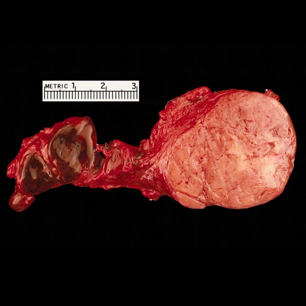 Carotid-body-tumour-gross-pathology.jpg 1,024 × 1,024; 81 KB
-
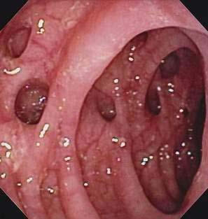 Diverticulosis1.jpg 297 × 313; 29 KB
-
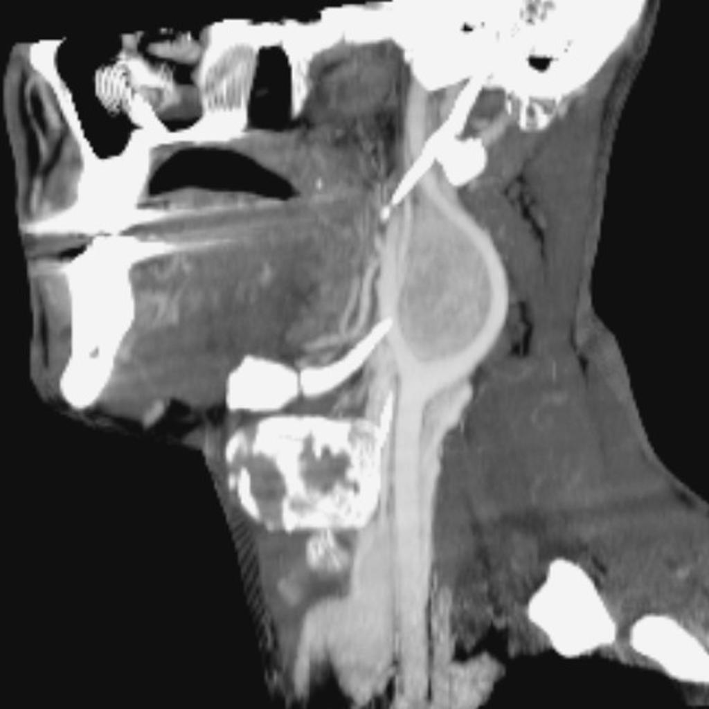 Carotid-body-tumour-1 (1).jpg 1,024 × 1,024; 60 KB
-
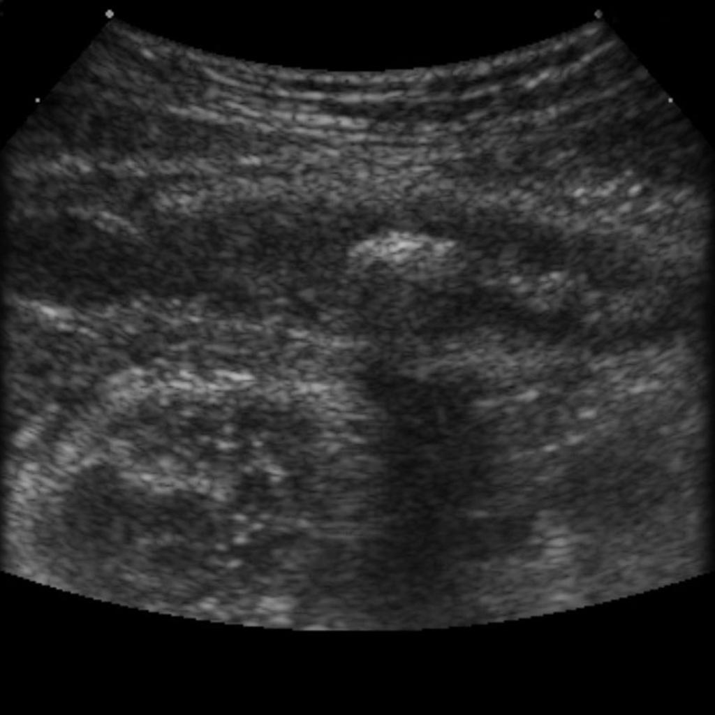 Appendicolith-on-ultrasound.jpg 1,024 × 1,024; 76 KB
-
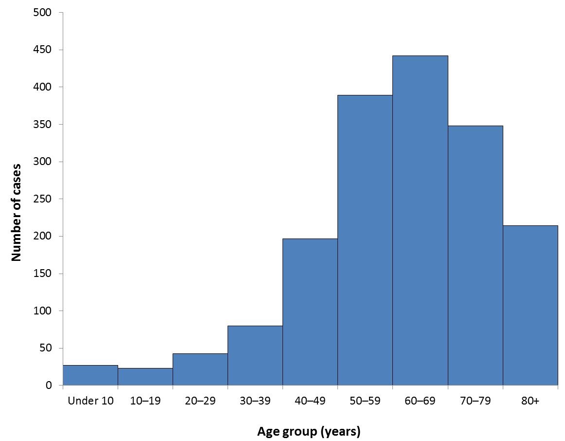 Reported cases by age 2013.jpg 1,162 × 906; 65 KB
-
 Villarreal.png 300 × 300; 120 KB
-
 Pulmonary-hamartoma-4-1.png 1,424 × 1,300; 1.14 MB
-
 Serous carcinoma - omentum 3 -- very high mag.jpg 256 × 171; 21 KB
-
 256px-Mucinous lmp ovarian tumour intermed mag.jpg 256 × 171; 22 KB
-
 Pulmonary-hamartoma-4-2.png 1,156 × 1,272; 810 KB
-
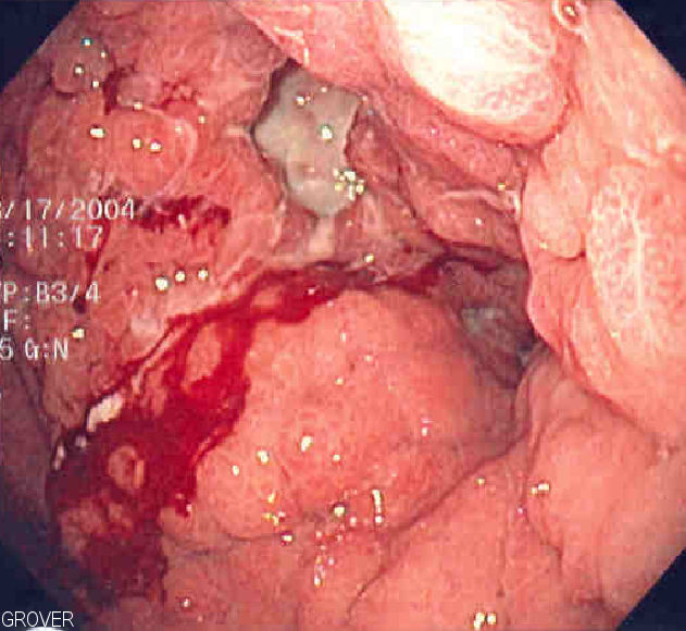 Linitis plastica1.jpg 631 × 580; 83 KB
-
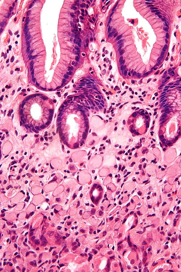 Gastric signet ring cell carcinoma.jpg 600 × 900; 173 KB
-
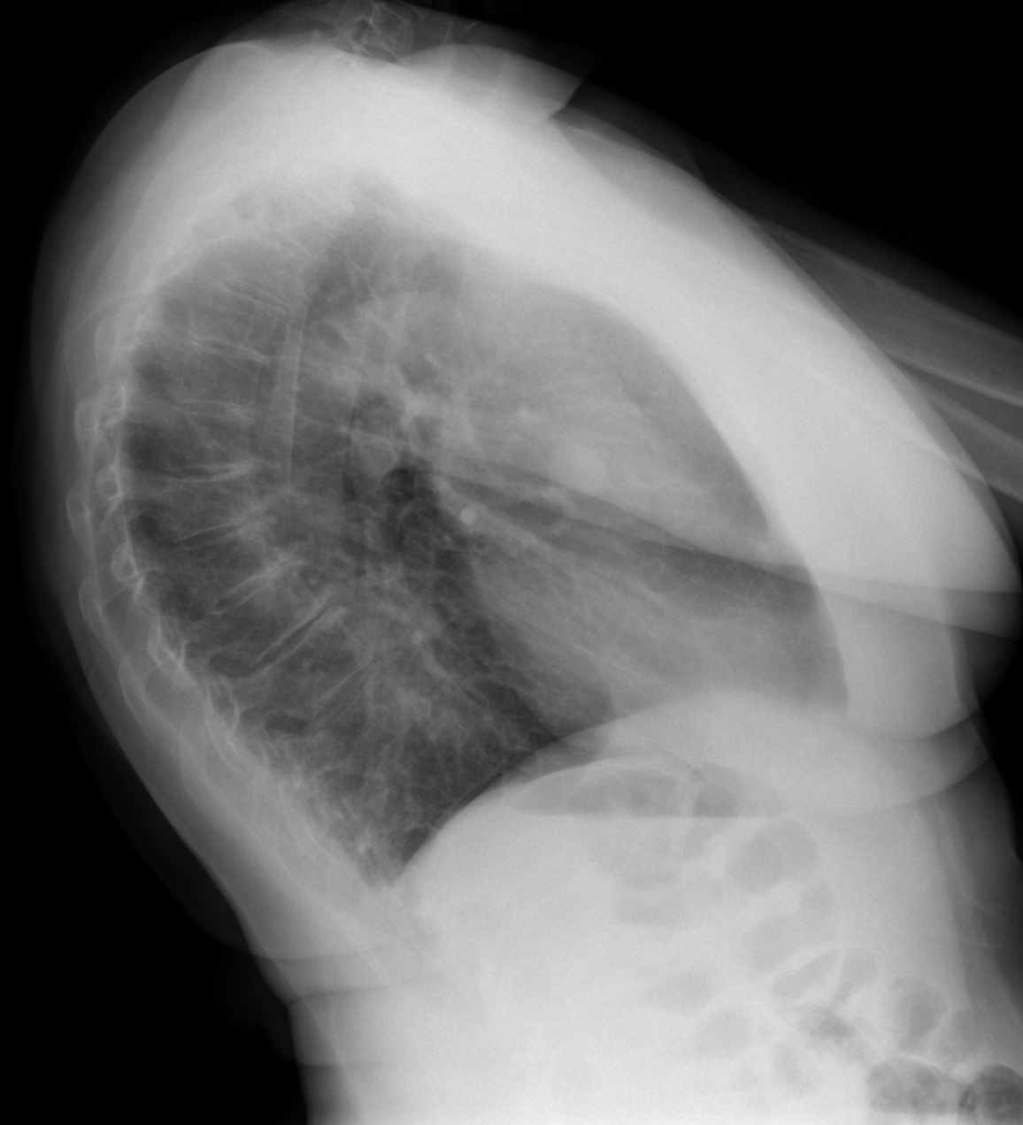
 Linitis-plastica.jpg 909 × 1,024; 75 KB
-
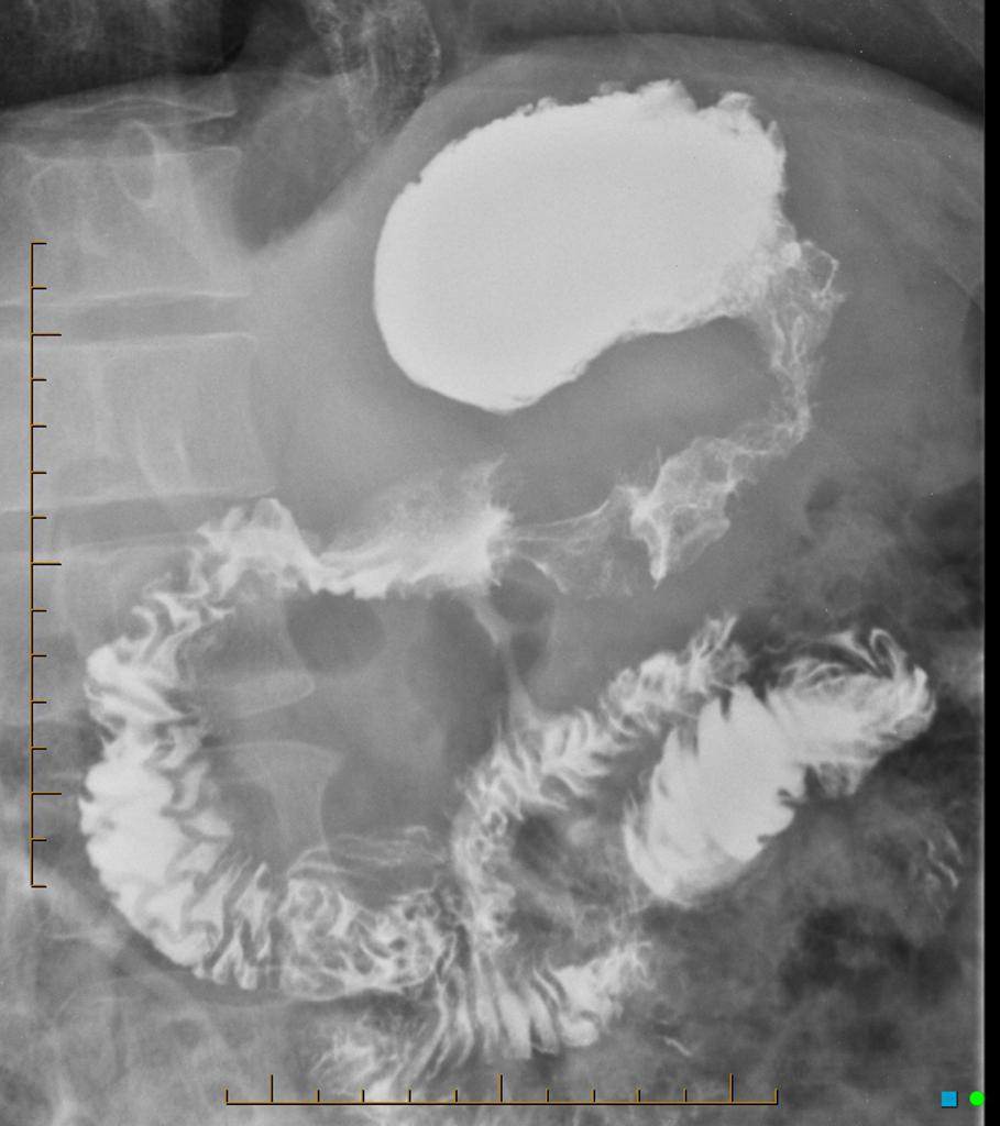
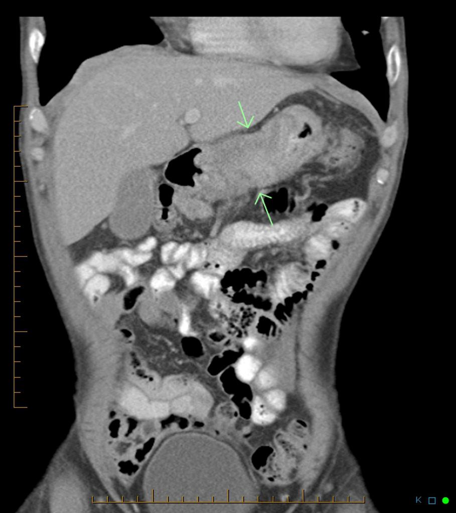 Linitis-plastica CT (2).jpg 909 × 1,024; 83 KB
-
 Thummathati.png 300 × 300; 140 KB
-
 Microscopic features of atrt 1.PNG 956 × 786; 858 KB
-
 800px-Colon diverticulosis whole slide.jpg 800 × 559; 104 KB
-
 Mirdula.png 300 × 300; 130 KB
-
 Mirdula Sharma.png 300 × 300; 130 KB
-
 CT image of atypical teratoid rhabdoid tumor 1.jpg 630 × 630; 44 KB
-
 Biliary-cystadenoma.jpg 1,024 × 884; 88 KB
-
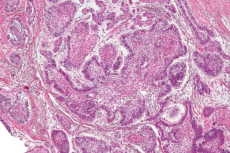 800px-Ameloblastoma - intermed mag (1).jpg 800 × 533; 214 KB
-
 Rectus-abdominis-muscle-desmoid-tumour.jpg 1,024 × 1,024; 53 KB
-
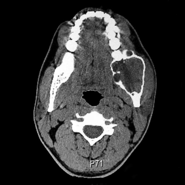 Ameloblastoma CT.jpg 630 × 630; 47 KB
-
 Axial bone marrow ameloblastoma 1.jpg 630 × 630; 18 KB
-
 Axial liver window ameloblastoma.jpg 609 × 630; 32 KB
-
 Coronal liver window ameloblastoma.jpg 630 × 602; 26 KB
-
 Axial non contrast ameloblastoma.jpg 587 × 630; 28 KB
-
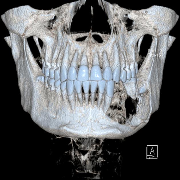 3D rendered volume.jpg 630 × 630; 56 KB
-
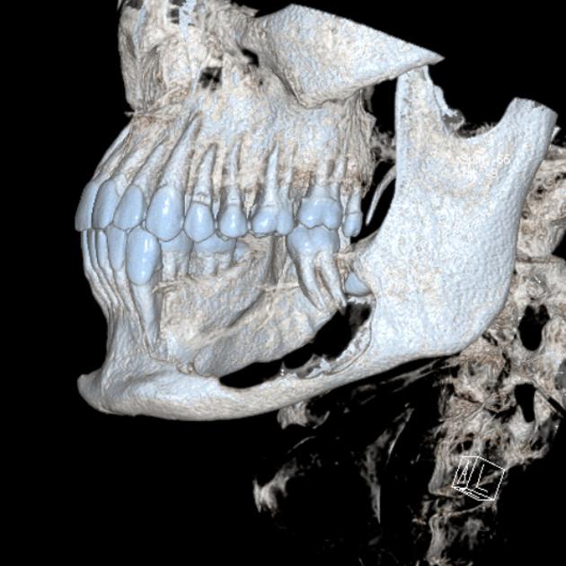 3 d ameloblastoma.jpg 630 × 630; 56 KB
-
 3D ameloblastoma.jpg 630 × 630; 57 KB
-
 Coronal C+ delayed ameloblastoma.jpg 630 × 534; 31 KB
-
 Coronal C + delayed ameloblastoma.jpg 630 × 534; 31 KB
-
 Axial C+ delayed ameloblastoma.jpg 630 × 630; 31 KB
-
 Bone window 1 ameloblastoma.jpg 630 × 630; 59 KB
-
 Bone window ameloblastoma.jpg 630 × 513; 36 KB
-
 Sagittal C+ delayed ameloblastoma.jpg 630 × 493; 28 KB
-
 C + delayed ameloblastoma.jpg 630 × 493; 38 KB
-
 AXIAL c+ ARTERIAL PHASE ameloblastoma.jpg 630 × 630; 29 KB
-
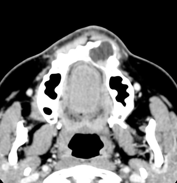
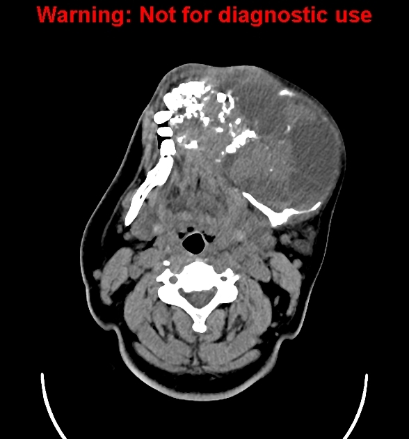 Axial C- soft tissue window.jpg 588 × 630; 110 KB
-
 Axial C+ soft tissue window.jpg 588 × 630; 131 KB
-
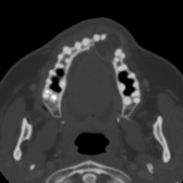
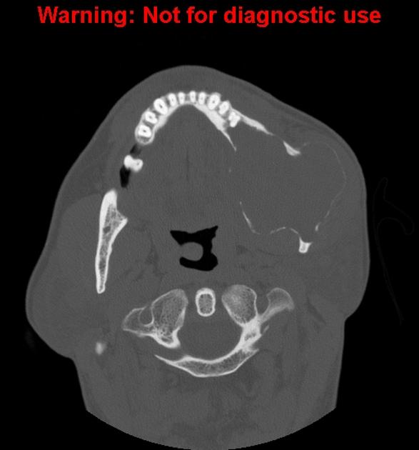
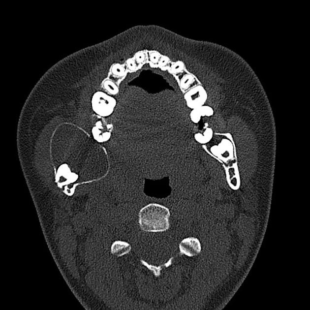
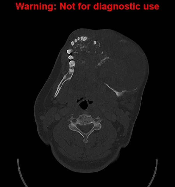 Axial bone window ameloblastoma.jpg 588 × 630; 102 KB
-
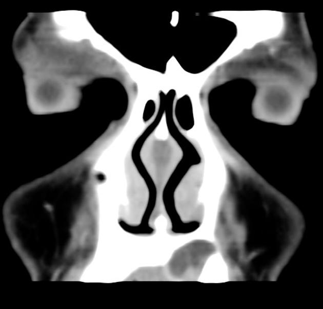
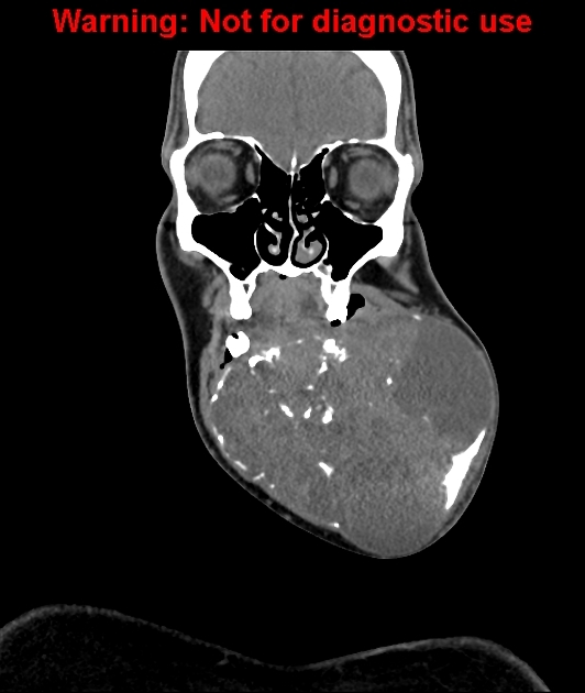 Coronal soft tissue window ameloblastoma.jpg 532 × 630; 100 KB
-
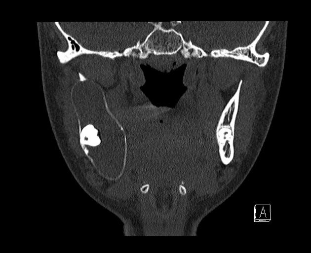
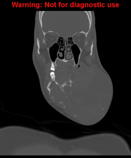 Coronal bone window ameloblastoma.jpg 521 × 630; 76 KB
-
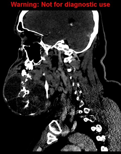 Saggital soft tissue window ameloblastoma.jpg 498 × 630; 158 KB
-
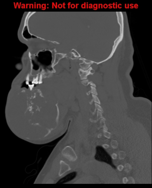 Saggital bone window ameloblastoma.jpg 511 × 630; 103 KB
-
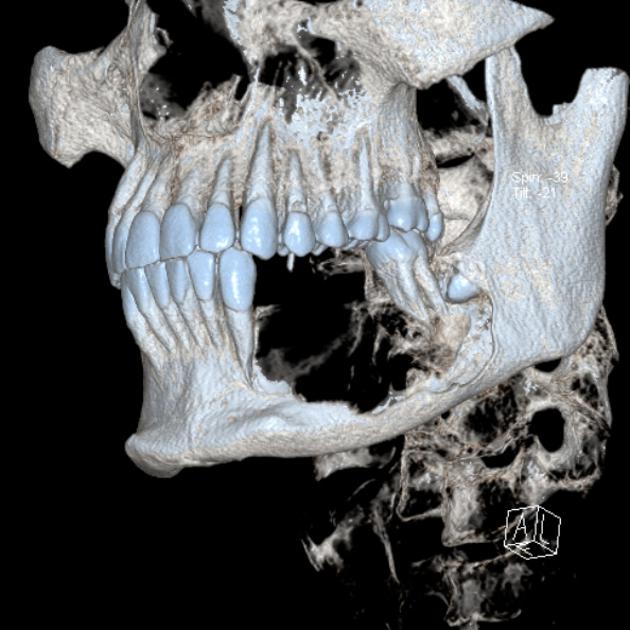
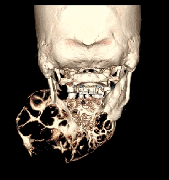 3 d amelo.jpg 593 × 630; 82 KB
-
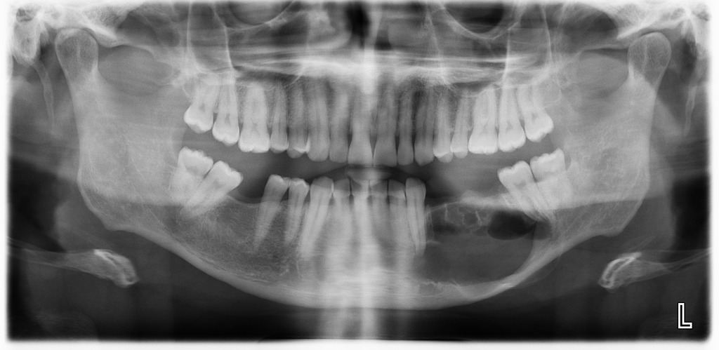 X ray ameloblastoma.jpeg 1,024 × 499; 90 KB


.jpg)


.jpg)


.jpg)


