Unused files
Jump to navigation
Jump to search
The following files exist but are not embedded in any page. Please note that other websites may link to a file with a direct URL, and so may still be listed here despite being in active use.
Showing below up to 50 results in range #24,651 to #24,700.
-
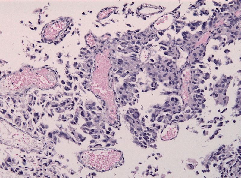 800px-PTPR papillary tumor pinealis HE microscopic image 2.jpg 800 × 593; 130 KB
-
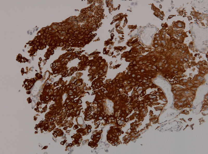 Ihcptpr1.jpg 800 × 593; 107 KB
-
 Ihcptpr2.jpg 472 × 351; 181 KB
-
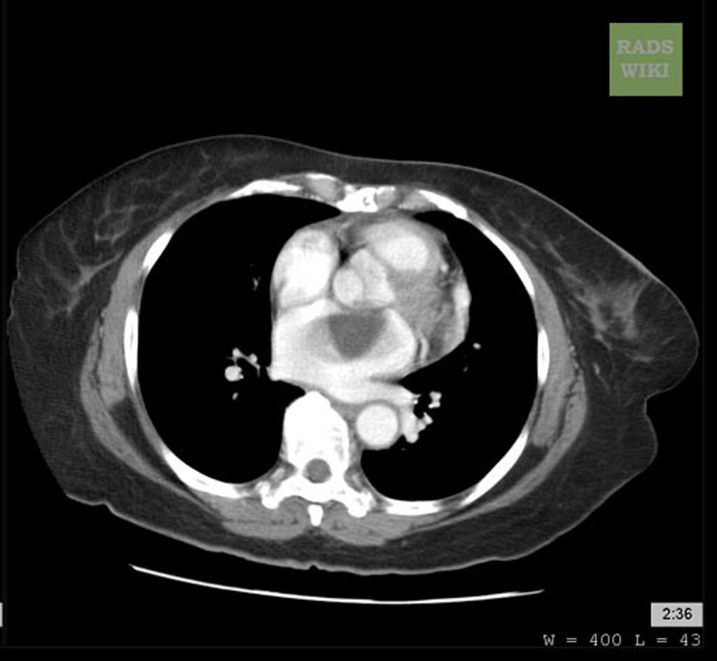 Cardiac-myxoma-1.jpg 1,024 × 944; 62 KB
-
 Axial C+ Arterial phase.jpeg 598 × 630; 58 KB
-
 Coronal C+ arterial phase.jpeg 461 × 630; 56 KB
-
 Axial C+ delayed.jpg 630 × 630; 35 KB
-
 Coronal C+ delayed.jpg 630 × 630; 39 KB
-
 Saggital C+ delayed.jpg 630 × 630; 41 KB
-
 Axial C+.jpeg 580 × 630; 50 KB
-
 Coronal C+.jpeg 502 × 630; 31 KB
-
 Axial bone window.jpeg 580 × 630; 38 KB
-
 Axial bone marrow.jpeg 484 × 630; 45 KB
-
 Bone window.jpeg 580 × 630; 38 KB
-
 Coronal T1 C+ fat sat.jpeg 630 × 630; 43 KB
-
 Axial T1 C+ fat saturated.jpeg 630 × 630; 43 KB
-
 Lynch syndrome overview.jpg 640 × 427; 92 KB
-
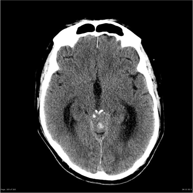
 Pineoblastoma ct image 1.jpg 1,024 × 1,024; 58 KB
-
 Pineoblastoma ct image 2.jpg 630 × 630; 40 KB
-
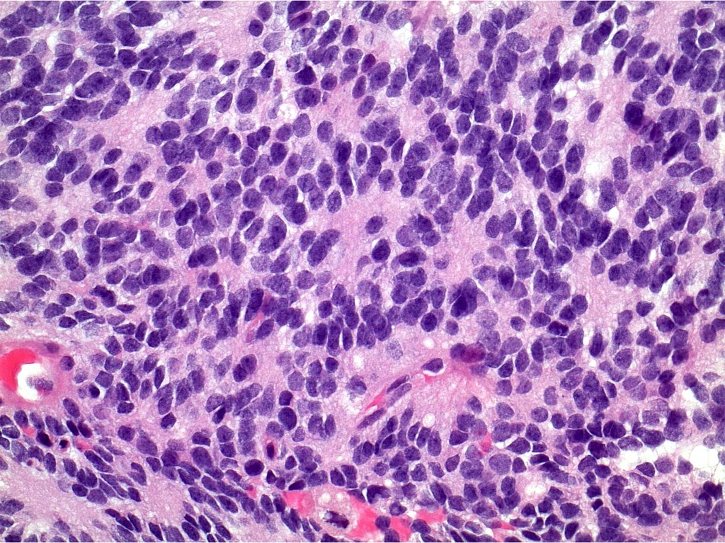
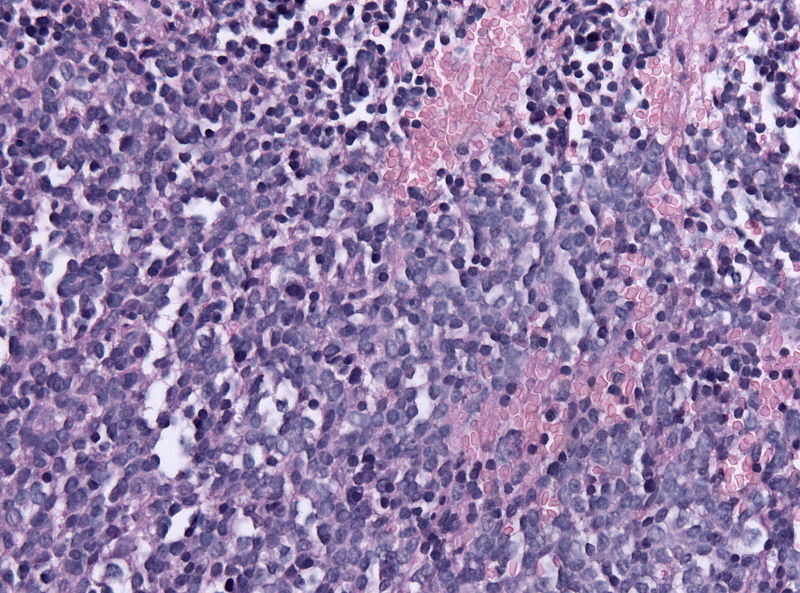 Microscopic image of pineoblastoma 1.jpg 800 × 593; 155 KB
-
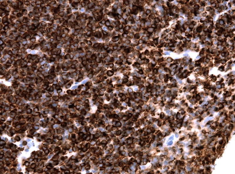 800px-Pineoblastoma neurofilament.jpg 800 × 593; 148 KB
-
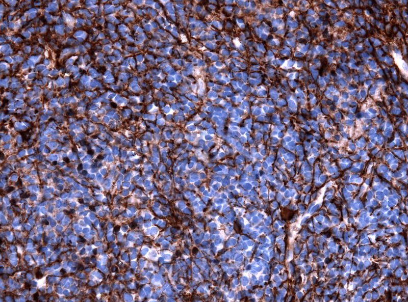 800px-Pineoblastoma gfap.jpg 800 × 593; 164 KB
-
 Microscopic image of pineoblastoma 2.jpg 1,024 × 767; 353 KB
-
 Pineoblastoma ct image 3.jpg 630 × 630; 44 KB
-
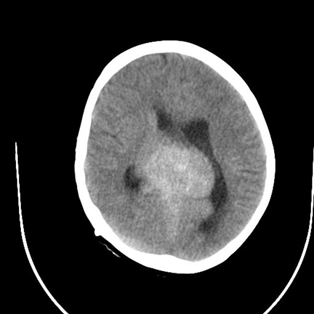 Pineoblastoma ct image 4.jpg 630 × 630; 42 KB
-
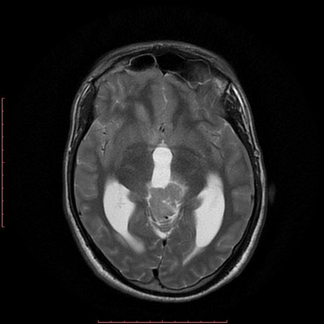
 MRI image of pineoblastoma 1.jpg 630 × 630; 23 KB
-
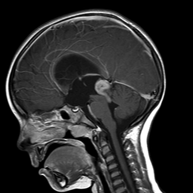 MRI image of pineoblastoma 2.jpg 630 × 630; 39 KB
-
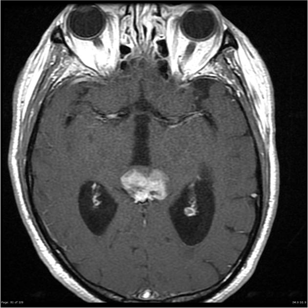 MRI image of pineoblastoma 3.jpg 630 × 630; 52 KB
-
 Muir-torre micropathology.jpg 640 × 427; 100 KB
-
 MRI image of pineoblastoma 4.jpg 630 × 630; 31 KB
-
 Lynchsyndrome grosspathology.jpg 567 × 223; 79 KB
-
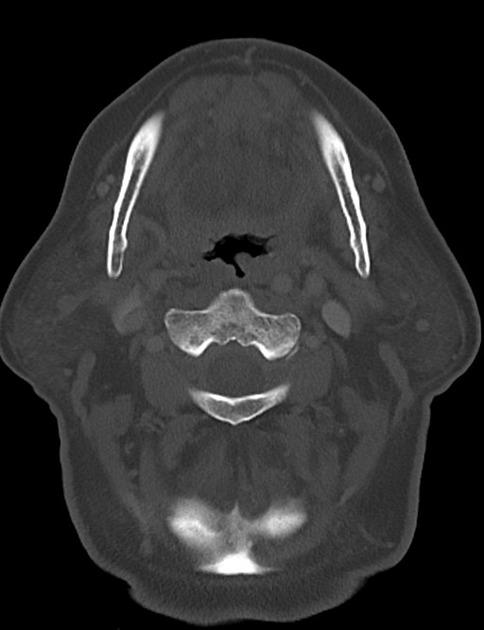
 CT image of pineal germinoma 1.jpg 630 × 630; 37 KB
-
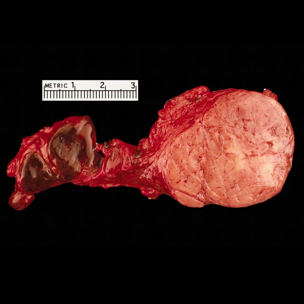 Carotid-body-tumour-gross-pathology.jpg 1,024 × 1,024; 81 KB
-
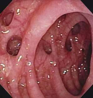 Diverticulosis1.jpg 297 × 313; 29 KB
-
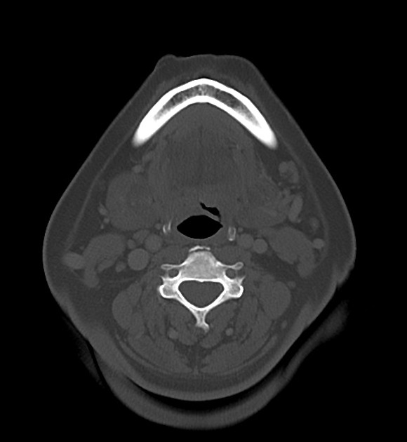
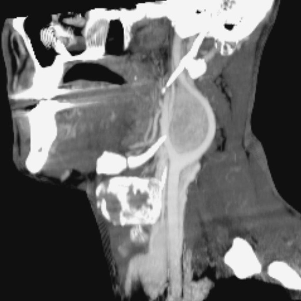 Carotid-body-tumour-1 (1).jpg 1,024 × 1,024; 60 KB
-
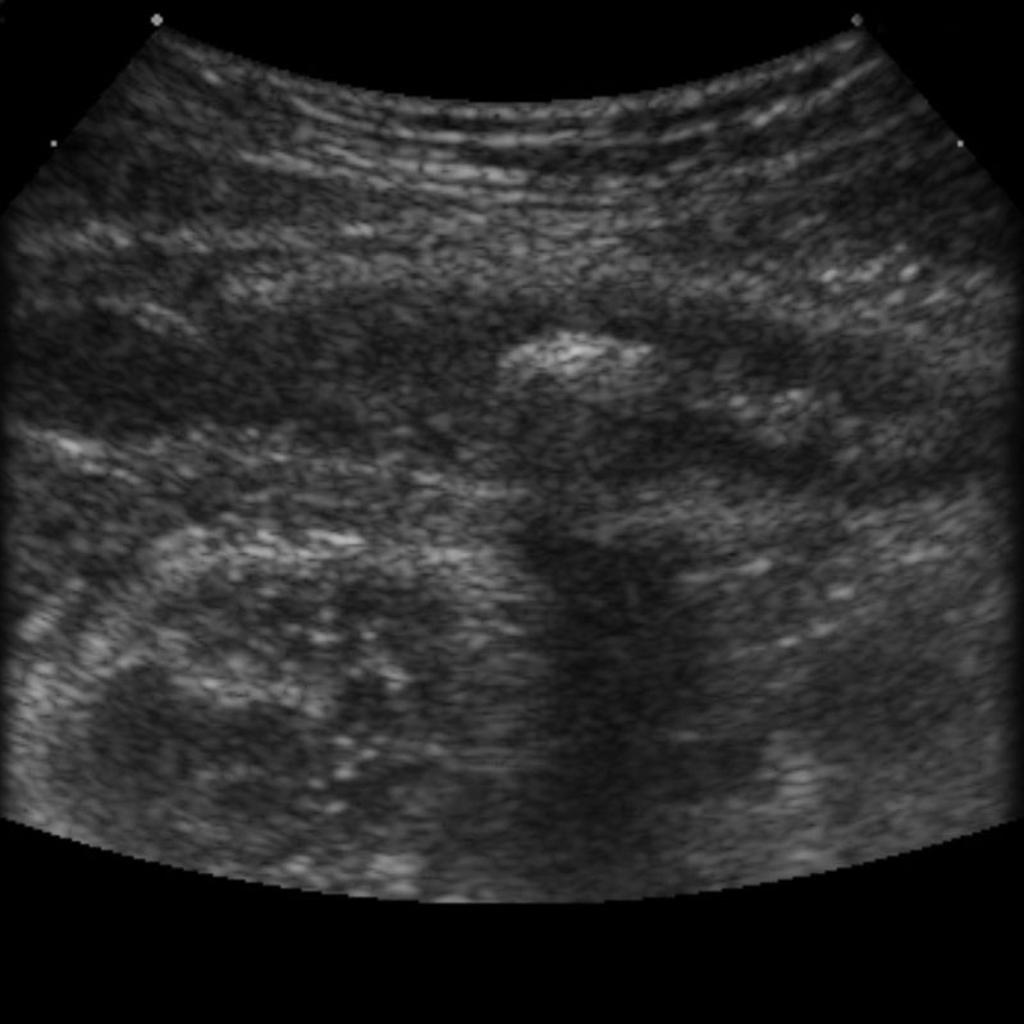 Appendicolith-on-ultrasound.jpg 1,024 × 1,024; 76 KB
-
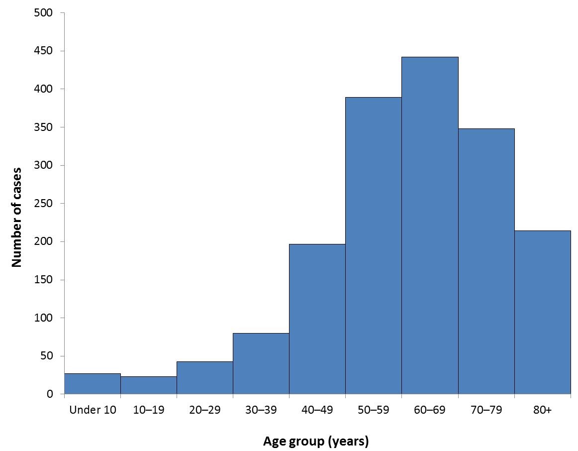 Reported cases by age 2013.jpg 1,162 × 906; 65 KB
-
 Villarreal.png 300 × 300; 120 KB
-
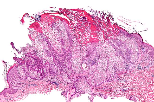
 Pulmonary-hamartoma-4-1.png 1,424 × 1,300; 1.14 MB
-
 Serous carcinoma - omentum 3 -- very high mag.jpg 256 × 171; 21 KB
-
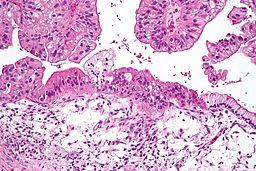 256px-Mucinous lmp ovarian tumour intermed mag.jpg 256 × 171; 22 KB
-
 Pulmonary-hamartoma-4-2.png 1,156 × 1,272; 810 KB
-
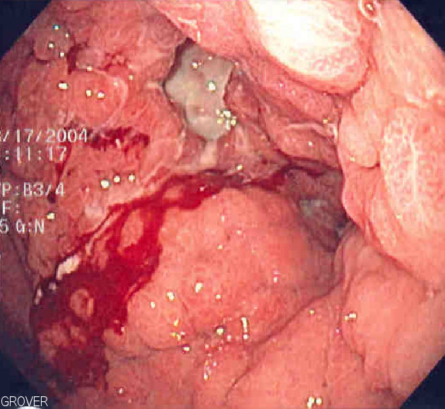 Linitis plastica1.jpg 631 × 580; 83 KB
-
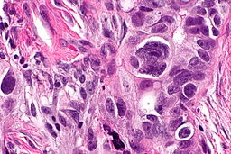
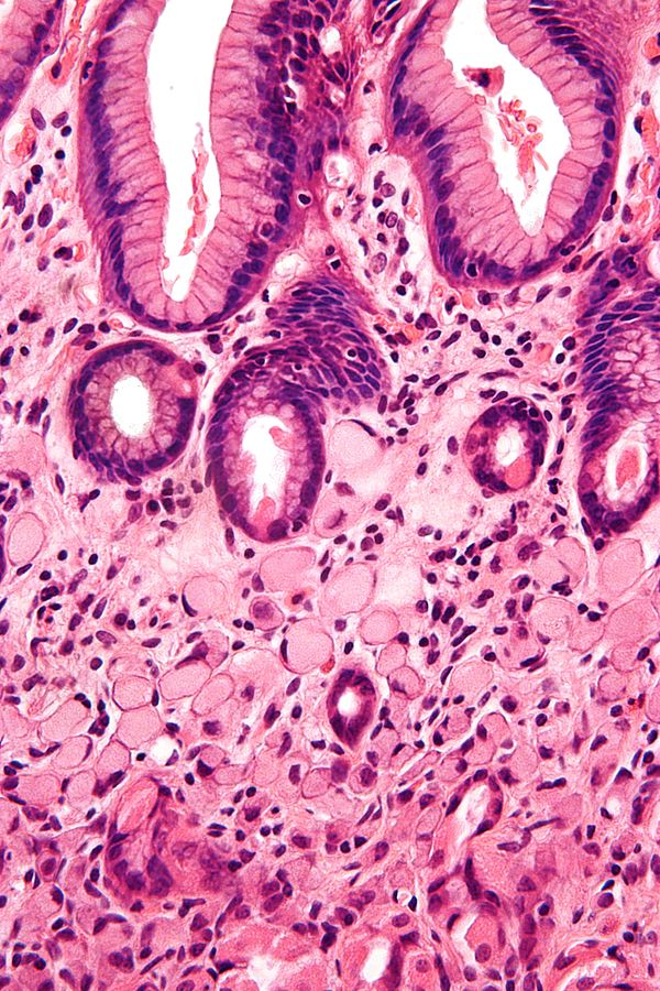 Gastric signet ring cell carcinoma.jpg 600 × 900; 173 KB
-
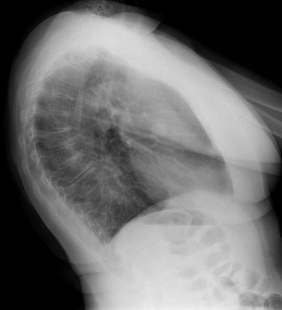
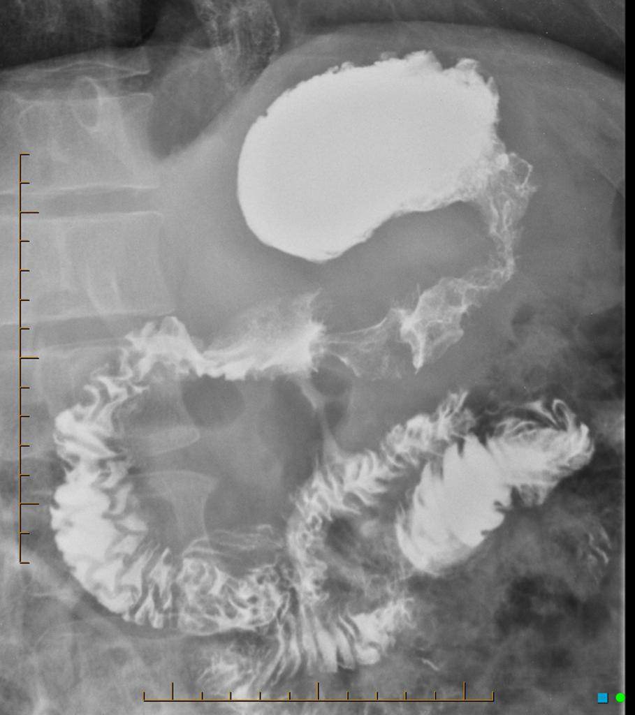 Linitis-plastica.jpg 909 × 1,024; 75 KB
-
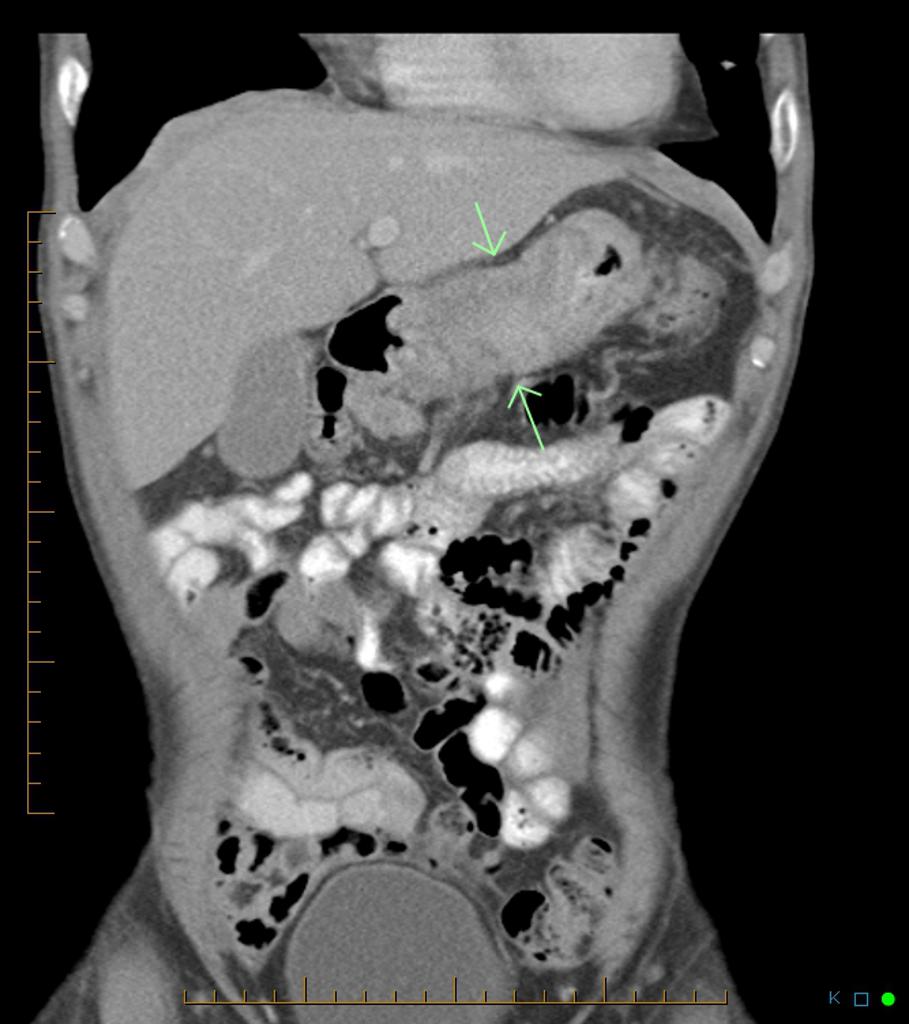 Linitis-plastica CT (2).jpg 909 × 1,024; 83 KB
-
 Thummathati.png 300 × 300; 140 KB
-
 Microscopic features of atrt 1.PNG 956 × 786; 858 KB
-
 800px-Colon diverticulosis whole slide.jpg 800 × 559; 104 KB
-
 Mirdula.png 300 × 300; 130 KB


.jpg)


.jpg)


