Unused files
Jump to navigation
Jump to search
The following files exist but are not embedded in any page. Please note that other websites may link to a file with a direct URL, and so may still be listed here despite being in active use.
Showing below up to 50 results in range #24,621 to #24,670.
-
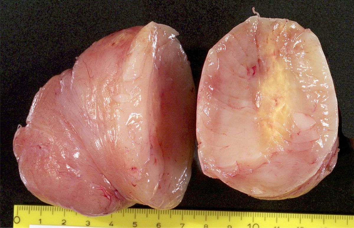 Neurofibroma1.jpg 1,200 × 772; 151 KB
-
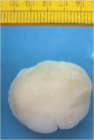 Neurofibroma2.jpg 302 × 448; 63 KB
-
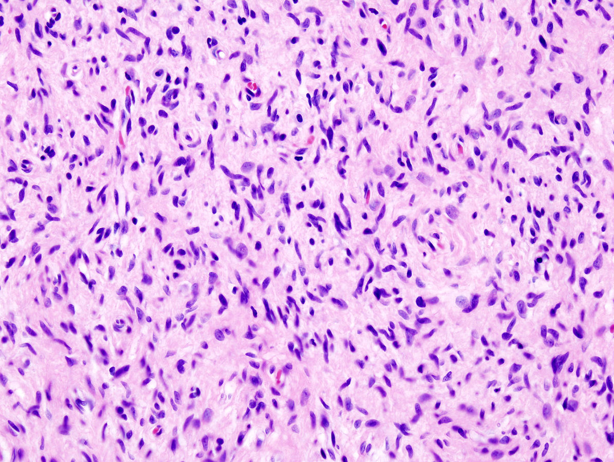 Neurofibroma3.jpg 1,194 × 899; 324 KB
-
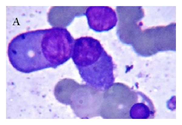 1271407.fig.001a.jpg 600 × 411; 116 KB
-
 1 Rouleaux formation.fig.001a.jpg 600 × 411; 116 KB
-
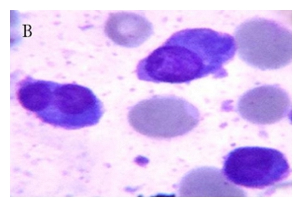 2 Uni-binucleated.fig.001b.jpg 600 × 411; 102 KB
-
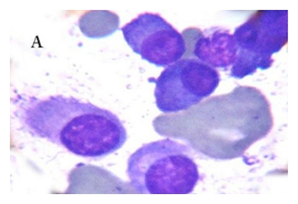 3 PC.fig.002a.jpg 600 × 411; 101 KB
-
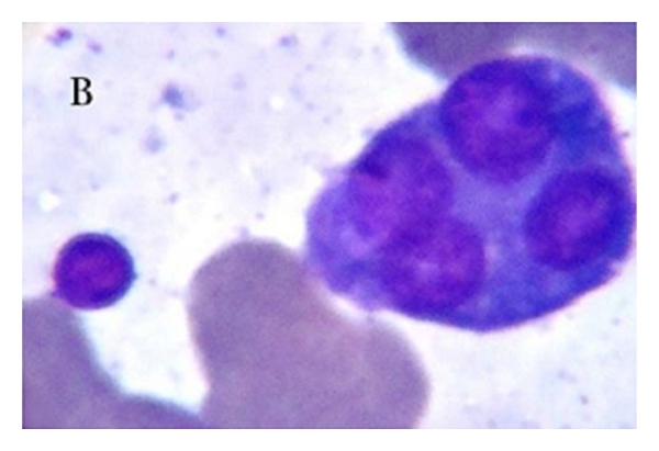 4 PC.fig.002b.jpg 600 × 411; 101 KB
-
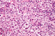 Dysgerminoma.jpg 218 × 145; 17 KB
-
 Schwannoma1.jpg 1,200 × 800; 429 KB
-
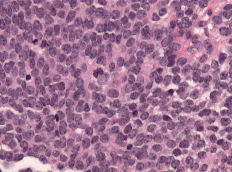
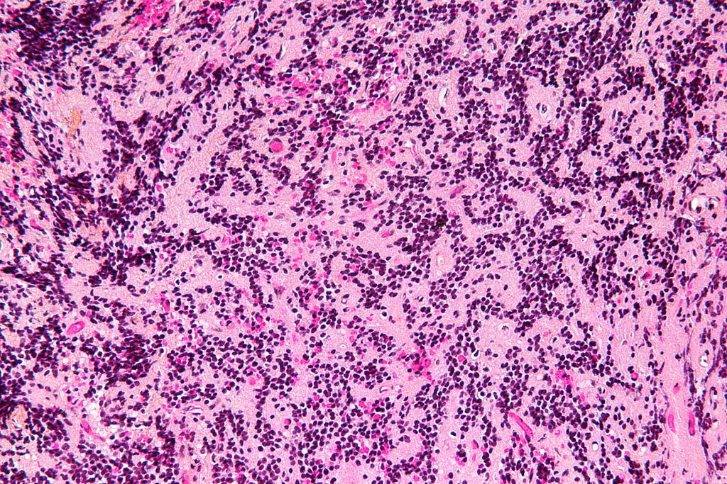 Pineocytoma - high mag.jpg 1,024 × 683; 330 KB
-
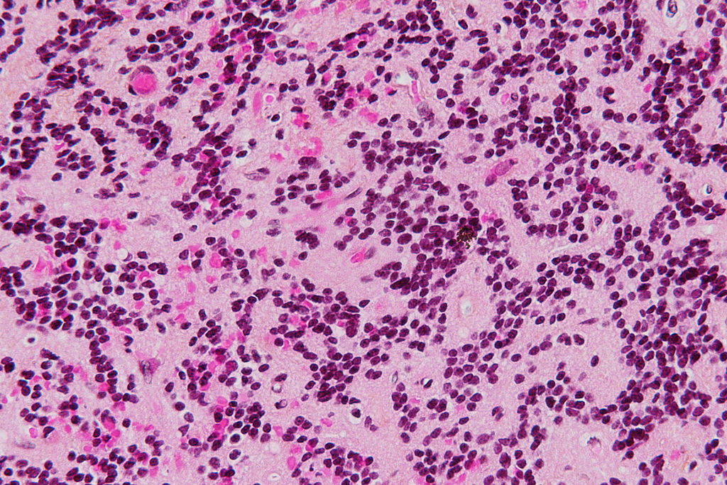 Pineocytoma - very high mag.jpg 1,024 × 683; 249 KB
-
 CT image pineocytoma 1.jpg 1,024 × 1,024; 58 KB
-
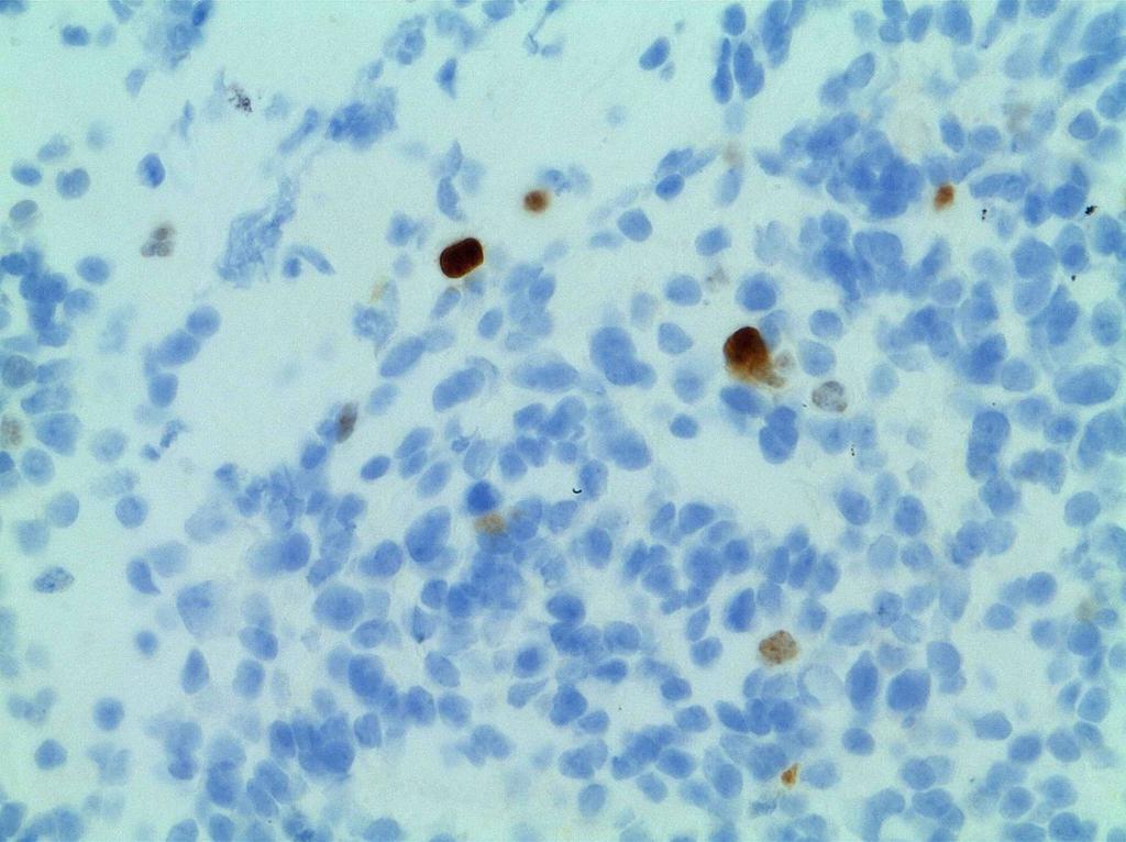
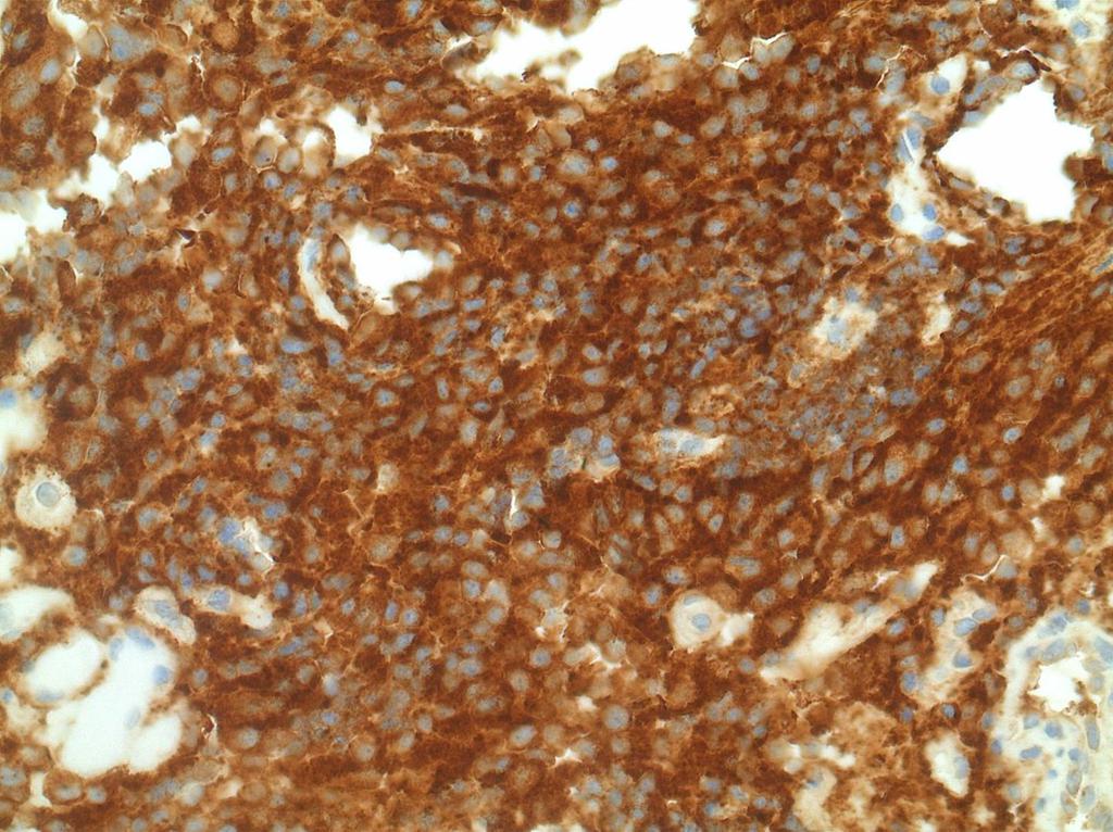 Ihc1.jpg 1,024 × 766; 154 KB
-
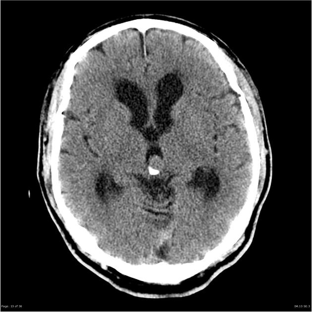 CT image pineocytoma 3.jpg 630 × 630; 50 KB
-
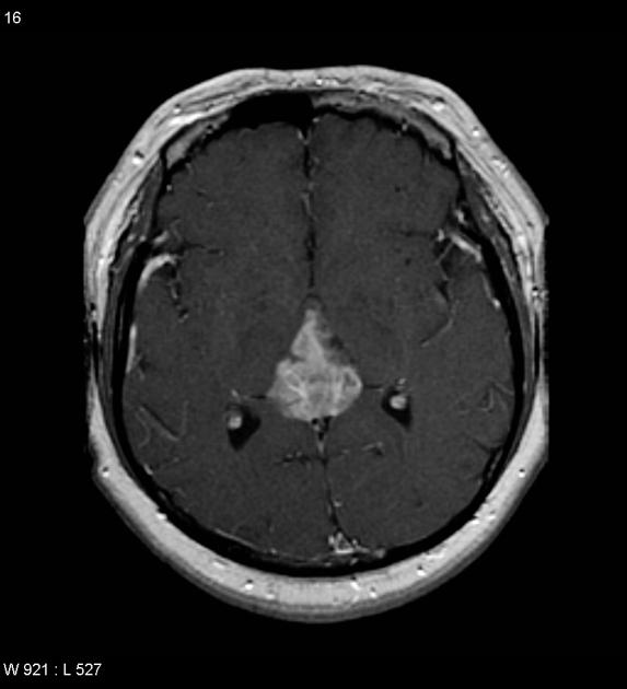 MRI image of pineocytoma 1.jpg 573 × 630; 28 KB
-
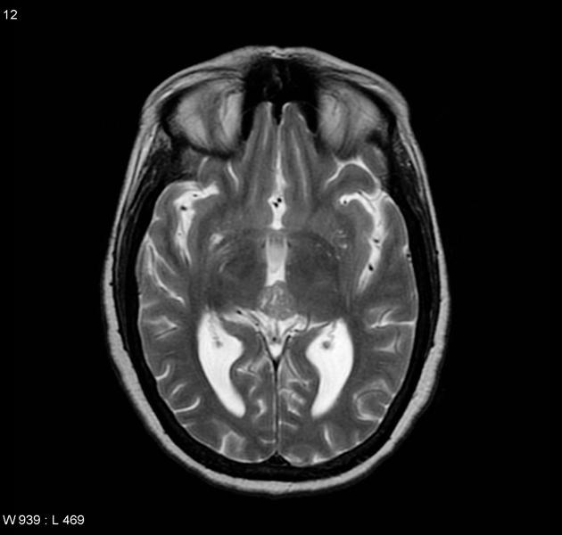 MRI image of pineocytoma 3.jpg 630 × 600; 30 KB
-
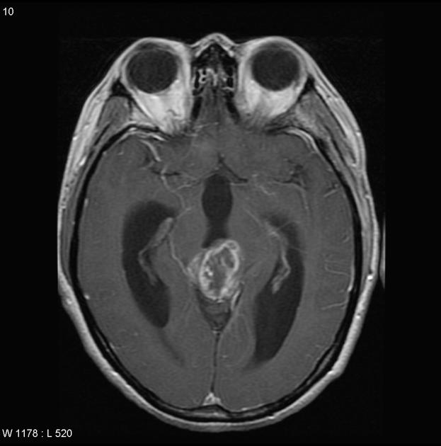 MRI image of pineocytoma 4.jpg 623 × 630; 32 KB
-
 Ihc2.jpg 1,024 × 766; 98 KB
-
 Edit2 2.5x3.5.jpg 500 × 573; 76 KB
-
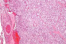 218px-Carotid body tumour 2 low mag.jpg 218 × 145; 16 KB
-
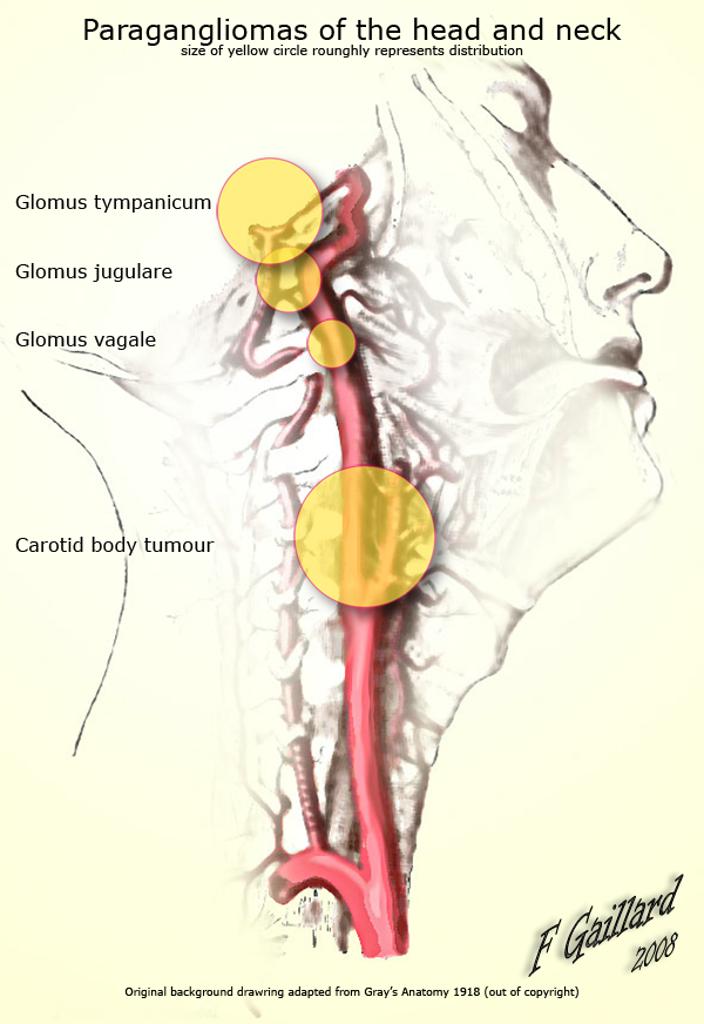 Distribution-of-paragangliomas.jpg 704 × 1,024; 65 KB
-
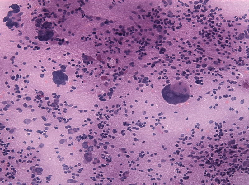
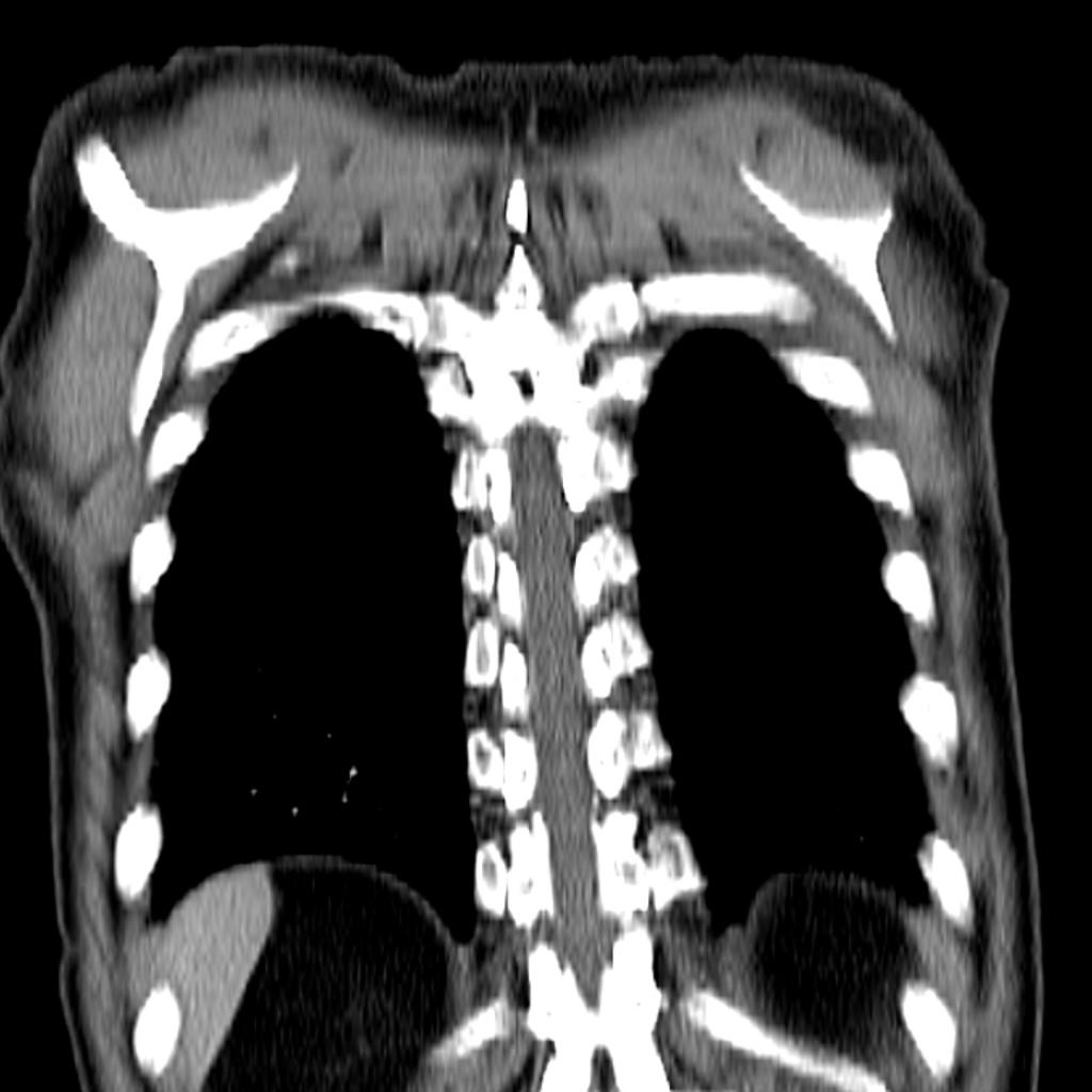 Small-cell-lung-cancer.jpg 1,024 × 1,024; 75 KB
-
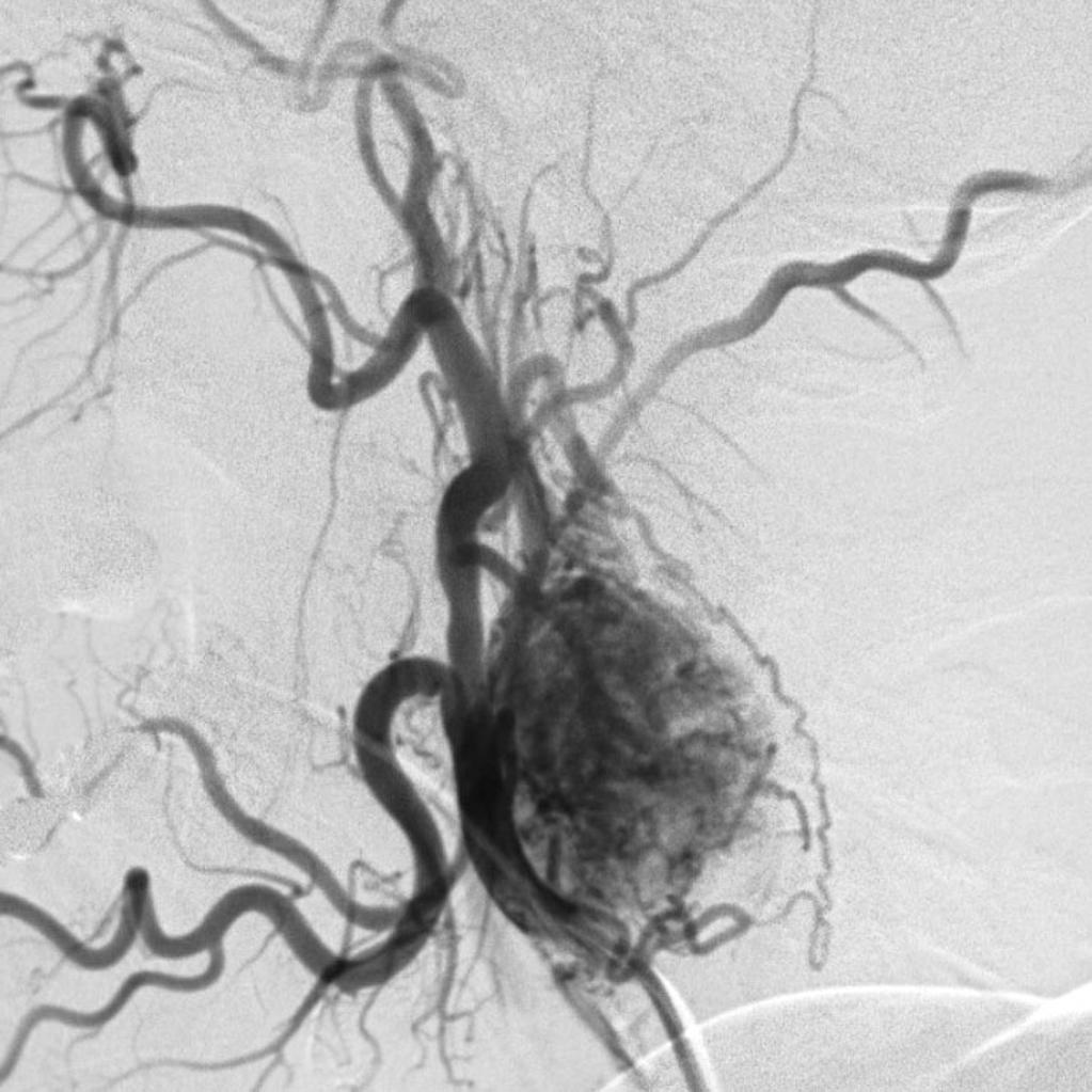 Carotid-body-tumour-on-angiography.jpg 1,023 × 1,023; 128 KB
-
 Small-cell-lung-cancer-1-2.jpg 1,024 × 1,024; 55 KB
-
-
-
-

-
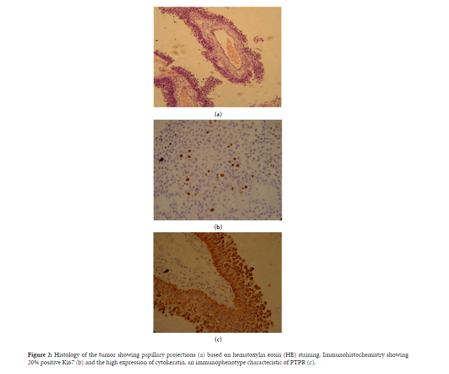 Papillary Tumor of the Pineal Region microscopic image 1.PNG 883 × 736; 390 KB
-
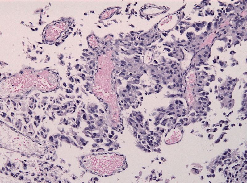 800px-PTPR papillary tumor pinealis HE microscopic image 2.jpg 800 × 593; 130 KB
-
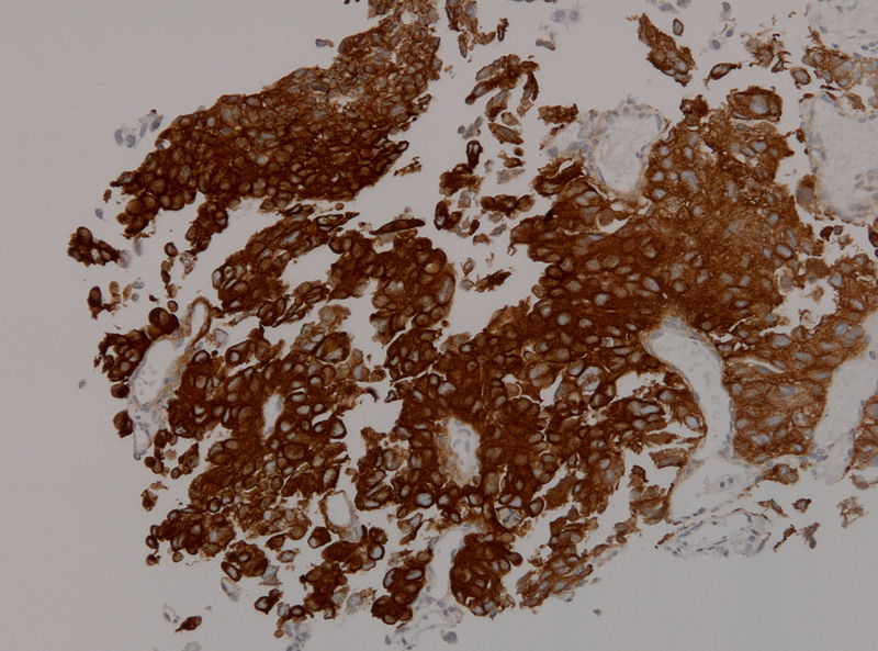 Ihcptpr1.jpg 800 × 593; 107 KB
-
 Ihcptpr2.jpg 472 × 351; 181 KB
-
 Cardiac-myxoma-1.jpg 1,024 × 944; 62 KB
-
 Axial C+ Arterial phase.jpeg 598 × 630; 58 KB
-
 Coronal C+ arterial phase.jpeg 461 × 630; 56 KB
-
 Axial C+ delayed.jpg 630 × 630; 35 KB
-
 Coronal C+ delayed.jpg 630 × 630; 39 KB
-
 Saggital C+ delayed.jpg 630 × 630; 41 KB
-
 Axial C+.jpeg 580 × 630; 50 KB
-
 Coronal C+.jpeg 502 × 630; 31 KB
-
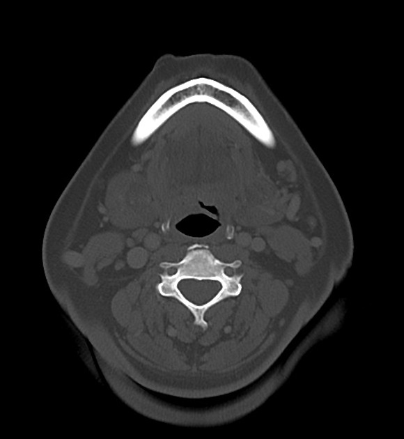 Axial bone window.jpeg 580 × 630; 38 KB
-
 Axial bone marrow.jpeg 484 × 630; 45 KB
-
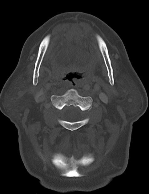
 Bone window.jpeg 580 × 630; 38 KB
-
 Coronal T1 C+ fat sat.jpeg 630 × 630; 43 KB
-
 Axial T1 C+ fat saturated.jpeg 630 × 630; 43 KB
-
 Lynch syndrome overview.jpg 640 × 427; 92 KB
-
 Pineoblastoma ct image 1.jpg 1,024 × 1,024; 58 KB
-
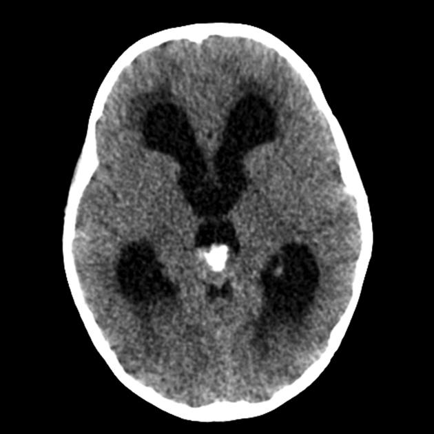 Pineoblastoma ct image 2.jpg 630 × 630; 40 KB
-
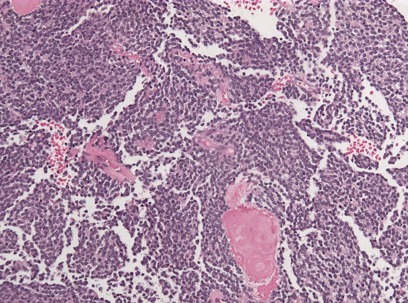
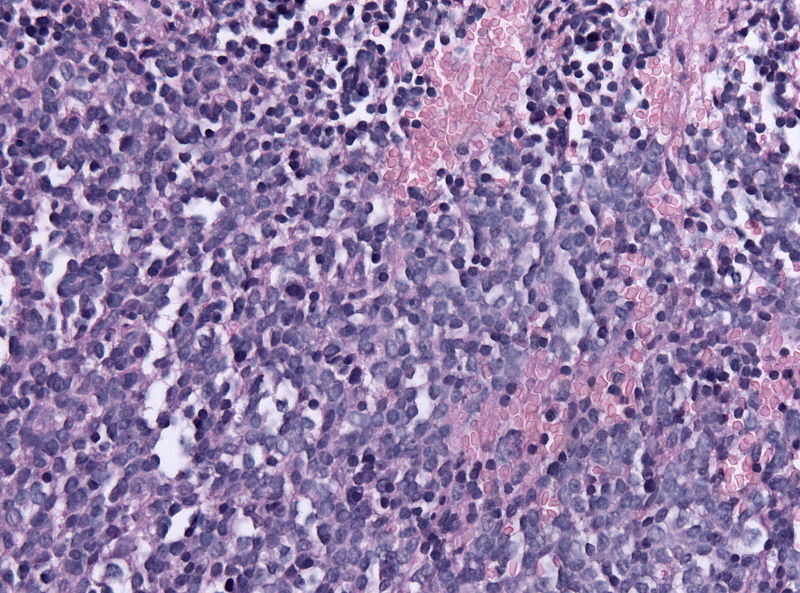 Microscopic image of pineoblastoma 1.jpg 800 × 593; 155 KB


