Unused files
Jump to navigation
Jump to search
The following files exist but are not embedded in any page. Please note that other websites may link to a file with a direct URL, and so may still be listed here despite being in active use.
Showing below up to 50 results in range #24,601 to #24,650.
-
 796px-Papillary cystadenoma lymphomatosum2.jpg 796 × 600; 165 KB
-
 800px-Adenoid cystic carcinoma - high mag.jpg 800 × 533; 183 KB
-
 800px-Basal cell adenocarcinoma - parotid gland - high mag.jpg 800 × 533; 159 KB
-
 800px-Epithelial-myoepithelial carcinoma - high mag (1).jpg 800 × 533; 158 KB
-
 800px-Salivary duct carcinoma -a- low mag.jpg 800 × 533; 201 KB
-
 Mucoepidermoid carcinoma (2) HE stain (1).jpg 600 × 452; 384 KB
-
 Pleomorphic adenoma (1) parotid gland.jpg 600 × 452; 164 KB
-
 800px-Acinic cell carcinoma - high mag.jpg 800 × 533; 176 KB
-
 Cerebellar medulloblastoma (1) in adult.jpg 500 × 376; 109 KB
-
 1600px-Medulloblastoma with rosettes.jpg 1,600 × 1,186; 403 KB
-
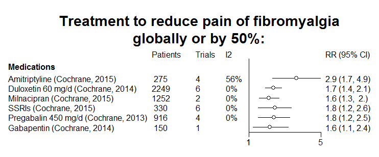 Fibromyalgia treatment.png 742 × 308; 20 KB
-
 Blobbogram of treatment of chronic low back pain.png 967 × 858; 63 KB
-
 Waldenstroms-macroglobulinaemia.jpg 1,024 × 1,024; 70 KB
-
 Essential thrombocythemia.jpg 600 × 452; 86 KB
-
 Axial T1 C1+ fat.jpg 616 × 630; 25 KB
-
 Coronal T1 C1+ pleomorphic adenoma.jpg 616 × 630; 23 KB
-
 Thyroid MedullaryCarcinoma 216 PA.JPG 800 × 600; 170 KB
-
 Sagittal noncontrast CT parotid lipoma.jpg 630 × 450; 69 KB
-
 Thyroid MedullaryCarcinoma MP CTR (2).jpg 800 × 600; 131 KB
-
 Teratoma.jpg 1,280 × 887; 398 KB
-
 Neurofibroma1.jpg 1,200 × 772; 151 KB
-
 Neurofibroma2.jpg 302 × 448; 63 KB
-
 Neurofibroma3.jpg 1,194 × 899; 324 KB
-
 1271407.fig.001a.jpg 600 × 411; 116 KB
-
 1 Rouleaux formation.fig.001a.jpg 600 × 411; 116 KB
-
 2 Uni-binucleated.fig.001b.jpg 600 × 411; 102 KB
-
 3 PC.fig.002a.jpg 600 × 411; 101 KB
-
 4 PC.fig.002b.jpg 600 × 411; 101 KB
-
 Dysgerminoma.jpg 218 × 145; 17 KB
-
 Schwannoma1.jpg 1,200 × 800; 429 KB
-
 Pineocytoma - high mag.jpg 1,024 × 683; 330 KB
-
 Pineocytoma - very high mag.jpg 1,024 × 683; 249 KB
-
 CT image pineocytoma 1.jpg 1,024 × 1,024; 58 KB
-
 Ihc1.jpg 1,024 × 766; 154 KB
-
 CT image pineocytoma 3.jpg 630 × 630; 50 KB
-
 MRI image of pineocytoma 1.jpg 573 × 630; 28 KB
-
 MRI image of pineocytoma 3.jpg 630 × 600; 30 KB
-
 MRI image of pineocytoma 4.jpg 623 × 630; 32 KB
-
 Ihc2.jpg 1,024 × 766; 98 KB
-
 Edit2 2.5x3.5.jpg 500 × 573; 76 KB
-
 218px-Carotid body tumour 2 low mag.jpg 218 × 145; 16 KB
-
 Distribution-of-paragangliomas.jpg 704 × 1,024; 65 KB
-
 Small-cell-lung-cancer.jpg 1,024 × 1,024; 75 KB
-
 Carotid-body-tumour-on-angiography.jpg 1,023 × 1,023; 128 KB
-
 Small-cell-lung-cancer-1-2.jpg 1,024 × 1,024; 55 KB
-
-
-
-

-
 Papillary Tumor of the Pineal Region microscopic image 1.PNG 883 × 736; 390 KB


.jpg)

_HE_stain_(1).jpg)
_parotid_gland.jpg)

_in_adult.jpg)


.jpg)


