Unused files
Jump to navigation
Jump to search
The following files exist but are not embedded in any page. Please note that other websites may link to a file with a direct URL, and so may still be listed here despite being in active use.
Showing below up to 50 results in range #24,521 to #24,570.
-
 Oligoastroct.jpg 819 × 1,024; 71 KB
-
 Axial T1 choroidal melanoma.jpg 630 × 630; 40 KB
-
 Axial T1 C + fat.jpg 630 × 630; 42 KB
-
 Coronal T1 choroidal melanoma.jpg 630 × 630; 41 KB
-
 Axial T1 C+ fat.jpg 630 × 630; 40 KB
-

-
 Coronal C+ delayed orbital malignant melanoma.jpg 630 × 589; 35 KB
-
 Axial C+ delayed orbital malignant melanoma.jpg 630 × 589; 31 KB
-
 800px-Laryngeal squamous carcinoma -- intermed mag.jpg 800 × 533; 183 KB
-
 800px-Laryngeal squamous carcinoma -- high mag.jpg 800 × 533; 151 KB
-
 800px-Laryngeal squamous carcinoma -- very high mag.jpg 800 × 533; 129 KB
-
 Orbital malignant melanoma Axial C+ delayed.jpg 630 × 589; 31 KB
-
 Axial C delayed choroidal malignant melanoma.png 630 × 589; 151 KB
-
 Coronal C delayed orbital malignant melanoma.png 630 × 589; 180 KB
-
 Pilocytic astrocytoma microscopic.jpg 1,024 × 683; 222 KB
-
 Follicular thyroid cancer.jpg 600 × 450; 77 KB
-
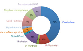
 Pilocytic astrocytoma location.jpg 567 × 558; 76 KB
-
 MRI pilocytic astrocytoma types.jpg 567 × 552; 76 KB
-
 Pilocytic astrocytoma micro 2.png 1,144 × 858; 1.21 MB
-
 Pilocytic astrocytoma micro 4.jpeg 1,017 × 736; 154 KB
-
 Immunohistochemistry of pilocytic astrocytoma Ki-67.jpeg 998 × 789; 234 KB
-
 CT 1.jpg 600 × 226; 39 KB
-
 CT VIPoma1.png 600 × 226; 127 KB
-
 CT VIPoma.jpg 600 × 450; 54 KB
-
 Giant-hepatic-haemangiomata.jpg 912 × 1,024; 62 KB
-
 PET CT.jpg 600 × 873; 93 KB
-
 Histology.jpg 600 × 223; 64 KB
-
 Giant-hepatic-haemangiomata (2).jpg 912 × 1,024; 82 KB
-
 Intra operative.jpg 2,998 × 2,317; 346 KB
-
 Surgical specimen.jpg 3,013 × 2,266; 306 KB
-
 Laryngeal-cancer-1 (1).jpg 1,024 × 1,024; 77 KB
-
 Pilocytic Micro.jpg 724 × 512; 333 KB
-
 Pilocytic astrocytoma endothelial proliferations.jpg 2,080 × 1,542; 940 KB
-
 Rosenthal fibers.jpg 721 × 512; 359 KB
-
 Trans-glottic-squamous-cell-carcinoma-1.jpg 1,024 × 1,024; 57 KB
-
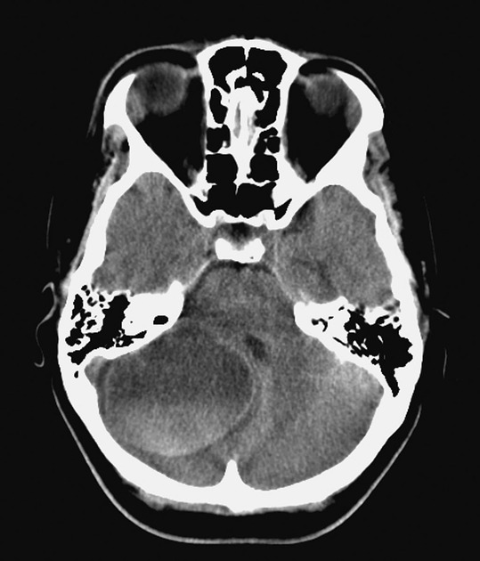 Pilocytic astrocytoma CT scan with hemorrhage.jpeg 539 × 630; 62 KB
-
 Location of pilocytic astrocytoma.PNG 292 × 180; 44 KB
-
 Follicular thyroid cancer 01.png 337 × 252; 176 KB
-
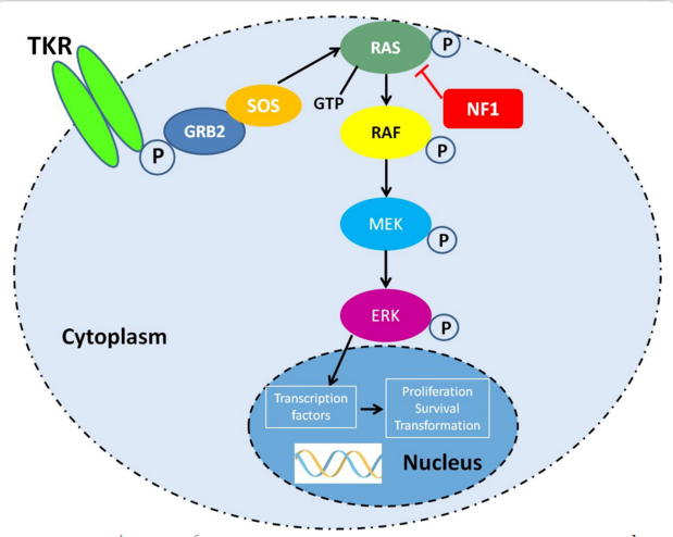 Genetic pathway pilocytic astrocytoma.PNG 619 × 494; 195 KB
-
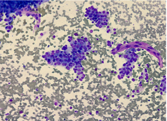
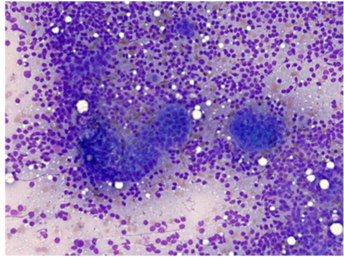 Lymph node FNAC metastatic follicular carcinoma.png 344 × 257; 225 KB
-
 Lymphnode FNAC metastatic follicular carcinoma 02.png 342 × 258; 203 KB
-
 FNAC lymph node metastatic follicular carcinoma 03.png 337 × 243; 177 KB
-
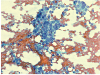
 Metastatic follicular carcinoma in the bone.png 338 × 245; 215 KB
-
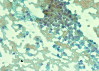
 Metastatic follicular cancer in the bone.png 342 × 254; 200 KB
-
 Unstimulated I-131.png 280 × 331; 50 KB
-
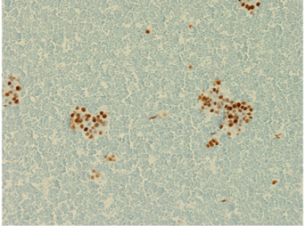
 Cervical metastasis of follicular thyroid cancer.png 325 × 235; 65 KB
-
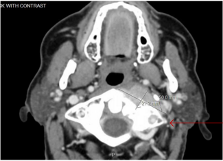
 CT Juvenile Pilocytic Astrocytoma.jpg 547 × 411; 64 KB
-
 CT scan of pilocytic astrocytoma.jpg 547 × 288; 59 KB
-
 AbbyCone.jpg 640 × 853; 95 KB
-
 MRI pilocytic astrocytoma 1.jpg 547 × 150; 27 KB


.jpg)


.jpg)


