Unused files
Jump to navigation
Jump to search
The following files exist but are not embedded in any page. Please note that other websites may link to a file with a direct URL, and so may still be listed here despite being in active use.
Showing below up to 50 results in range #24,481 to #24,530.
-
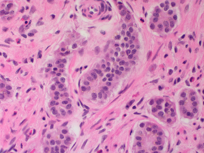
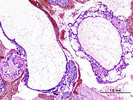
 Medulloepithelioma Histology.jpg 945 × 702; 1.24 MB
-
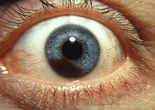 Iris melanoma.jpg 303 × 215; 117 KB
-
 Retinoblastoma fundoscopy.jpeg 630 × 630; 43 KB
-
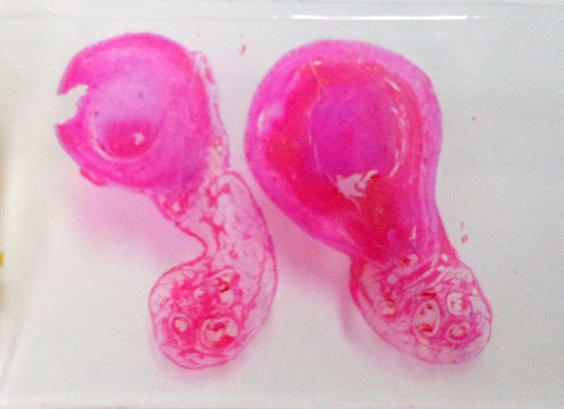 800px-Appendix Carcinoid Torsion 1X PA.JPG 800 × 580; 55 KB
-
 Muazam.png 300 × 300; 164 KB
-
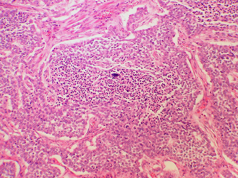
 Ewings sarcoma histology.jpg 640 × 427; 117 KB
-
 Ewings sarcoma histology 2.jpg 640 × 427; 142 KB
-
 800px-Appendix Carcinoid HP 14BR---.jpg 800 × 600; 123 KB
-
 800px-Appendix Carcinoid Necrosis PA.JPG 800 × 600; 240 KB
-
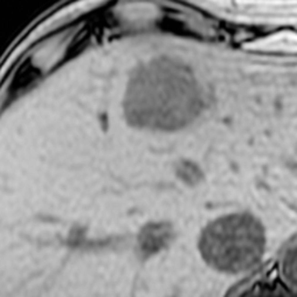 Axial T1 out of phase hepatic adenoma.jpg 1,024 × 1,024; 55 KB
-
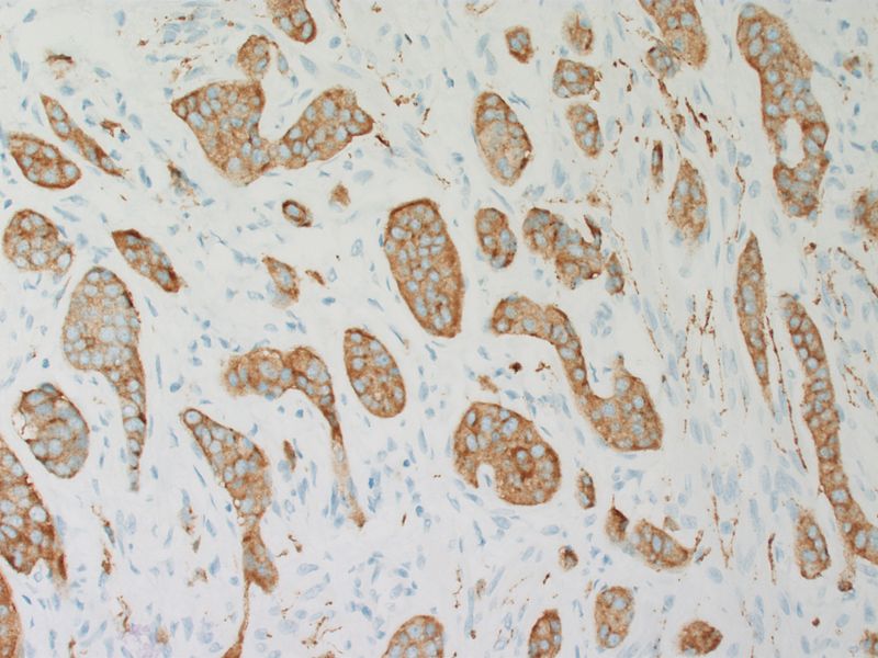 800px-Appendix Carcinoid Synaptophysin 14BR---.jpg 800 × 600; 86 KB
-
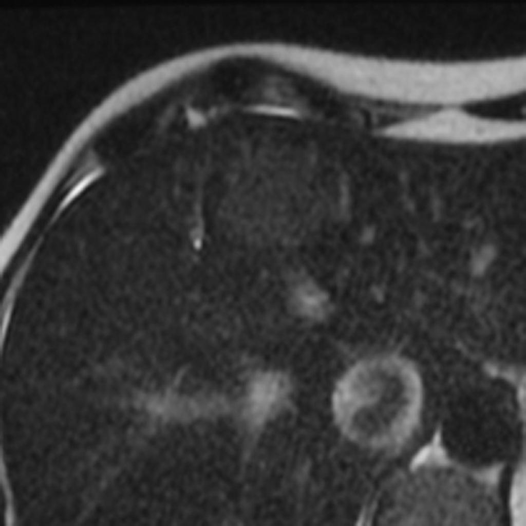 Hepatic-adenoma-T2 fat sat.jpg 1,024 × 1,024; 55 KB
-
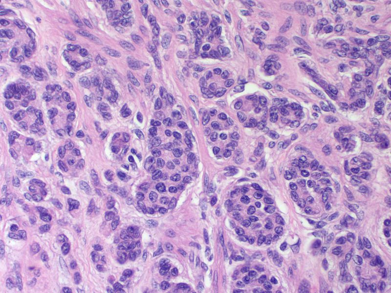 800px-Appendix Carcinoid HP CTR.jpg 800 × 600; 115 KB
-
 Papillary thyroid cancer.jpeg 456 × 630; 103 KB
-
 Papillary thyroid cancer(MRI).jpg 630 × 630; 96 KB
-
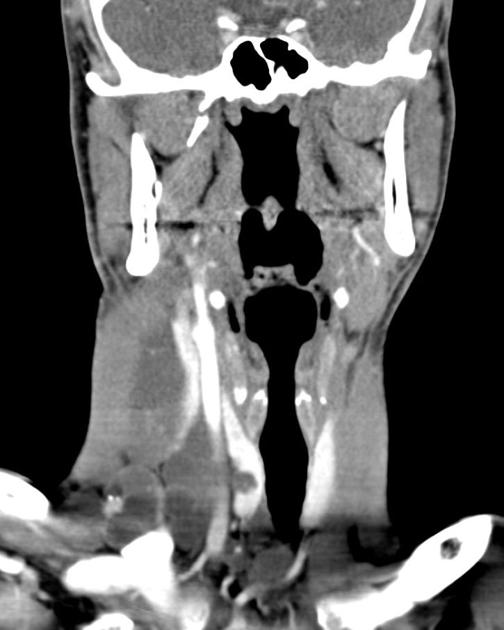 Coronal liver window thyroid metastasis.jpg 504 × 630; 34 KB
-
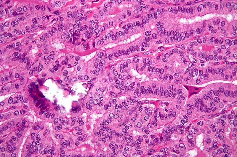
 Papillary thyroid cancer orphan annie eye nucleus.jpg 630 × 502; 64 KB
-
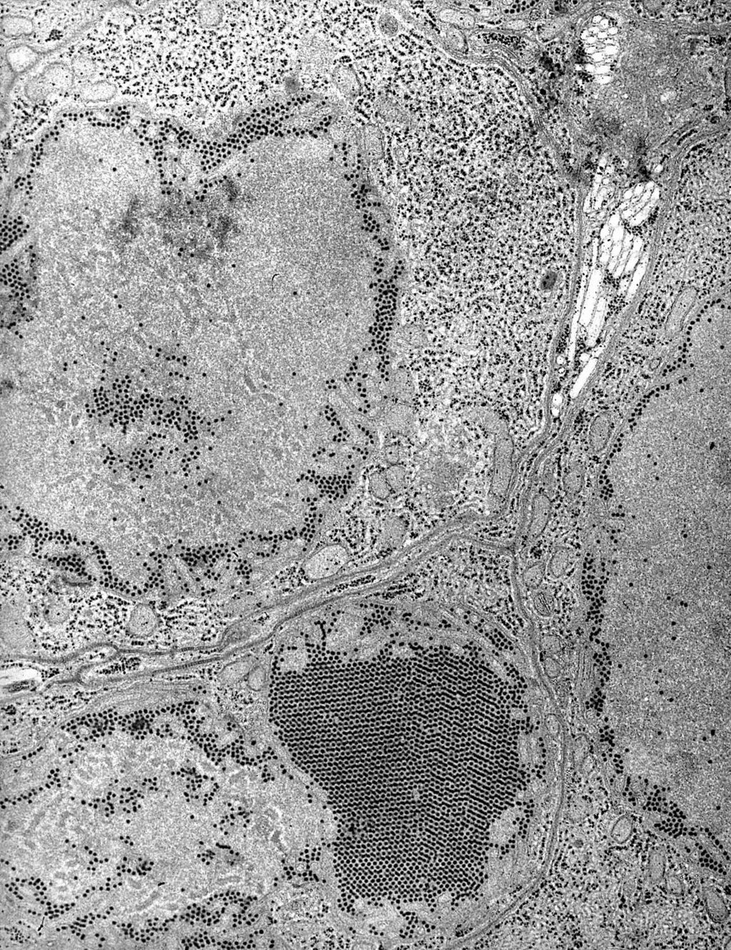 St. Louis Encephalitis (SLE) virus EM PHIL 1871 lores.JPG 1,024 × 1,331; 499 KB
-
 PTC 02.jpg 800 × 533; 155 KB
-
 Thyroid PapillaryCarcinoma CribriformMorularVariant04.jpg 800 × 600; 75 KB
-
 PTC 10.jpg 800 × 533; 140 KB
-
 Papillary thyroid carcinoma oncocytic variant -- high mag.jpg 800 × 533; 131 KB
-
 Papillary thyroid carcinoma oncocytic variant -- very high mag.jpg 800 × 533; 116 KB
-
 PTC 01.jpg 800 × 533; 164 KB
-
 193px-Hydatidiform mole (1) complete type.jpg 193 × 145; 17 KB
-
 753-11648-1-PB pilo11.jpg 422 × 323; 99 KB
-
 753-11649-1-PBpilo21234.jpg 429 × 326; 113 KB
-
 12342222 piloo.jpg 550 × 678; 102 KB
-
 Pilo123456.jpg 550 × 675; 99 KB
-
 G04nv32c04e.jpeg.gif 382 × 500; 144 KB
-
 G04nv32c10e.jpeg.gif 296 × 500; 92 KB
-
SVG test.svg 800 × 395; 11 KB
-

-
 Pilo CT cytic1234.gif 351 × 500; 138 KB
-
 G04nv32g02b.jpeg.gif 345 × 500; 163 KB
-
 G04nv32g02c.jpeg.gif 493 × 500; 215 KB
-

-
-
-
 CT oligoastrocytoma 2.jpg 504 × 630; 42 KB
-
 Oligoastroct.jpg 819 × 1,024; 71 KB
-
 Axial T1 choroidal melanoma.jpg 630 × 630; 40 KB
-
 Axial T1 C + fat.jpg 630 × 630; 42 KB
-
 Coronal T1 choroidal melanoma.jpg 630 × 630; 41 KB
-
 Axial T1 C+ fat.jpg 630 × 630; 40 KB
-
-
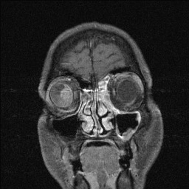
 Coronal C+ delayed orbital malignant melanoma.jpg 630 × 589; 35 KB
-
 Axial C+ delayed orbital malignant melanoma.jpg 630 × 589; 31 KB
-
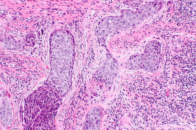 800px-Laryngeal squamous carcinoma -- intermed mag.jpg 800 × 533; 183 KB
-
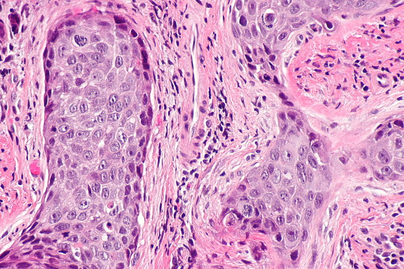 800px-Laryngeal squamous carcinoma -- high mag.jpg 800 × 533; 151 KB


.jpg)


_virus_EM_PHIL_1871_lores.JPG)


_complete_type.jpg)


