Unused files
Jump to navigation
Jump to search
The following files exist but are not embedded in any page. Please note that other websites may link to a file with a direct URL, and so may still be listed here despite being in active use.
Showing below up to 50 results in range #24,451 to #24,500.
-
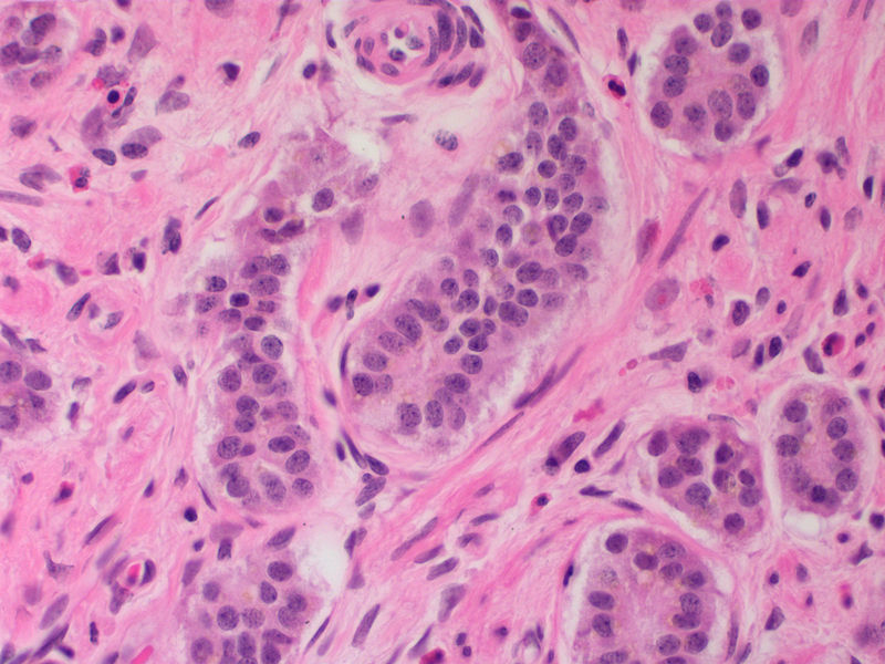
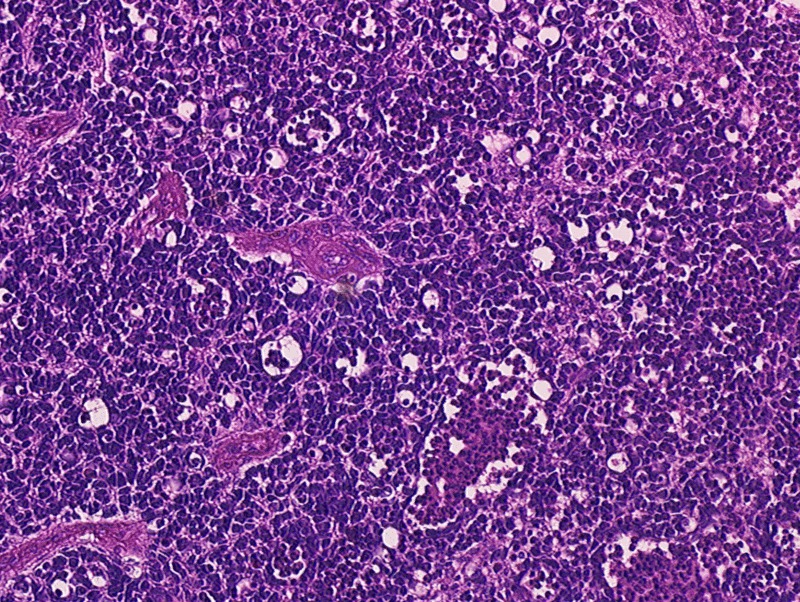 Retinoblastoma Pathology (2).jpg 800 × 602; 326 KB
-
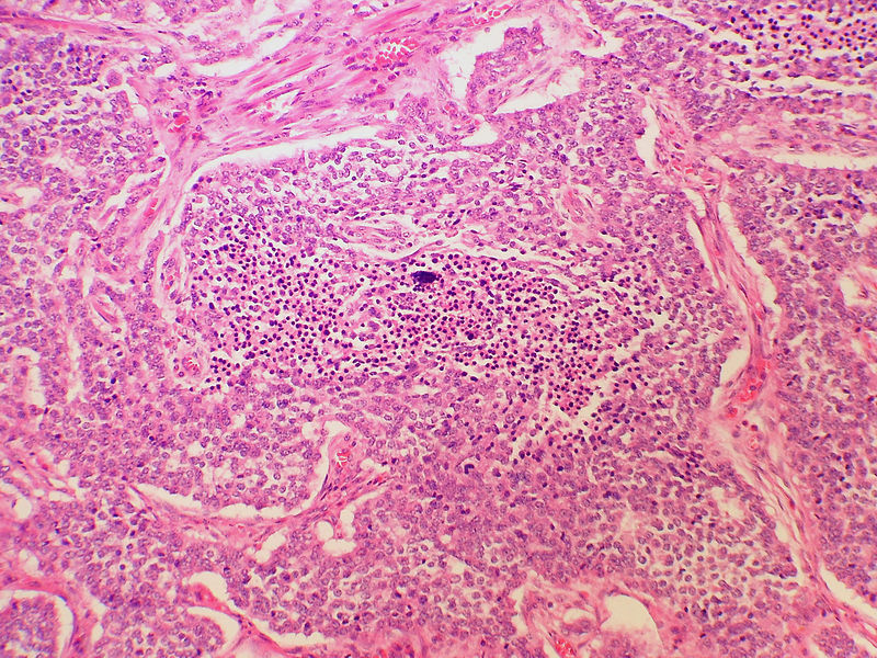
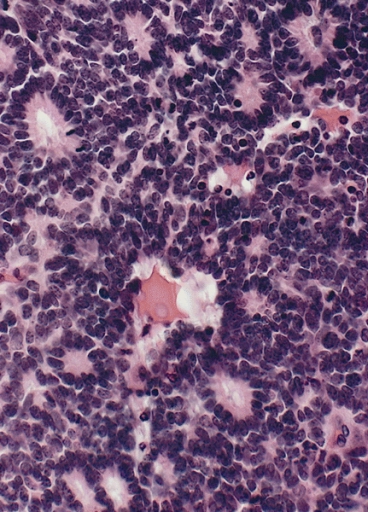 Flexner- Wintersteiner Rosettes in Retinoblastoma.jpg 368 × 512; 192 KB
-
-
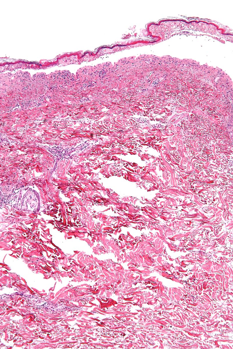 800px-Confluent epidermal necrosis - low mag.jpg 800 × 1,200; 328 KB
-
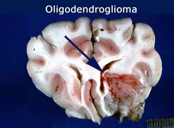 Oligodendroglioma gross 2.jpg 350 × 257; 33 KB
-
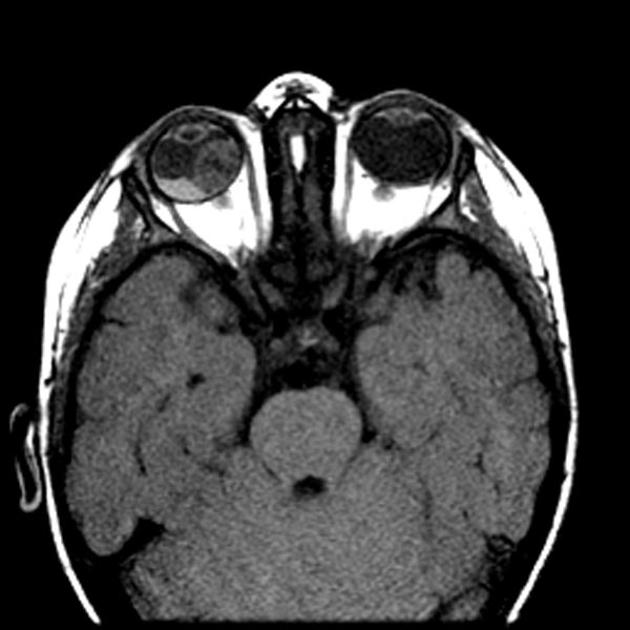 Axial MRI scan.jpg 630 × 630; 52 KB
-
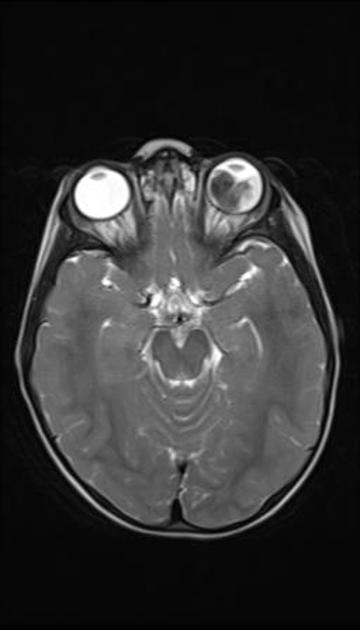 T2 MRI of retinoblastoma.jpg 360 × 630; 18 KB
-
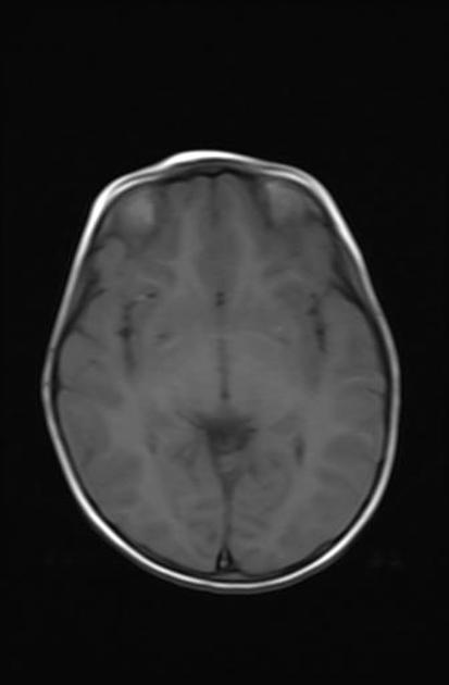 T1 MRI of retinoblastoma.jpg 413 × 630; 13 KB
-
 T1 C+ MRI retinoblastoma.jpg 630 × 630; 26 KB
-
 T1 C+ MRI of retinoblastoma.jpg 630 × 630; 42 KB
-
 CLL MRI T1 C+.jpg 578 × 630; 37 KB
-
 Diagram showing stage T2 thyroid cancer CRUK 258.png 342 × 221; 29 KB
-
-
 1752-1947-5-402-1.jpg 600 × 204; 43 KB
-
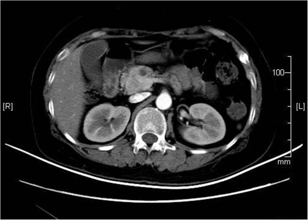
 NEM111.jpg 533 × 368; 223 KB
-
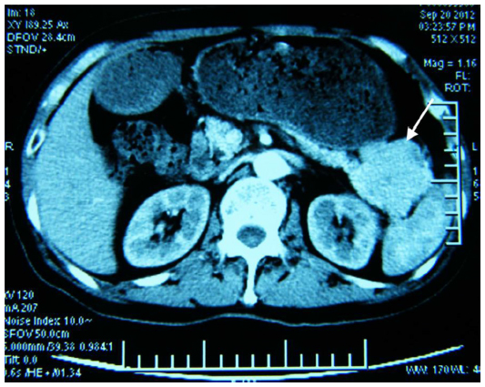 NEM23.jpg 533 × 425; 257 KB
-
 Scintigraphy.jpg 600 × 250; 32 KB
-
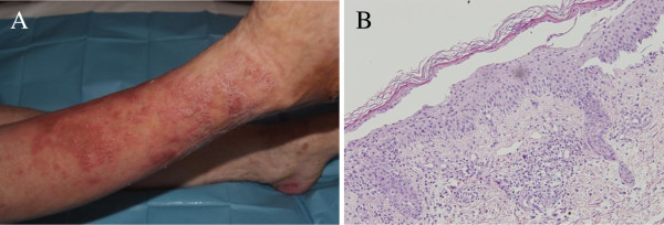
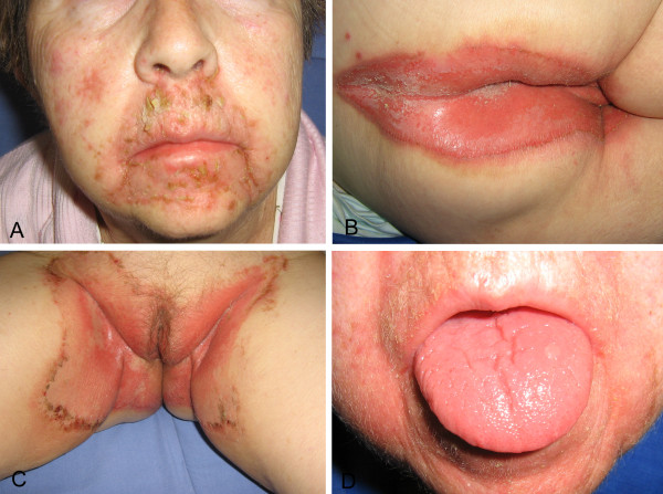
 Glucagonoma'.jpg 600 × 331; 136 KB
-
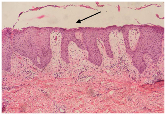
 Glucagonoma1.jpg 600 × 447; 88 KB
-
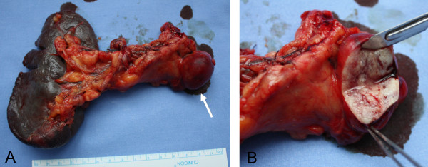 Gross1.jpg 600 × 234; 53 KB
-
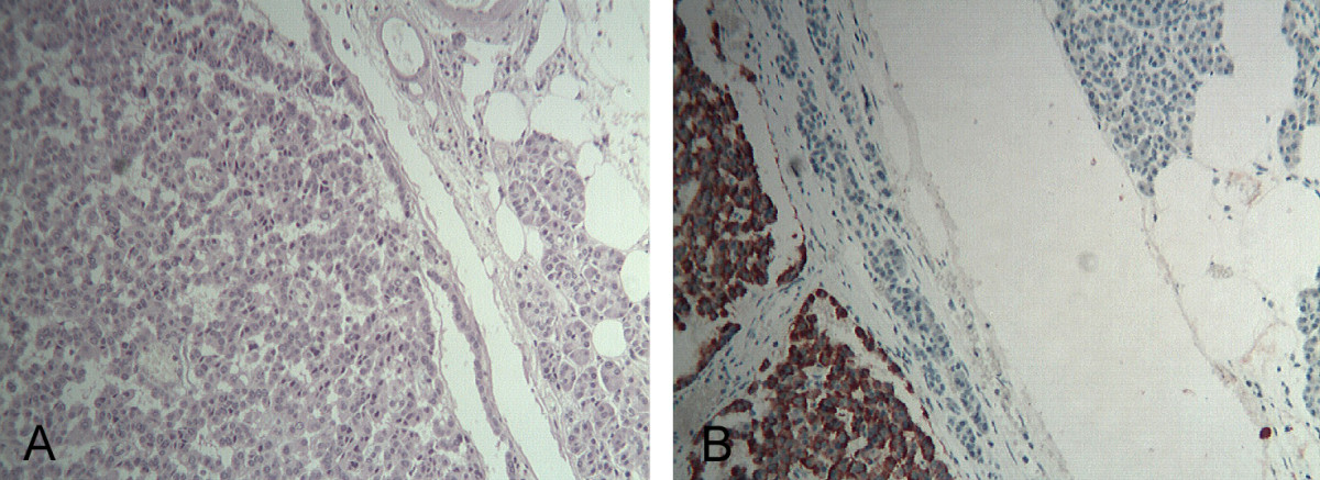 Histo.jpg 1,200 × 437; 220 KB
-
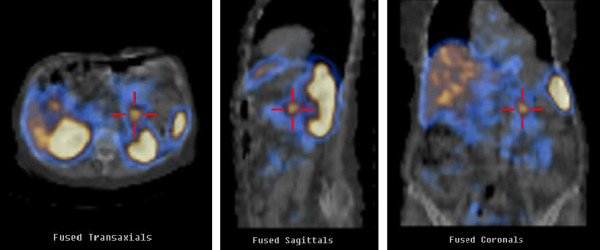
 Spect.jpg 1,133 × 541; 122 KB
-
 Incidence 2015.PNG 789 × 211; 22 KB
-
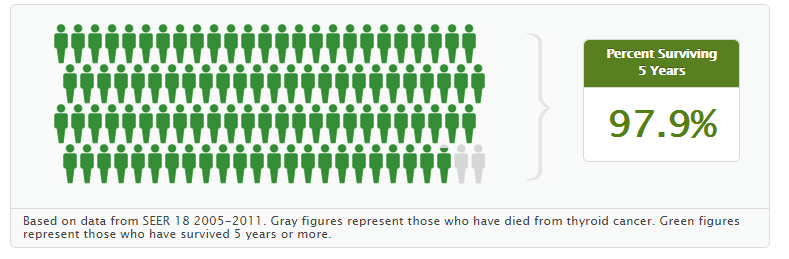 5 year survival rate of thyroid cancer.PNG 789 × 259; 19 KB
-
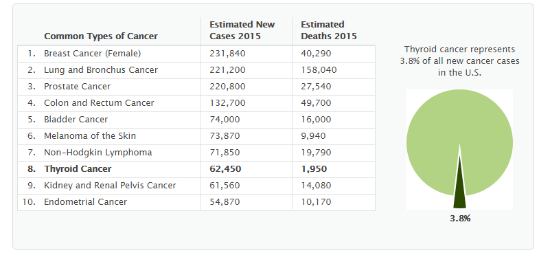
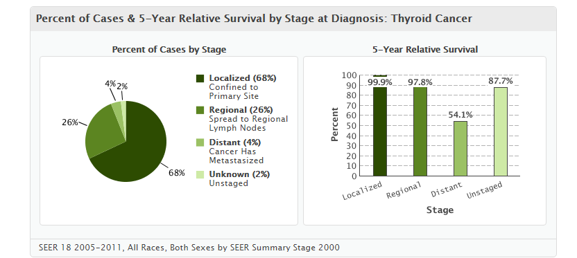 5 year survival pie chart for thyroid cancer.PNG 832 × 373; 37 KB
-
 Prevalence thyroid cancer.PNG 785 × 368; 27 KB
-
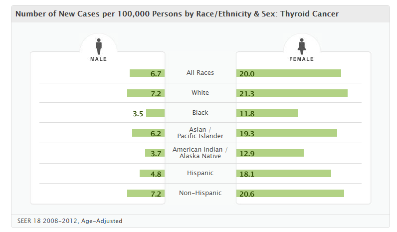 Gender thyroid cancer.PNG 796 × 465; 31 KB
-
 Mortality Thyroid cancer.PNG 787 × 409; 29 KB
-
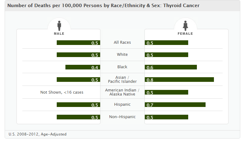 Thyroid cancer mortality gender based.PNG 787 × 458; 29 KB
-
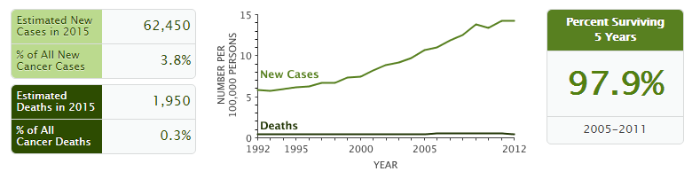
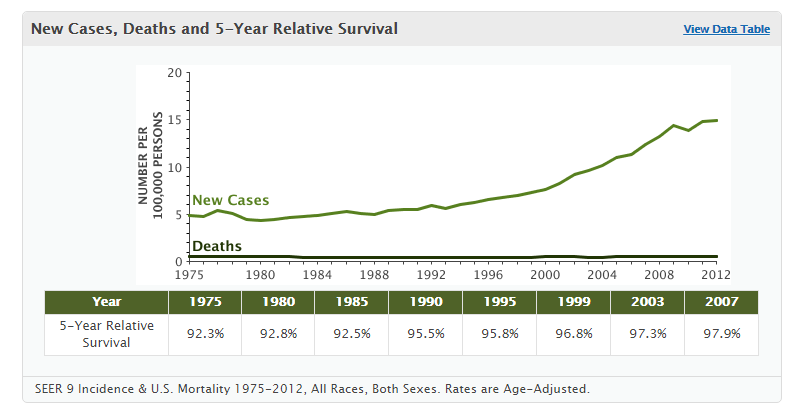 New cases, deaths and 5 year relative survival.PNG 799 × 414; 27 KB
-
 Medulloepithelioma Histology.jpg 945 × 702; 1.24 MB
-
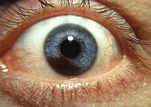 Iris melanoma.jpg 303 × 215; 117 KB
-
 Retinoblastoma fundoscopy.jpeg 630 × 630; 43 KB
-
 800px-Appendix Carcinoid Torsion 1X PA.JPG 800 × 580; 55 KB
-
 Muazam.png 300 × 300; 164 KB
-
 Ewings sarcoma histology.jpg 640 × 427; 117 KB
-
 Ewings sarcoma histology 2.jpg 640 × 427; 142 KB
-
 800px-Appendix Carcinoid HP 14BR---.jpg 800 × 600; 123 KB
-
 800px-Appendix Carcinoid Necrosis PA.JPG 800 × 600; 240 KB
-
 Axial T1 out of phase hepatic adenoma.jpg 1,024 × 1,024; 55 KB
-
 800px-Appendix Carcinoid Synaptophysin 14BR---.jpg 800 × 600; 86 KB
-
 Hepatic-adenoma-T2 fat sat.jpg 1,024 × 1,024; 55 KB
-
 800px-Appendix Carcinoid HP CTR.jpg 800 × 600; 115 KB
-
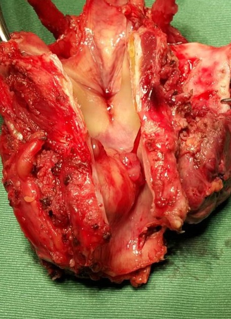 Papillary thyroid cancer.jpeg 456 × 630; 103 KB
-
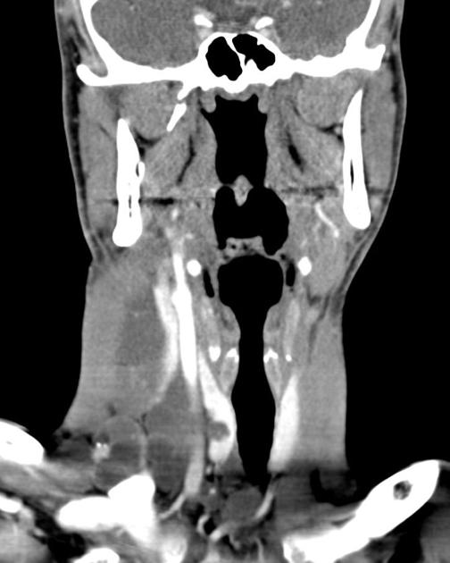
 Papillary thyroid cancer(MRI).jpg 630 × 630; 96 KB
-
 Coronal liver window thyroid metastasis.jpg 504 × 630; 34 KB
-
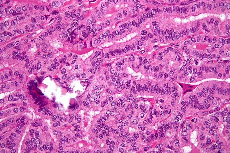
 Papillary thyroid cancer orphan annie eye nucleus.jpg 630 × 502; 64 KB
-
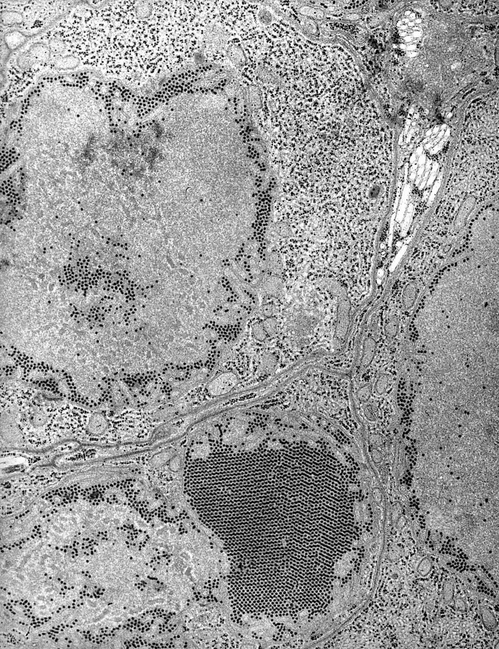 St. Louis Encephalitis (SLE) virus EM PHIL 1871 lores.JPG 1,024 × 1,331; 499 KB
-
 PTC 02.jpg 800 × 533; 155 KB
-
 Thyroid PapillaryCarcinoma CribriformMorularVariant04.jpg 800 × 600; 75 KB
.jpg)


.jpg)


_virus_EM_PHIL_1871_lores.JPG)

