Unused files
Jump to navigation
Jump to search
The following files exist but are not embedded in any page. Please note that other websites may link to a file with a direct URL, and so may still be listed here despite being in active use.
Showing below up to 50 results in range #24,421 to #24,470.
-
 Sergekorjian-circle.png 300 × 300; 371 KB
-
 Gross pituitary adenoma A..jpg 182 × 254; 52 KB
-
 Coronal T1+ C.jpg 627 × 630; 24 KB
-
 Carloslopez.png 300 × 300; 167 KB
-
 Ashleymedeiros.png 300 × 300; 168 KB
-
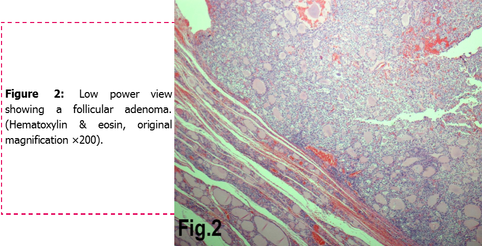 Follicular adenoma.png 943 × 481; 864 KB
-
 120px-Adrenal Neuroblastoma VascularInvasion MP CTR.jpg 120 × 90; 6 KB
-
 Molecular subtypes of medulloblastoma.png 600 × 500; 180 KB
-
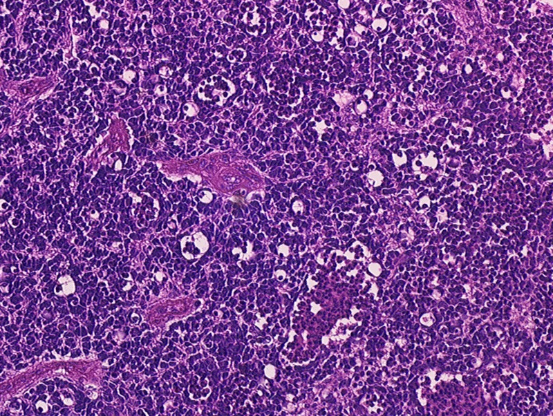
 Medulloblsatoma.JPG 500 × 376; 109 KB
-
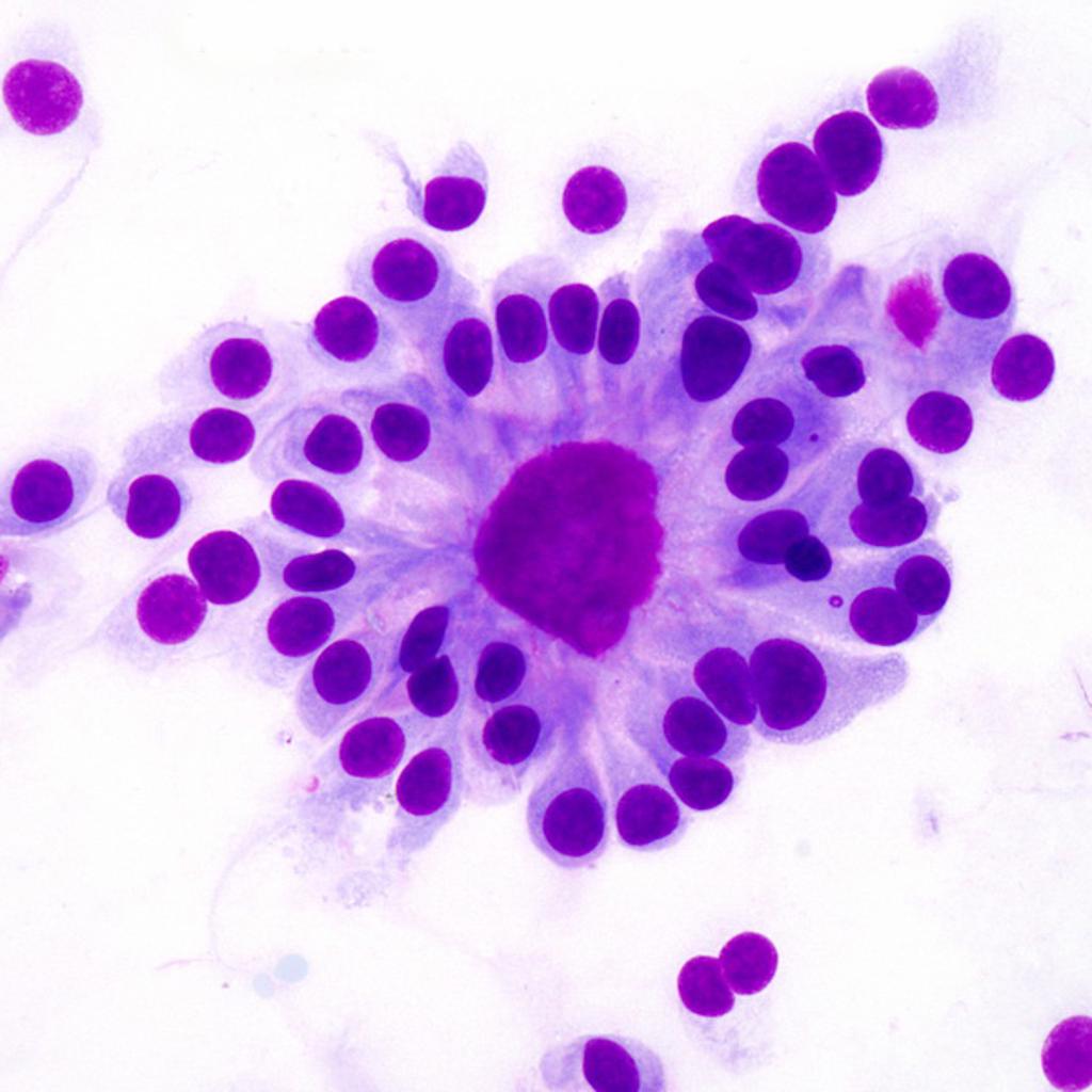 Ependymoma-true-ependymal-rosettes.jpg 1,024 × 1,024; 95 KB
-
 Micropathology.jpg 600 × 226; 155 KB
-
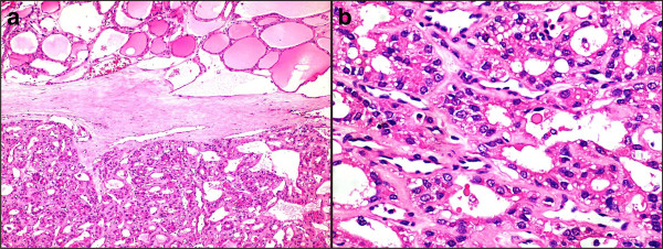
 Microfollicular stain.jpg 600 × 451; 222 KB
-
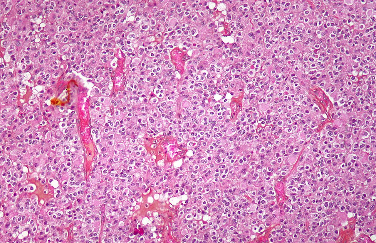
 Thyroid adenoma.jpg 1,024 × 768; 97 KB
-
 Ependymoma-lateral-ventricle C+.jpg 1,024 × 1,024; 75 KB
-
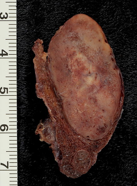 Follicular Adenoma of the Thyroid Gland.jpg 440 × 599; 64 KB
-
 Oligodendroglioma1 low mag.jpg 1,280 × 829; 415 KB
-
 Oligodendroglioma1 high mag.jpg 1,024 × 683; 190 KB
-
 Anaplastic oligodendroglioma minigemistocytes.jpg 800 × 593; 106 KB
-
 IMG 1582.JPG 2,448 × 3,264; 666 KB
-
 Oligodendroglioma discrete invasion HE.jpg 2,080 × 1,542; 1.11 MB
-
 MAP2 anaplastic oligodendroglioma.jpg 2,080 × 1,542; 744 KB
-
 IDH1 R132H in anaplastic ologodendroglioma.jpg 2,080 × 1,542; 697 KB
-
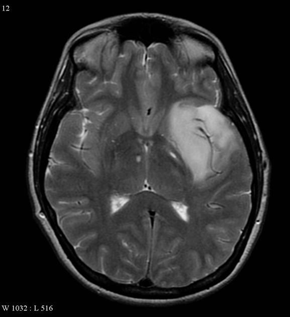 Oligodendroglioma MRI axial T2.jpg 577 × 630; 32 KB
-
 Oligodendroglioma coronal T1 C+.jpg 630 × 600; 29 KB
-
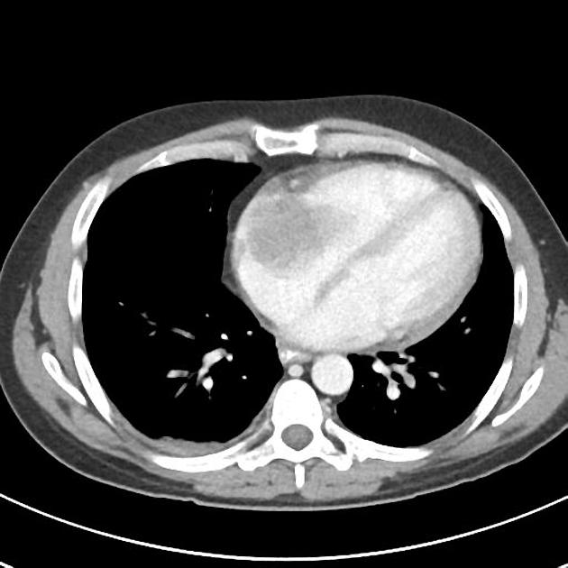 Burkitt lymphoma CT Chest 01.jpg 630 × 630; 38 KB
-
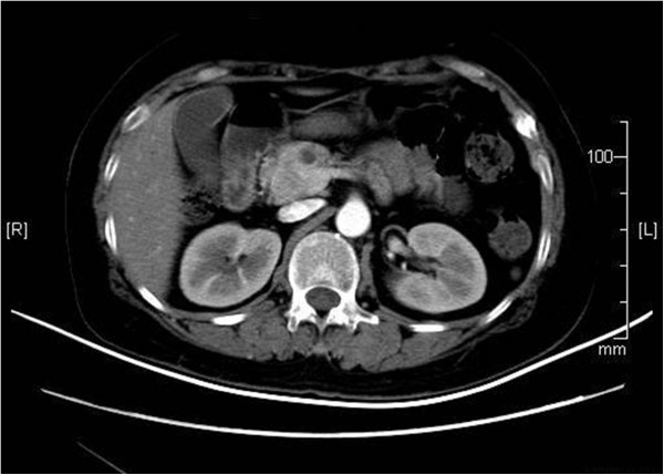
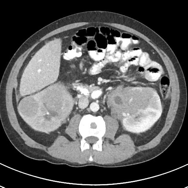 Burkitt's lymphoma CT abdomen 02.jpg 630 × 630; 47 KB
-
 MRI transeverse neuroblastoma.jpg 630 × 630; 19 KB
-
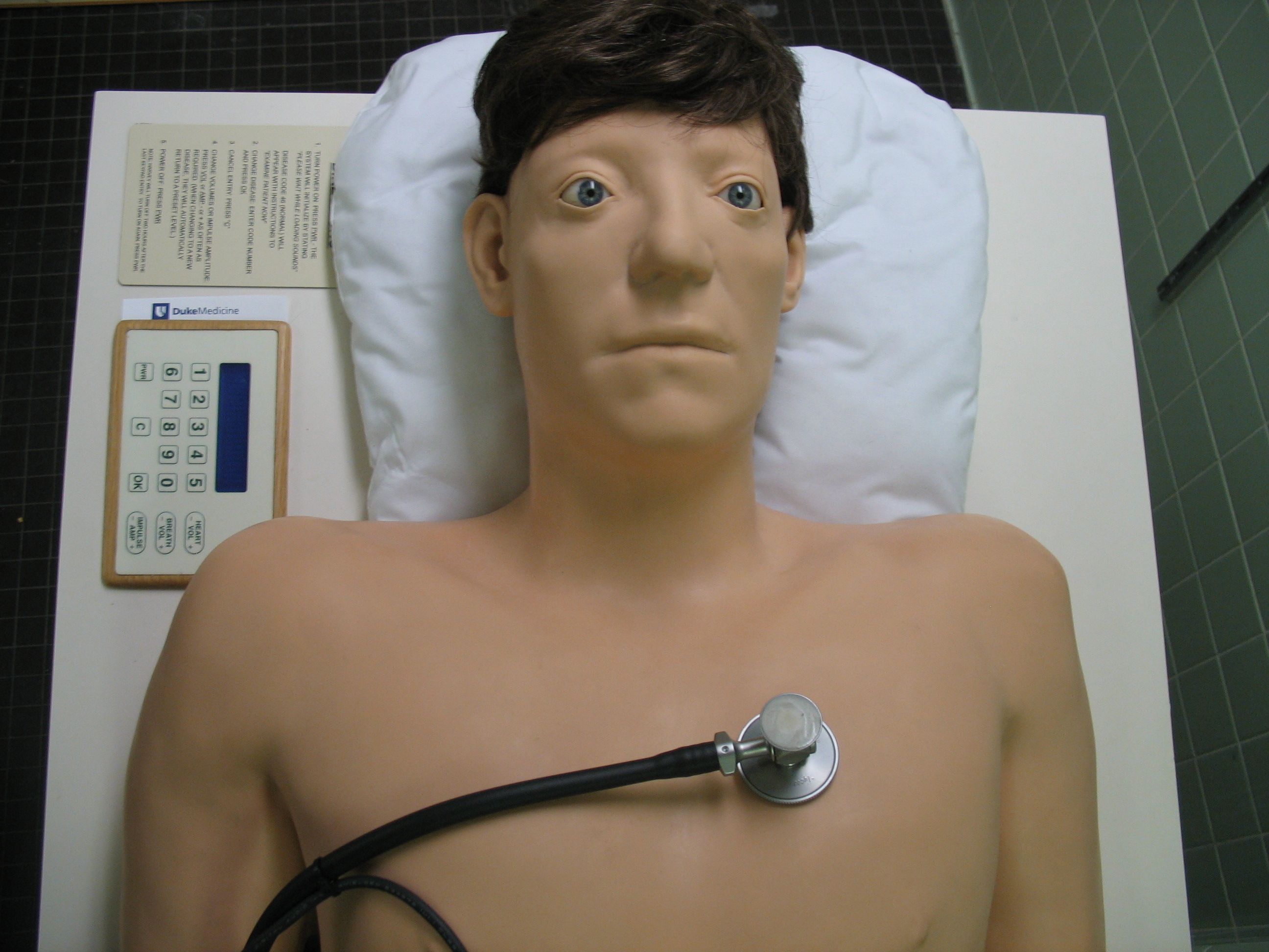 Harvey front.jpg 2,592 × 1,944; 427 KB
-
 Chris heart sounds model.png 1,000 × 562; 594 KB
-
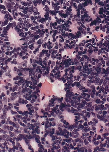
 Retinoblastoma Pathology.GIF 800 × 602; 562 KB
-
 Retinoblastoma Pathology (2).jpg 800 × 602; 326 KB
-
 Flexner- Wintersteiner Rosettes in Retinoblastoma.jpg 368 × 512; 192 KB
-
-
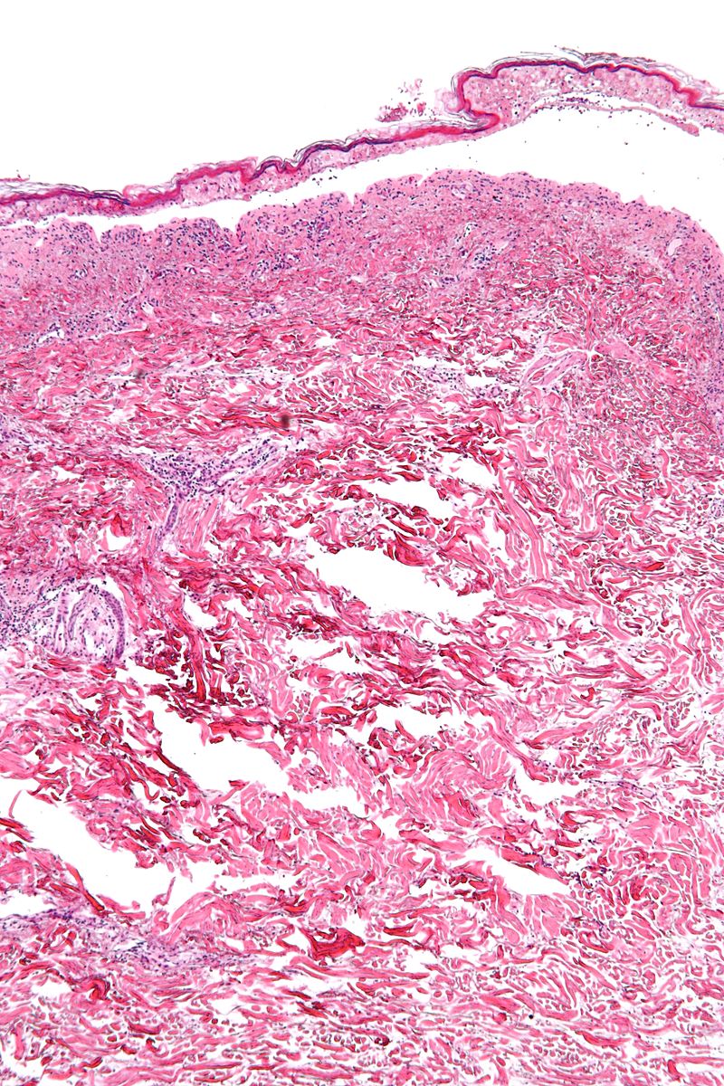 800px-Confluent epidermal necrosis - low mag.jpg 800 × 1,200; 328 KB
-
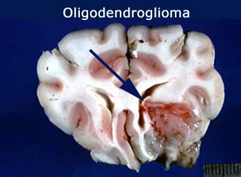 Oligodendroglioma gross 2.jpg 350 × 257; 33 KB
-
 Axial MRI scan.jpg 630 × 630; 52 KB
-
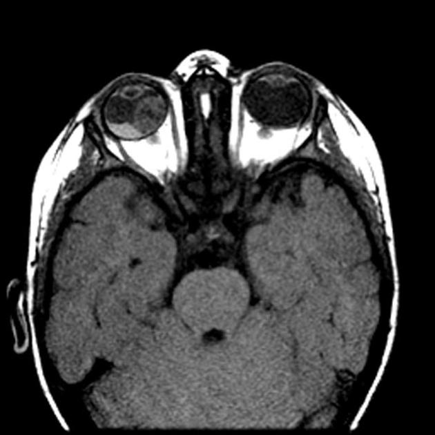
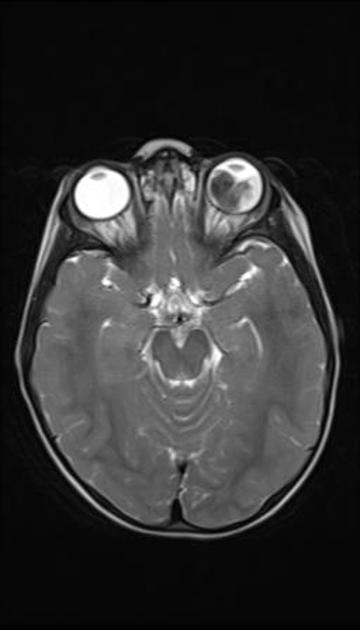 T2 MRI of retinoblastoma.jpg 360 × 630; 18 KB
-
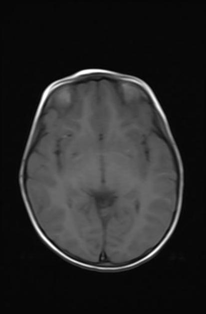 T1 MRI of retinoblastoma.jpg 413 × 630; 13 KB
-
 T1 C+ MRI retinoblastoma.jpg 630 × 630; 26 KB
-
 T1 C+ MRI of retinoblastoma.jpg 630 × 630; 42 KB
-
 CLL MRI T1 C+.jpg 578 × 630; 37 KB
-
 Diagram showing stage T2 thyroid cancer CRUK 258.png 342 × 221; 29 KB
-
-
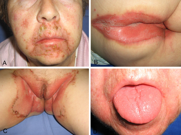
 1752-1947-5-402-1.jpg 600 × 204; 43 KB
-
 NEM111.jpg 533 × 368; 223 KB
-
 NEM23.jpg 533 × 425; 257 KB
-
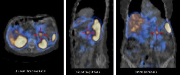 Scintigraphy.jpg 600 × 250; 32 KB
-
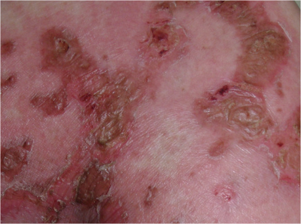
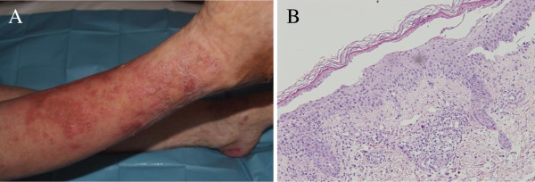
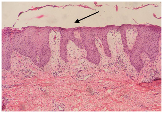
 Glucagonoma'.jpg 600 × 331; 136 KB
-
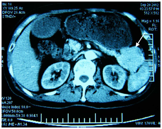
 Glucagonoma1.jpg 600 × 447; 88 KB
-
 Gross1.jpg 600 × 234; 53 KB


.jpg)


