Unused files
Jump to navigation
Jump to search
The following files exist but are not embedded in any page. Please note that other websites may link to a file with a direct URL, and so may still be listed here despite being in active use.
Showing below up to 50 results in range #24,371 to #24,420.
-
 MRI meningioma with vascular pedicle Dr Frank Gaillard.jpg 427 × 442; 21 KB
-
 MRI meningioma anaplastic Dr Frank Gaillard.jpg 410 × 442; 20 KB
-
 Angiography meningioma Dr Bruno Di Muzio.JPG 442 × 408; 17 KB
-
 Mucosal neuromas in MEN 2.jpg 600 × 457; 148 KB
-
 Metastasis in MEN 2.jpg 600 × 334; 116 KB
-
 Medulloblsatoma 2.JPG 120 × 90; 9 KB
-
 Medulloblastoma 3.jpg 120 × 89; 6 KB
-
 Medulloblastoma 4.jpg 120 × 89; 5 KB
-
 Medulloblastoma 5.jpg 120 × 89; 5 KB
-
 Medulloblastoma 6.jpg 120 × 89; 6 KB
-
 Medulloblastoma 7.jpg 120 × 85; 5 KB
-
 Medulloblastoma 8.jpg 120 × 89; 6 KB
-
 Medulloblastoma 9.jpg 120 × 89; 7 KB
-
 Medulloblastoma Areas of geographic necrosis..jpg 120 × 89; 6 KB
-
-
 Medulloblastoma Partial MAP2 immunoreactivity..jpg 120 × 89; 6 KB
-
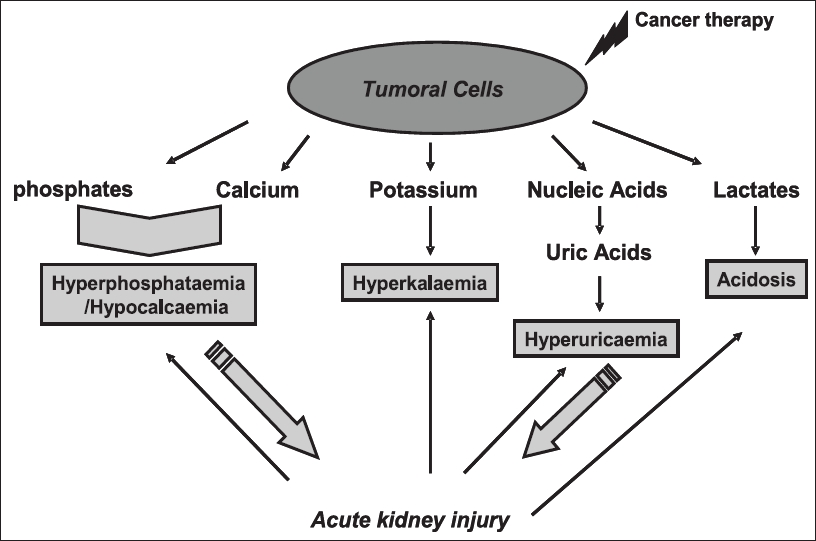 TLS-kidney injury.jpeg 816 × 541; 120 KB
-
 Medulloblastoma gross pathology Dr Frank Gaillard.jpg 442 × 442; 31 KB
-
 Osteosarcoma (2).jpg 1,024 × 1,024; 94 KB
-
 Lung carcinoid low mag.jpg 800 × 533; 215 KB
-
 Lung carcinoid intermed mag.jpg 800 × 533; 185 KB
-
 Lung carcinoid very high mag.jpg 800 × 533; 132 KB
-
 800px-Lung carcinoid - high mag.jpg 800 × 533; 169 KB
-
 Left-piriform-fossa-mass-likely-scc (3).jpg 1,020 × 1,024; 58 KB
-
 Scc.jpg 320 × 213; 35 KB
-
 Melanoma.JPG 320 × 240; 29 KB
-
 BCC.jpg 320 × 213; 30 KB
-
 Osteosarcoma-conventional-histology (1).jpg 1,024 × 1,024; 151 KB
-
 Gastrinoma.jpeg 600 × 600; 73 KB
-
 Osteoid formation.jpg 640 × 427; 144 KB
-
 Liver-MRI-ZES.png 475 × 305; 118 KB
-
 ZES-MRI22png .png 474 × 311; 173 KB
-
 ZES-MRI3png copy.tiff 383 × 359; 540 KB
-
 ZES-MRI3.png 385 × 376; 167 KB
-
 Osteosarcoma femur.jpg 1,024 × 1,024; 52 KB
-
 Osteosarcoma-distal-femur MRI.jpg 1,024 × 1,024; 65 KB
-
 Osteosarcoma MRI T2.jpg 1,024 × 1,024; 69 KB
-
 Osteosarcoma-distal-femur MRI T1c.jpg 1,024 × 1,024; 50 KB
-
 Pancreatic insulinoma Histology 1.JPG 500 × 376; 114 KB
-
 SRS1.jpg 750 × 774; 63 KB
-
 SRS2.jpg 750 × 781; 72 KB
-
 SRS3.jpg 353 × 361; 94 KB
-
 SRS4.jpg 263 × 270; 29 KB
-
 PITUITARY ADENOMAS.jpg 756 × 682; 117 KB
-
 Pothuru.png 300 × 300; 173 KB
-
 Chouturi.png 300 × 300; 170 KB
-
 Alkateb.png 300 × 300; 145 KB
-
 Mediastinitis CT 1.jpg 300 × 218; 13 KB
-
 Shanshancen.png 300 × 300; 369 KB
-
 Haideri.png 300 × 300; 169 KB


.jpg)


.jpg)


.jpg)


