Unused files
Jump to navigation
Jump to search
The following files exist but are not embedded in any page. Please note that other websites may link to a file with a direct URL, and so may still be listed here despite being in active use.
Showing below up to 50 results in range #24,321 to #24,370.
-
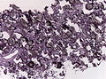
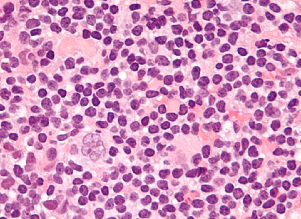 Popcorn cell in nodular lymphocyte predominant Hodgkin lymphoma.jpg 1,024 × 745; 147 KB
-
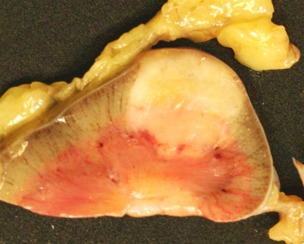
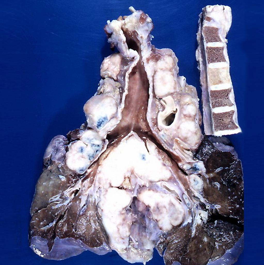 Hodgkin's lymphoma Gross Pathology.jpg 1,023 × 1,024; 143 KB
-
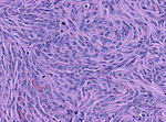
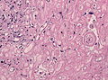
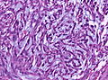
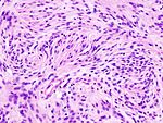 Grade 1 Meningioma.jpg 150 × 113; 9 KB
-
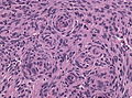
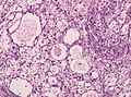
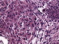
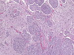 Grade 2 menigioma.jpg 150 × 111; 6 KB
-
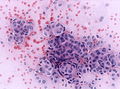
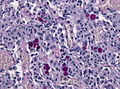
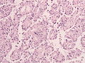
 Grade 3 menigioma.jpg 150 × 111; 7 KB
-
 Meningeotheliomatous meningeoma 1.jpg 120 × 89; 6 KB
-
 Meningothelioal Meningioma 3.jpg 120 × 89; 5 KB
-
 Meningothelial Meningioma showing 2.jpg 120 × 90; 5 KB
-
 Meningothelial Meningioma 4.jpg 120 × 80; 5 KB
-
 Meningioma fibromatous variant.jpg 120 × 90; 6 KB
-
 Meningiom fibrous variant2.JPG 120 × 90; 6 KB
-
 Miningioma (2) transitional type.jpg 119 × 90; 14 KB
-
 Psammomatous meningioma.jpg 120 × 89; 6 KB
-
 Angiomatous meningioma.jpg 120 × 89; 5 KB
-
 Microcystic meningeoma.jpg 120 × 89; 6 KB
-
 Secretory meningioma PAS.jpg 120 × 89; 6 KB
-
 Chordoid meningoma.jpg 120 × 89; 7 KB
-
 Alcian blue chordoid meningioma.jpg 120 × 89; 5 KB
-
 Rhabdoid meningioma.jpg 120 × 89; 6 KB
-
 Papillary meningioma.jpg 120 × 89; 5 KB
-
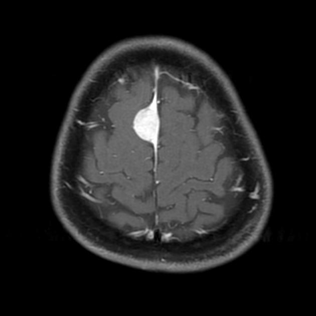
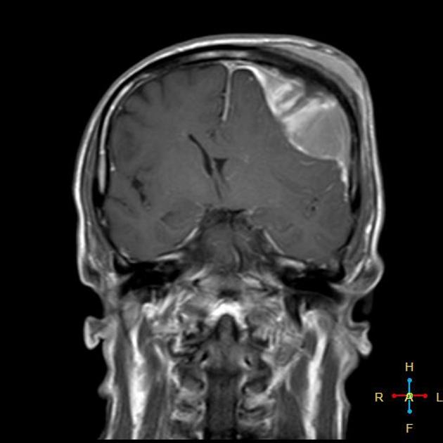
 Coronal T1 C+ .jpg 627 × 630; 24 KB
-
 Allaham.png 300 × 300; 98 KB
-
 Coronal T1+C.jpg 627 × 630; 24 KB
-
 Angiomyolipoma gross.JPG 1,024 × 822; 81 KB
-
 Renal-angiomyolipoma-8 (5).jpg 1,024 × 1,024; 136 KB
-
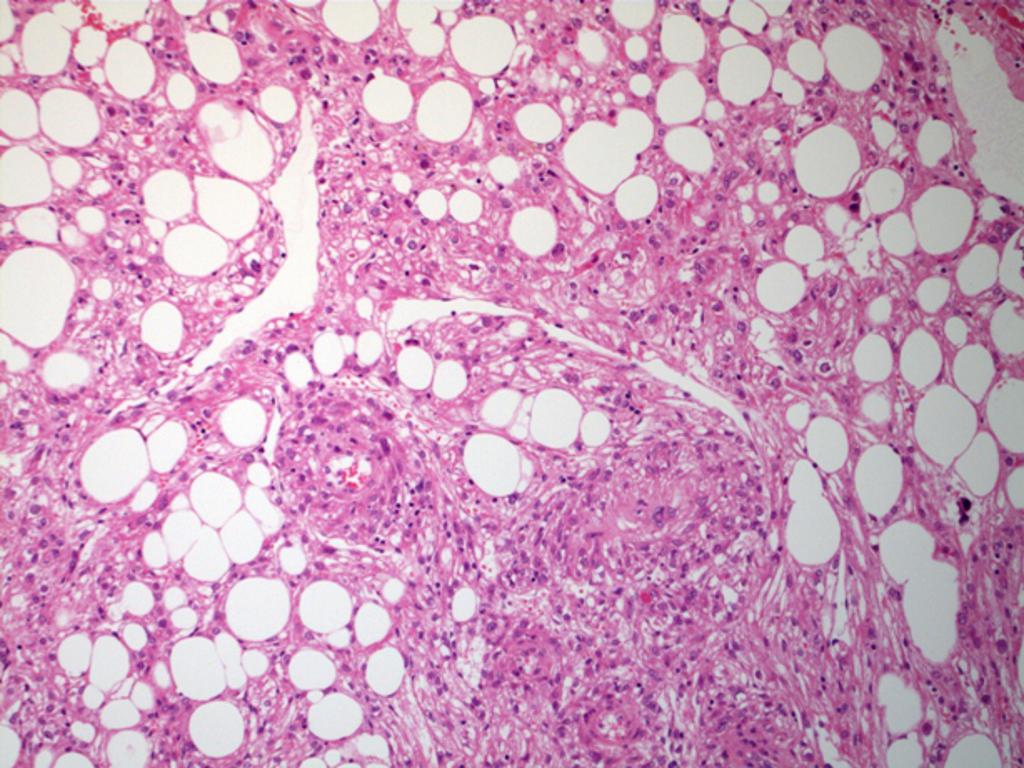 Angiomyolipoma-7 (1).jpg 1,024 × 768; 143 KB
-
 Coronal T1 C+ Gliomatosis cerebri.jpg 630 × 630; 98 KB
-
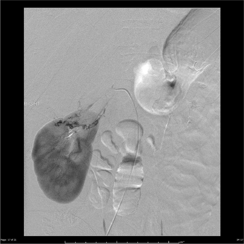
 Retroperitoneal-haemorrhage-from-renal-angiomyolipoma.jpg 1,024 × 848; 91 KB
-
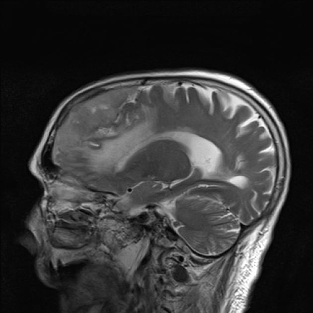
 Saggital T1 C+.jpg 623 × 630; 44 KB
-
 Post surgical folow up.png 1,000 × 662; 27 KB
-
 Prevalence MEN 2.jpg 600 × 473; 49 KB
-
 CT scan lfactory meningioma.jpg 630 × 630; 46 KB
-
 CT scan of cerbellopontine angle meningioma.jpg 596 × 630; 35 KB
-
 CT scan Sphenoid wing meningioma.jpg 483 × 630; 36 KB
-
 CT scan of cerbral convexity meningioma.jpg 630 × 504; 31 KB
-
 CT scan calcified meningioma.jpeg 630 × 507; 48 KB
-
 CT falx meningioma.jpg 1,024 × 1,024; 93 KB
-
 MRI meningioma spoke wheel appearance.jpg 609 × 630; 32 KB
-
 Osteosarcoma gross pathology.jpg 1,024 × 1,024; 94 KB
-
 MRI convexity meningioma Dr Sajoscha Sorrentino.jpg 511 × 630; 21 KB
-
 Axial DWI.jpg 623 × 630; 23 KB
-
 MRI convexity meningioma.jpg 511 × 630; 21 KB
-
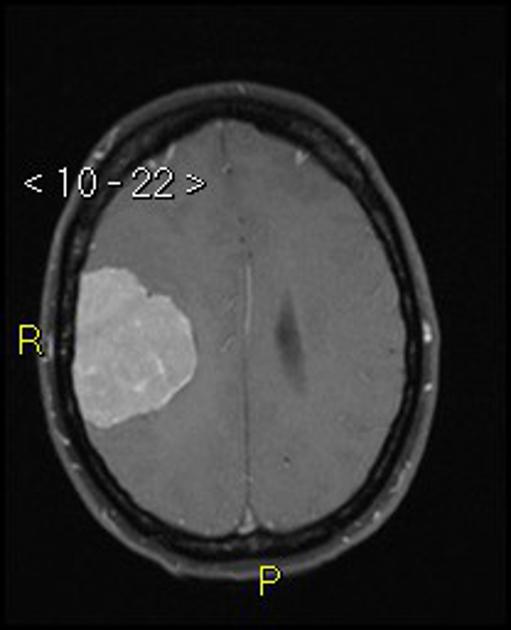
 MRI Atypical meningioma.jpg 442 × 442; 42 KB
-
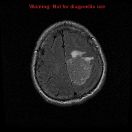
 MRI malignant anaplastic meningioma.jpg 630 × 630; 27 KB
-
 MRI olfactory meningioma Dr Frank Gaillard.jpg 504 × 630; 42 KB
-
 MRI meningioma cystic Dr Frank Gaillard.jpg 353 × 442; 15 KB
-
 MRI meningioma dural tail sign Dr Frank Gaillard.jpg 630 × 630; 30 KB
-
 MRI meningioma falx Dr Hani Al Salam.jpg 442 × 442; 25 KB
-
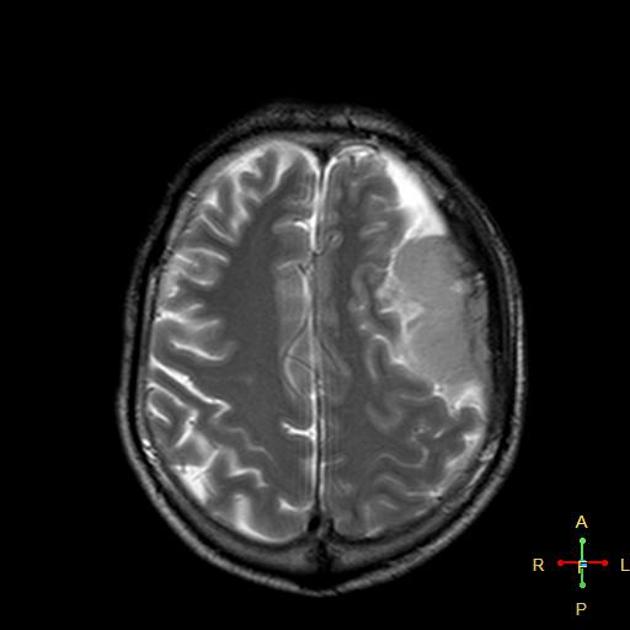
 MRI malignant meningioma Dr Ahmed Abd Rabou.jpg 630 × 630; 32 KB
-
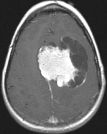
 MRI invasive meningioma Dr Bita Abbasi.jpeg 630 × 630; 36 KB


.jpg)
.jpg)


_transitional_type.jpg)