Unused files
Jump to navigation
Jump to search
The following files exist but are not embedded in any page. Please note that other websites may link to a file with a direct URL, and so may still be listed here despite being in active use.
Showing below up to 50 results in range #24,301 to #24,350.
-
 53704cf488a848544c186cae250f65 thumb.jpg 176 × 176; 5 KB
-
 MEN 2 treatment.png 1,001 × 680; 89 KB
-
 MIcroscopic pathology of medullary thyroid cancer .jpg 230 × 153; 20 KB
-
 Plexiform schwannoma with high magnifaction.jpg 120 × 98; 4 KB
-
 Psammomatous melanotic schwannoma.jpg 120 × 80; 5 KB
-
 Antoni A and B with high magnification.jpg 800 × 533; 150 KB
-
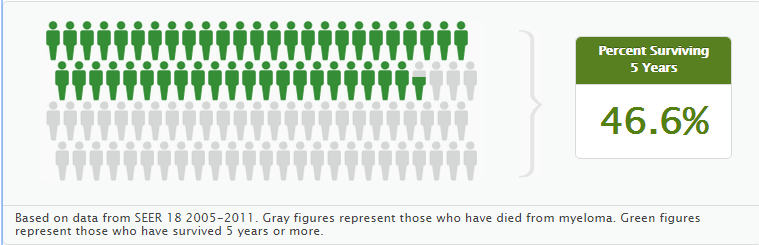 5 year survival rate.png 759 × 245; 24 KB
-
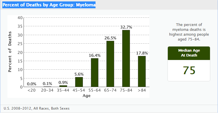 Percent of Deaths by Age Group Myeloma.png 760 × 396; 40 KB
-
 PJSIntussusception.jpg 739 × 504; 91 KB
-
 PJSpolyps.jpg 738 × 503; 142 KB
-
 PJSpolyps2.jpg 737 × 357; 100 KB
-
 Multi-ple myeloma.jpg 800 × 533; 121 KB
-
 CT acoustic neuroma.jpg 593 × 630; 31 KB
-
 Arikapudi.png 300 × 300; 111 KB
-
 Pheochromocytoma MRI 02.jpg 442 × 442; 25 KB
-
 Non-Hodgkin lymphoma chest-x ray.jpeg 1,024 × 802; 159 KB
-
 Hodgkin's Disease1.jpg 800 × 1,196; 105 KB
-
 Osteosarcoma distal femur pathology.jpg 1,024 × 1,024; 94 KB
-
 Reed-Sternberg lymphocyte.jpg 1,024 × 655; 70 KB
-
 Hodgkin lymphoma cytology large.jpg 1,024 × 753; 165 KB
-
 Popcorn cell in nodular lymphocyte predominant Hodgkin lymphoma.jpg 1,024 × 745; 147 KB
-
 Hodgkin's lymphoma Gross Pathology.jpg 1,023 × 1,024; 143 KB
-
 Grade 1 Meningioma.jpg 150 × 113; 9 KB
-
 Grade 2 menigioma.jpg 150 × 111; 6 KB
-
 Grade 3 menigioma.jpg 150 × 111; 7 KB
-
 Meningeotheliomatous meningeoma 1.jpg 120 × 89; 6 KB
-
 Meningothelioal Meningioma 3.jpg 120 × 89; 5 KB
-
 Meningothelial Meningioma showing 2.jpg 120 × 90; 5 KB
-
 Meningothelial Meningioma 4.jpg 120 × 80; 5 KB
-
 Meningioma fibromatous variant.jpg 120 × 90; 6 KB
-
 Meningiom fibrous variant2.JPG 120 × 90; 6 KB
-
 Miningioma (2) transitional type.jpg 119 × 90; 14 KB
-
 Psammomatous meningioma.jpg 120 × 89; 6 KB
-
 Angiomatous meningioma.jpg 120 × 89; 5 KB
-
 Microcystic meningeoma.jpg 120 × 89; 6 KB
-
 Secretory meningioma PAS.jpg 120 × 89; 6 KB
-
 Chordoid meningoma.jpg 120 × 89; 7 KB
-
 Alcian blue chordoid meningioma.jpg 120 × 89; 5 KB
-
 Rhabdoid meningioma.jpg 120 × 89; 6 KB
-
 Papillary meningioma.jpg 120 × 89; 5 KB
-
 Coronal T1 C+ .jpg 627 × 630; 24 KB
-
 Allaham.png 300 × 300; 98 KB
-
 Coronal T1+C.jpg 627 × 630; 24 KB
-
 Angiomyolipoma gross.JPG 1,024 × 822; 81 KB
-
 Renal-angiomyolipoma-8 (5).jpg 1,024 × 1,024; 136 KB
-
 Angiomyolipoma-7 (1).jpg 1,024 × 768; 143 KB
-
 Coronal T1 C+ Gliomatosis cerebri.jpg 630 × 630; 98 KB
-
 Retroperitoneal-haemorrhage-from-renal-angiomyolipoma.jpg 1,024 × 848; 91 KB
-
 Saggital T1 C+.jpg 623 × 630; 44 KB
-
 Post surgical folow up.png 1,000 × 662; 27 KB


.jpg)
.jpg)


_transitional_type.jpg)