Unused files
Jump to navigation
Jump to search
The following files exist but are not embedded in any page. Please note that other websites may link to a file with a direct URL, and so may still be listed here despite being in active use.
Showing below up to 50 results in range #24,221 to #24,270.
-
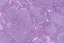 218px-Follicular lymphoma -- low mag.jpg 218 × 145; 14 KB
-
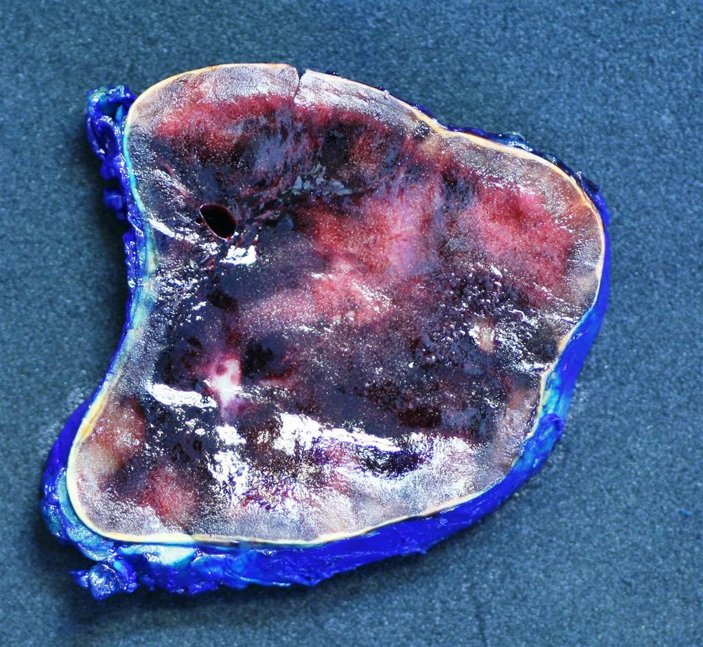
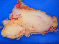 192px-Lymphoma macro.jpg 192 × 145; 11 KB
-
 Cervical Cancer Screening Guidelines for Average-Risk Women.jpg 1,393 × 1,389; 619 KB
-
 459px-Ependymom sag FLAIR.jpg 459 × 480; 29 KB
-
 Ependymoma.jpg 1,024 × 1,024; 149 KB
-
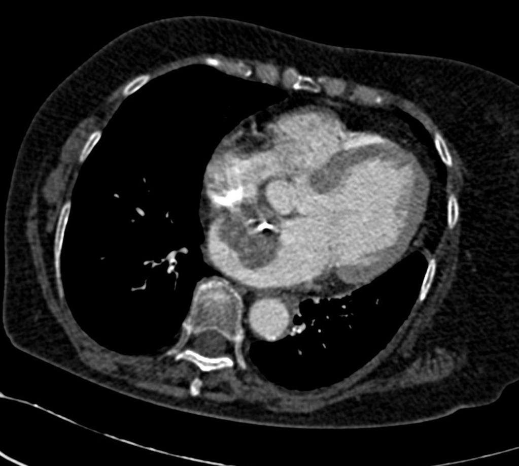 Left-atrial-myxoma.jpg 1,023 × 918; 70 KB
-
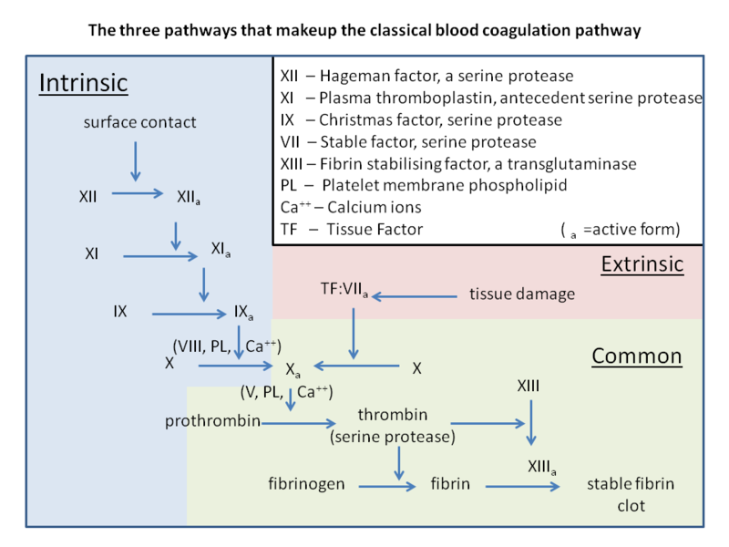 800px-Classical blood coagulation pathway.png 800 × 600; 124 KB
-
 Oncology Project Checklist v3.pdf ; 112 KB
-
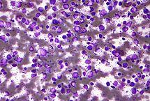 215px-Diffuse large B cell lymphoma - cytology low mag.jpg 215 × 145; 16 KB
-
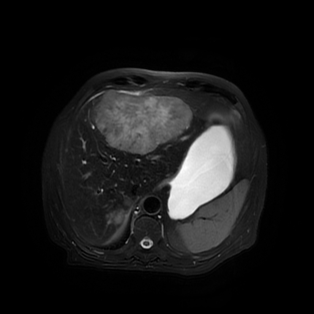 Hepatic-metastases-from-colon-carcinoma-triphasic-mri.jpg 1,024 × 1,024; 150 KB
-
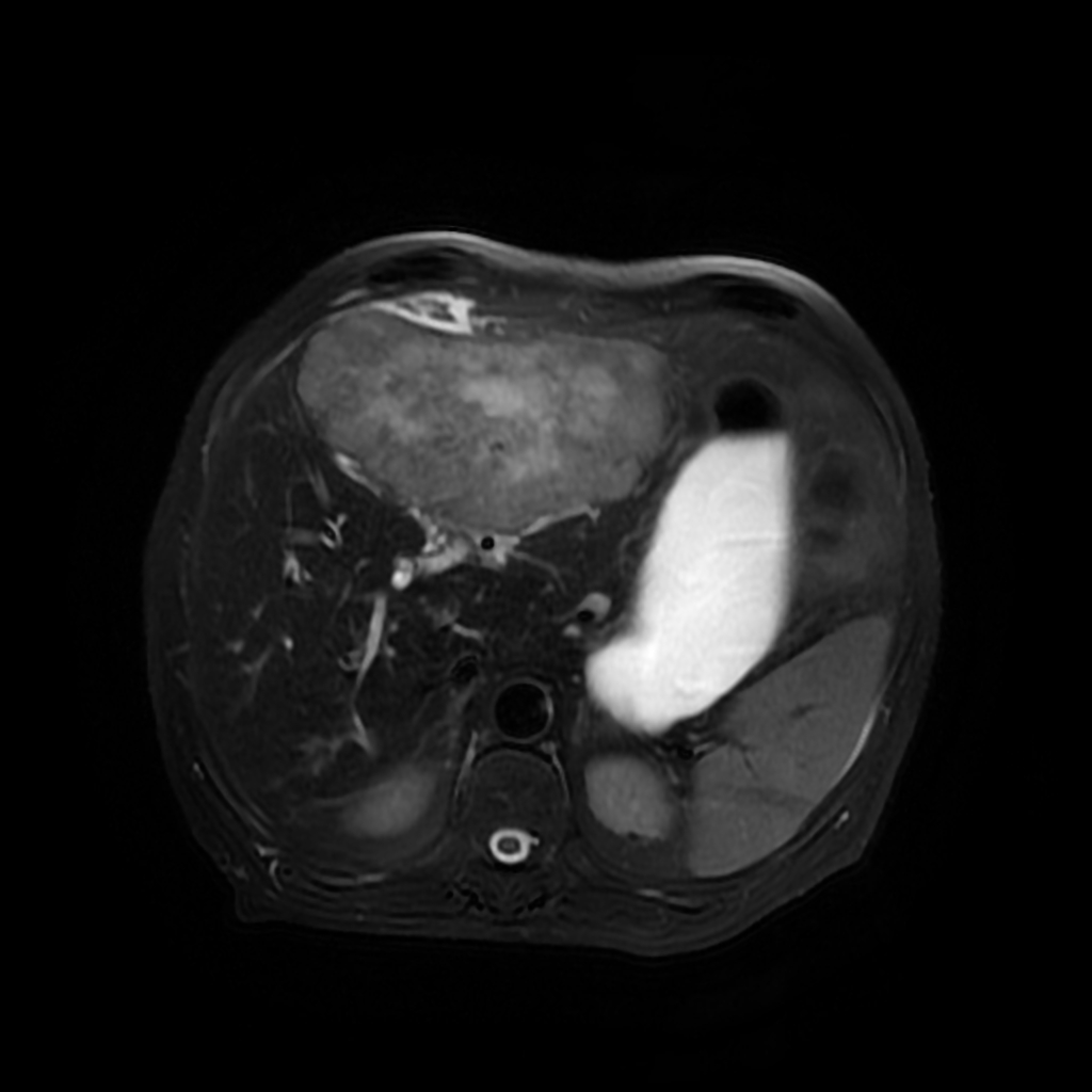 Hepatic-metastases-from-colon-carcinoma-triphasic-mri(1).jpg 1,024 × 1,024; 156 KB
-
WJG Colorectal Staging copy.svg 992 × 518; 190 KB
-
 638px-Myxoid liposarcoma (06).jpg 638 × 480; 67 KB
-
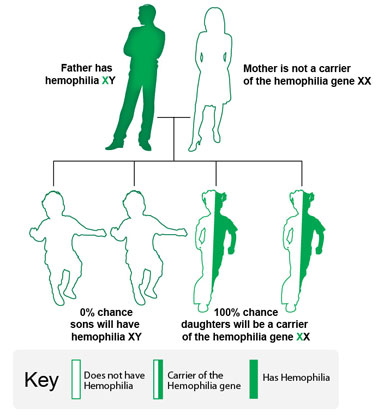
 Hemophilia-genes.jpg 358 × 417; 31 KB
-
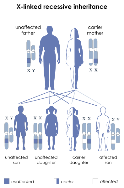 X-linked recessive.svg.png 395 × 600; 67 KB
-
 800px-Bone Chondrosarcoma Mesenchymal MP5 PA.JPG 800 × 597; 173 KB
-
 Layout v22.jpg 384 × 418; 32 KB
-
 Astrocytoma 1.jpg 800 × 1,155; 182 KB
-
 Layout v2.jpg 387 × 415; 33 KB
-
 Hemophilia-genes (1).jpg 358 × 417; 31 KB
-
 Layout v22 (1).jpg 384 × 418; 32 KB
-
 707px-Bone Chondrosarcoma Dedifferentiated PA copy.jpg 707 × 599; 187 KB
-
 800px-Bone Chondrosarcoma Dedifferentiated HP PA.jpg 800 × 600; 118 KB
-
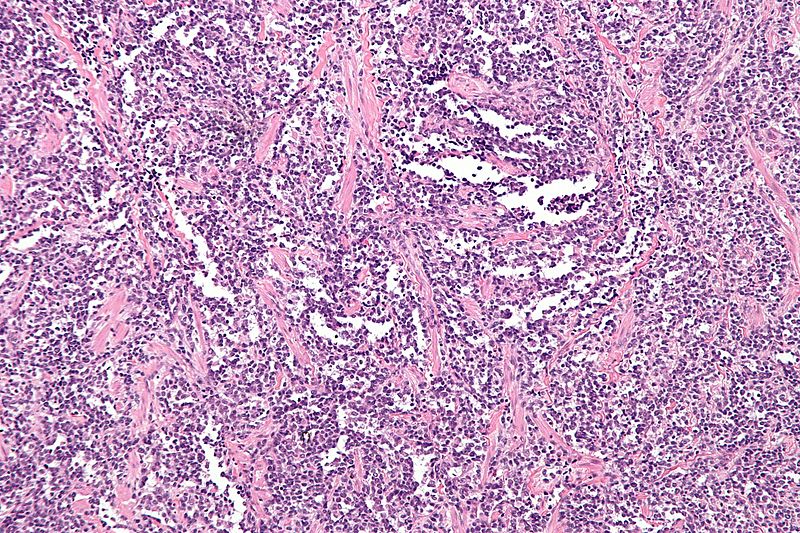
 Follicular lymphoma, spleen.jpg 459 × 512; 179 KB
-
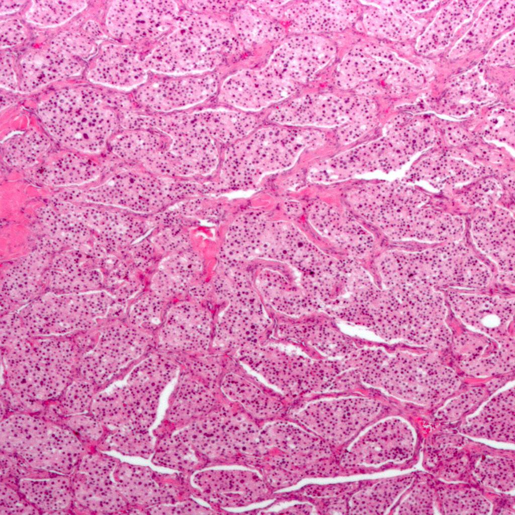 Carotid-body-tumour-histology.jpg 1,024 × 1,024; 242 KB
-
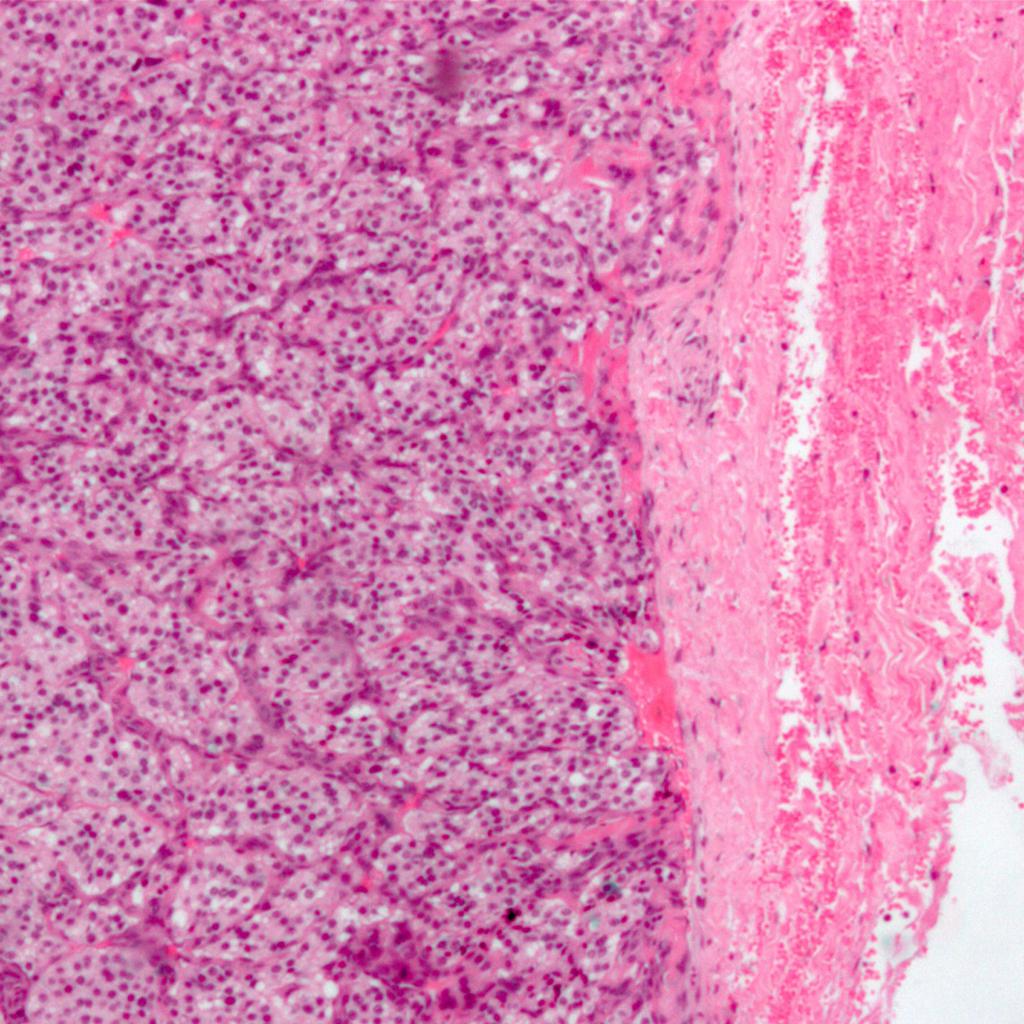 Carotid-body-tumour-histology (1).jpg 1,024 × 1,024; 187 KB
-
 218px-Carotid body tumour 2 intermed mag.jpg 218 × 145; 17 KB
-
 Images 266 (1).gif 480 × 417; 35 KB
-
 Images 267.gif 480 × 417; 31 KB
-
 320px-Endometrioid endometrial adenocarcinoma very high mag2.jpg 320 × 213; 31 KB
-
 Endometrioid endometrial adenocarcinoma very high mag.jpg 640 × 427; 96 KB
-
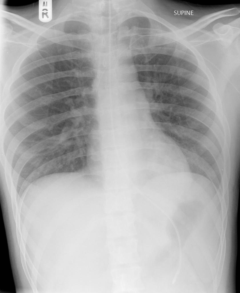 Normal-nasogastric-tube-position.jpg 840 × 1,024; 52 KB
-
 487px-Mrichondrosarcoma.jpg 487 × 480; 30 KB
-
 Mrichondrosarcoma.jpg 695 × 685; 93 KB
-
 800px-Alveolar rhabdomyosarcoma - very high mag.jpg 800 × 533; 129 KB
-
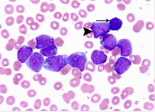 AMLS0.png 601 × 435; 418 KB
-
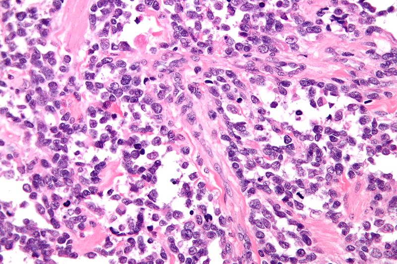
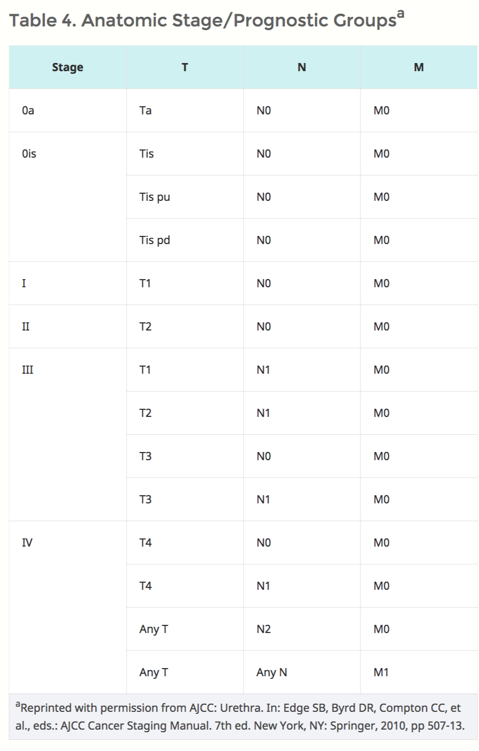
 Urethral Cancer1 .png 931 × 1,662; 381 KB
-
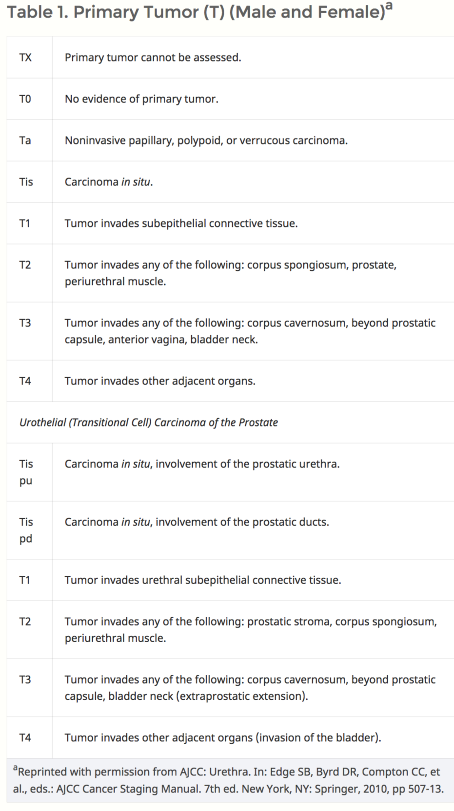
 Urethral Cancer2.png 942 × 565; 104 KB
-
 Urethral Cancer3.png 950 × 351; 84 KB
-
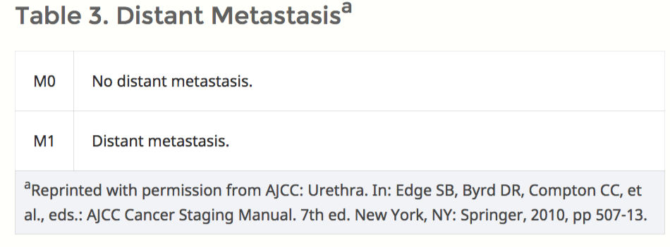
 Urethral Cancer4.png 942 × 1,477; 140 KB
-
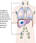
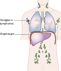 124px-Diagram showing stage 1 Hogkin's lymphoma CRUK 191.svg.png 124 × 145; 13 KB
-
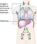 124px-Diagram showing stage 2 Hodgkin's lymphoma CRUK 208.svg.png 124 × 145; 14 KB
-
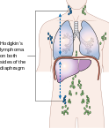 124px-Diagram showing stage 3 Hodgkin's lymphoma CRUK 221.svg.png 124 × 145; 14 KB
-
 124px-Diagram showing stage 4 Hodgkin's lymphoma CRUK 230.svg.png 124 × 145; 14 KB
-
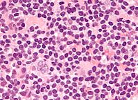
 197px-Hodgkin lymphoma cytology large.jpg 197 × 145; 14 KB
-
-
 Adrenal-myelolipoma-4.JPG 1,024 × 942; 212 KB
-
 Alveolar rhabdomyosarcoma - intermed mag.jpg 800 × 533; 226 KB
-
 Alveolar RMS.jpg 800 × 533; 226 KB
-
 IMG 20150522 210951.jpg 1,164 × 1,164; 502 KB


.jpg)
.jpg)


.jpg)
.jpg)


.jpg)

.gif)


