Uncategorized files
Jump to navigation
Jump to search
Showing below up to 50 results in range #10,781 to #10,830.
-
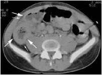
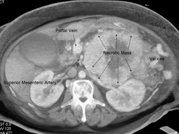
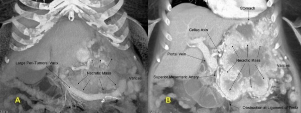
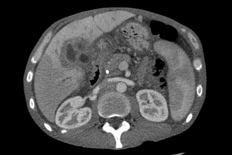
 CT Pancreatic cancer.png 512 × 413; 201 KB
-

-
 CT SCAN DISC HERNIATION.JPG 240 × 182; 13 KB
-
 CT Scan.JPG 630 × 472; 38 KB
-
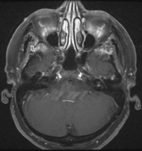
 CT Scan of Normal paranasal sinuses.gif 420 × 468; 86 KB
-
 CT Thickened peritoneum and ascites.png 552 × 495; 111 KB
-
 CT Typhilitis.jpg 203 × 150; 6 KB
-
 CT UBC.gif 1,280 × 720; 385 KB
-
 CT VIPoma.jpg 600 × 450; 54 KB
-
 CT VIPoma1.png 600 × 226; 127 KB
-
 CT abdomen - liver cirrhosis - 01.jpg 512 × 512; 25 KB
-
 CT acoustic neuroma.jpg 593 × 630; 31 KB
-
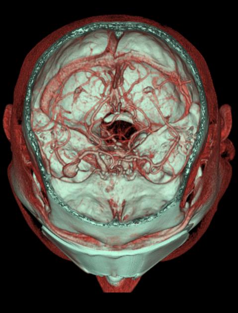 CT angiogram .jpg 479 × 630; 43 KB
-
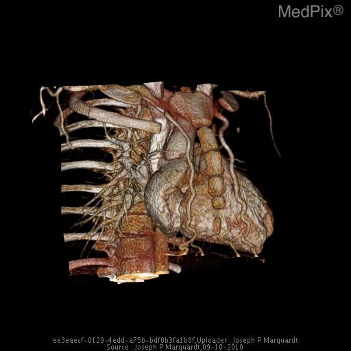
 CT angiography.gif 1,000 × 1,000; 854 KB
-
-
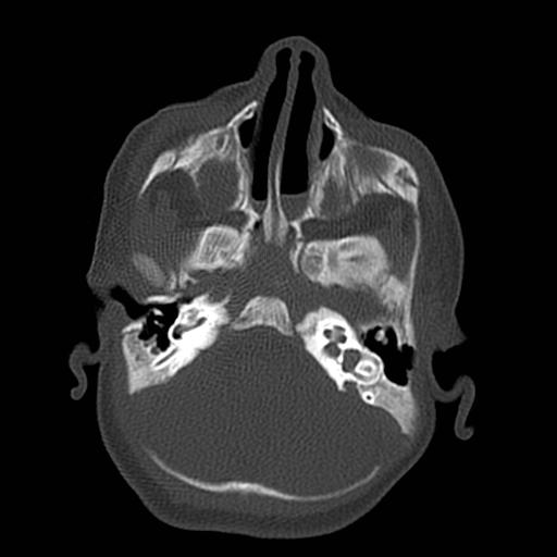 CT bilateral bony.jpg 512 × 512; 24 KB
-
 CT bing neel syn gif.gif 472 × 417; 127 KB
-
 CT bing neel syndrome.png 472 × 417; 169 KB
-
 CT chest pneumonia abscesses caverns effusions.jpg 512 × 512; 155 KB
-
 CT cholangioca.jpg 800 × 534; 45 KB
-
 CT chondrosarcoma.gif 1,280 × 720; 394 KB
-
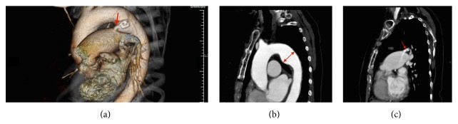
 CT dissection.png 220 × 187; 21 KB
-
 CT falx meningioma.jpg 1,024 × 1,024; 93 KB
-
 CT gastric cancer.gif 1,000 × 1,000; 1.48 MB
-
 CT gastroent.jpg 180 × 152; 6 KB
-
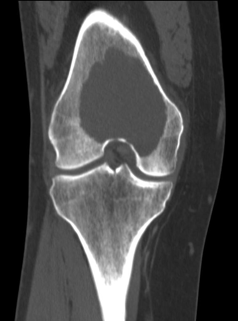 CT giant-cell-tumour-femur .jpg 760 × 1,024; 42 KB
-
 CT giant cell tumor.gif 720 × 960; 197 KB
-
 CT gif.gif 1,024 × 1,024; 884 KB
-
 CT image of a GIST tumor in the gastric cardia.jpg 682 × 512; 76 KB
-
 CT image of atypical teratoid rhabdoid tumor 1.jpg 630 × 630; 44 KB
-
 CT image of left side pleural effusion.jpg 800 × 478; 38 KB
-
 CT image of ovarian granulosa cell tumor.jpg 473 × 205; 29 KB
-
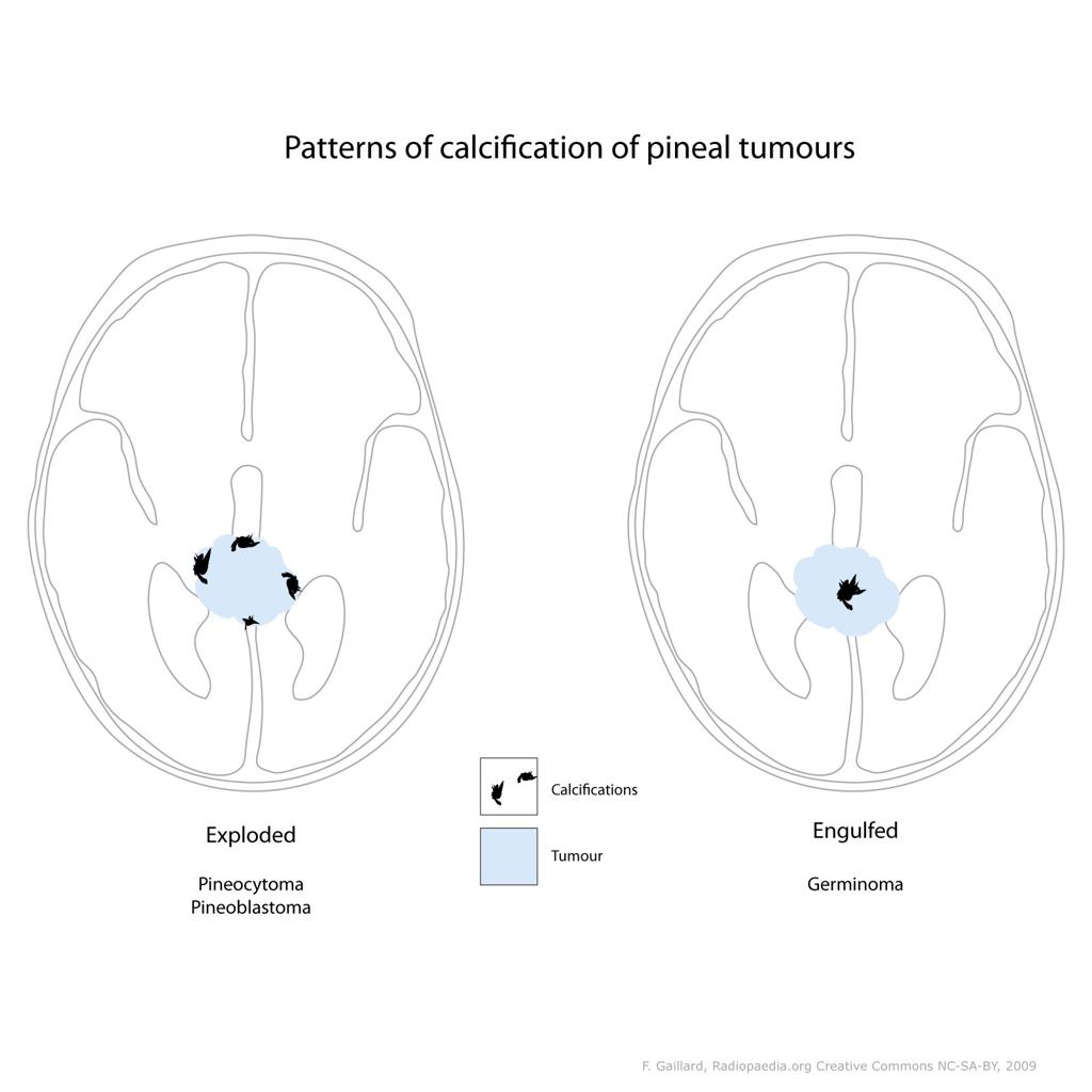
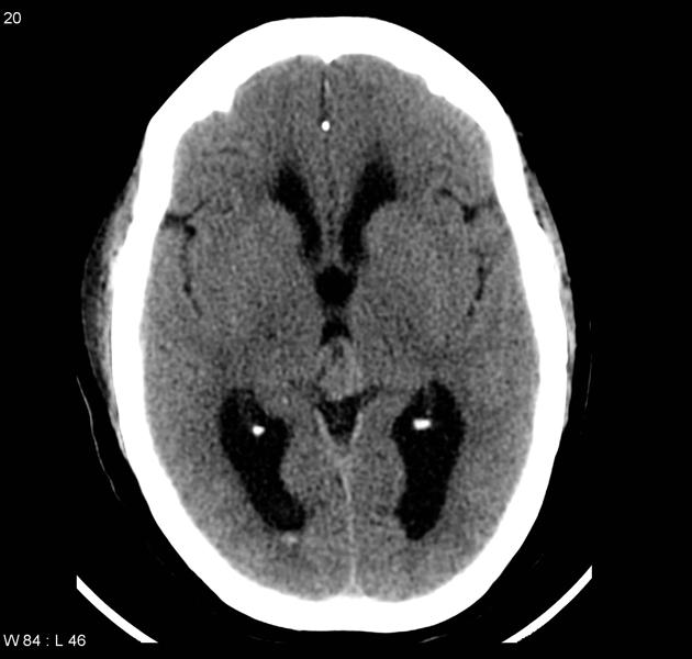
 CT image of pineal germinoma 1.jpg 630 × 630; 37 KB
-
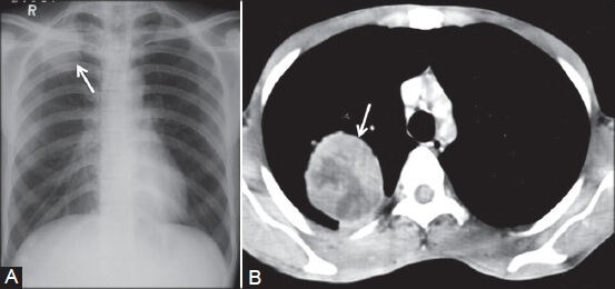 CT image of pleural fibroma.jpg 553 × 260; 24 KB
-
 CT image pineocytoma 1.jpg 1,024 × 1,024; 58 KB
-
 CT image pineocytoma 2.jpg 630 × 600; 35 KB
-
 CT image pineocytoma 3.jpg 630 × 630; 50 KB
-
 CT image pleural empyema.jpg 687 × 600; 54 KB
-
 CT image right side pleural effusion.jpg 800 × 572; 84 KB
-
 CT image showing the PDA.jpg 642 × 174; 24 KB
-
 CT lung.jpeg 256 × 256; 7 KB
-
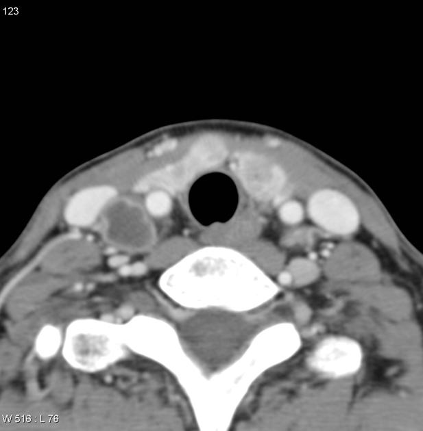 CT neck nodule.jpg 617 × 630; 26 KB
-
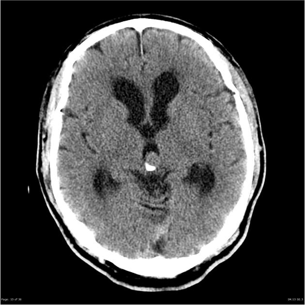
 CT noncontrast medulloblastoma Dr Mohammad Taghi Niknejad.jpg 442 × 442; 13 KB
-
 CT of brain of Mikael Häggström.png 800 × 570; 90 KB
-
 CT of brain of Mikael Häggström S3 I8.JPG 512 × 512; 37 KB
-
 CT of ground glass lung nodule.png 342 × 256; 98 KB
-
 CT of neurofibromatosis type 2.jpg 630 × 630; 30 KB
-

-
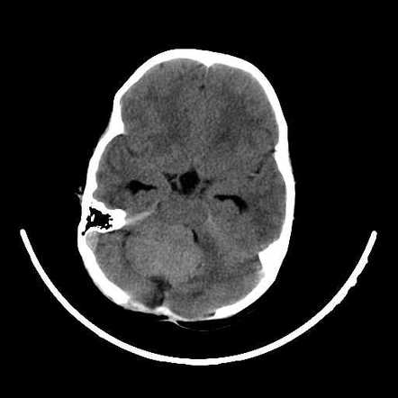
 CT of vertebrobasilar.jpg 391 × 442; 28 KB
-
 CT oligoastrocytoma 2.jpg 504 × 630; 42 KB


