Unused files
Jump to navigation
Jump to search
The following files exist but are not embedded in any page. Please note that other websites may link to a file with a direct URL, and so may still be listed here despite being in active use.
Showing below up to 50 results in range #10,681 to #10,730.
-
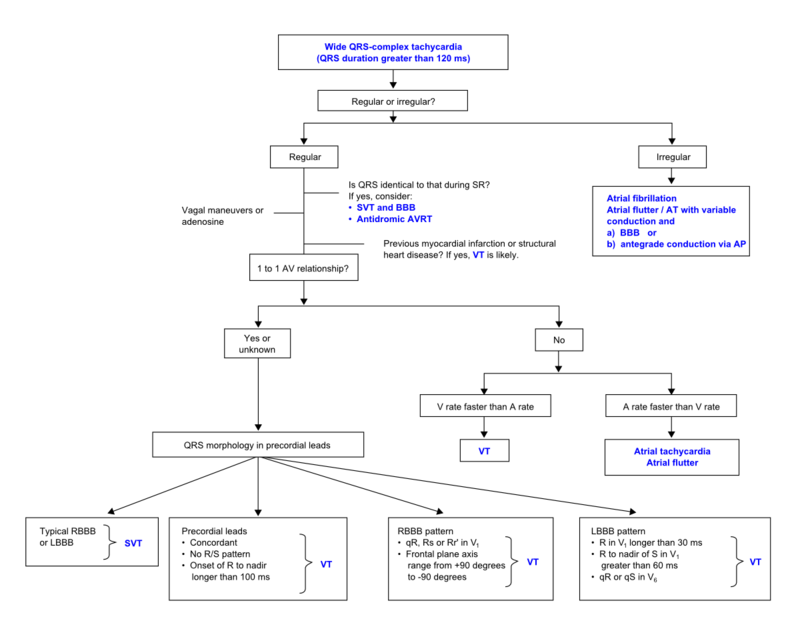 Wide complex tachycardia algorithm.png 800 × 633; 65 KB
-
 Vereckei algorithm.png 601 × 600; 26 KB
-
 Brugada algorithm.png 601 × 600; 39 KB
-
 Rhythm RBTBmorph nl.png 800 × 415; 16 KB
-
 Atrial flutter1.jpg 800 × 426; 155 KB
-
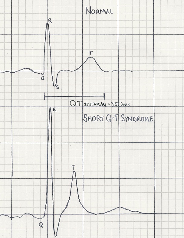 Short QT.jpg 608 × 787; 156 KB
-
 Baseline model 2.png 975 × 901; 62 KB
-
 Wellens' Warning.gif 384 × 194; 4.88 MB
-
 E334.jpg 800 × 426; 106 KB
-
 De-LAHB.png 679 × 599; 306 KB
-
 IntraventricularconductiondefectEKG.jpg 3,292 × 1,887; 5.78 MB
-
 Pacemaker with long AV interval.jpg 800 × 426; 43 KB
-
 Dual chamber pacemaker.jpg 800 × 373; 95 KB
-
 Atrial flutter2.jpg 3,004 × 1,599; 4.46 MB
-
 Pacemaker3.jpg 798 × 111; 22 KB
-
 Wolff-Parkinson-White syndrome10.jpg 3,004 × 1,599; 4.47 MB
-
 Time dependent capture pacemaker 2.jpg 800 × 175; 41 KB
-
 Atrial fibrillation 7.jpg 3,283 × 2,149; 3.19 MB
-
 Atrial fibrillation 17.jpg 3,283 × 4,004; 3.39 MB
-
 Filter settings.png 473 × 599; 275 KB
-
 Wctpresentation.jpg 3,004 × 1,599; 4.51 MB
-
 WCTadenosine.jpg 3,004 × 1,599; 4.48 MB
-
 Wcttosinusrhythm.jpg 3,004 × 1,599; 4.46 MB
-
 Wide complex tachycardia1.jpg 3,004 × 677; 445 KB
-
 LBBB with AMI.jpg 800 × 435; 66 KB
-
 Pacemaker 16.jpg 800 × 241; 62 KB
-
 Tachy brady syndrome.jpg 800 × 426; 71 KB
-
 Tdepointes.png 765 × 429; 135 KB
-
 Torsadesdepointes.jpg 800 × 495; 66 KB
-
 Atrial Infarction 1.png 800 × 536; 127 KB
-
 STdepression01.jpg 700 × 315; 45 KB
-
 PacemakerPotential1.jpg 3,292 × 1,887; 5.9 MB
-
 Non Q wave MI.jpg 3,004 × 1,599; 4.43 MB
-
 Premature Ventricular 2.png 800 × 179; 9 KB
-
 Premature Atrial Contraction 6.png 800 × 179; 9 KB
-
 LBBB13.jpg 800 × 426; 73 KB
-
 Paced atrial rhythm with a bipolar atrial pacemaker.jpg 3,004 × 1,599; 4.46 MB
-
 Sick sinus syndrome6.jpg 800 × 235; 31 KB
-
 Right bundle branch block 26.jpg 800 × 426; 150 KB
-
 Non Q MI.jpg 3,004 × 1,599; 4.46 MB
-
 Wide complex tachycardia2.jpg 3,004 × 1,599; 4.43 MB
-
 Cardiogenic shock.JPG 650 × 502; 63 KB
-
 Ebsteins anomaly.jpg 800 × 507; 58 KB
-
 Diastolic dysfunction ECHO.png 832 × 400; 125 KB
-
 Diastolic dysfunction ECHO2.png 1,650 × 1,107; 149 KB
-
 Displacement of tricspid valve toward RV apex in Ebsteins.JPG 468 × 342; 24 KB
-
 Coronary arteries.svg.png 512 × 294; 62 KB
-
 Regadenoson2.png 800 × 383; 18 KB
-
 Regorafenib.svg.png 512 × 157; 7 KB
-
 WLP.png 246 × 255; 94 KB


