Unused files
Jump to navigation
Jump to search
The following files exist but are not embedded in any page. Please note that other websites may link to a file with a direct URL, and so may still be listed here despite being in active use.
Showing below up to 50 results in range #10,601 to #10,650.
-
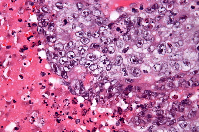
 Ganglioglioma-002.jpg 531 × 600; 30 KB
-
 Ganglioglioma-003.jpg 551 × 600; 34 KB
-
 Ganglioglioma-004.jpg 562 × 600; 32 KB
-
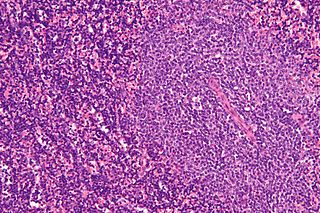 Chronic lymphocytic leukemia lymph node.jpg 320 × 213; 38 KB
-
 Placeholder.jpg 306 × 206; 2 KB
-
 Onychomadesis.png 315 × 372; 179 KB
-
 Onychophosis.png 153 × 69; 16 KB
-
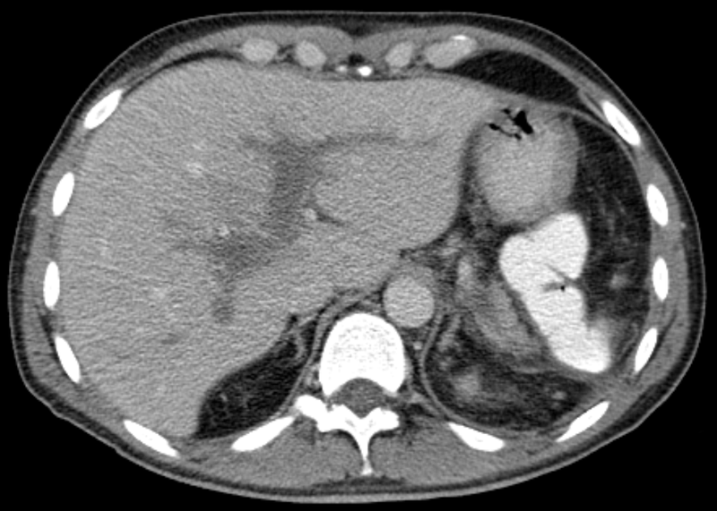
 Hepatorenal syndrome.jpg 800 × 533; 235 KB
-
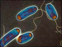 Legionella.jpg 203 × 152; 7 KB
-
 Pfortaderthrombose001.png 800 × 571; 248 KB
-
 Kavernoese Transformation nach Pfortaderthrombose 001.png 800 × 432; 219 KB
-
 Drug rash.JPG 226 × 281; 12 KB
-
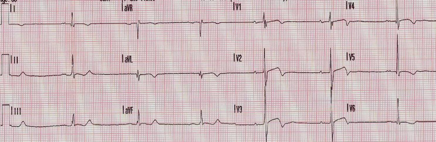
 Poor R wave progression1.jpeg 1,200 × 624; 116 KB
-
 Hirnbiopsie menigiosis.jpg 639 × 599; 210 KB
-
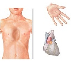 Marfansyndrome.jpg 239 × 210; 14 KB
-
 Sachin Tendulkar 14.jpg 1,024 × 768; 139 KB
-
 Optimized-ship.JPG 1,200 × 896; 122 KB
-
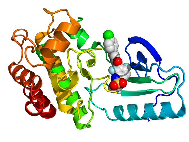 3OG7.png 800 × 600; 285 KB
-
 Telangectasia.jpg 248 × 231; 7 KB
-
 Dupuytren's2010.JPG 800 × 505; 57 KB
-
 Caput medusae1.jpg 600 × 450; 93 KB
-
 Hoap.png 553 × 385; 128 KB
-
 Esophageal varices.jpg 482 × 499; 36 KB
-
 800px-Embryonal carcinoma - very high mag - cropped.jpg 800 × 533; 132 KB
-
 Skin Tumors-P6251257.jpg 800 × 600; 221 KB
-
 800px-Giant cell tumour of bone - high mag.jpg 800 × 533; 192 KB
-
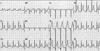
 Arrythmia.jpg 638 × 355; 32 KB
-
 Hyperkalemia2.jpg.png 365 × 577; 19 KB
-
 QT Prolongation1.jpg 750 × 545; 48 KB
-
 QT Prolongation2.jpg 250 × 309; 14 KB
-
 Prognostic.jpg 330 × 319; 15 KB
-
 SOB.jpg 551 × 261; 24 KB
-
 Wellen sign in proximal LAD.jpg 1,488 × 485; 146 KB
-
 Pericarditis.jpg 424 × 218; 21 KB
-
 Complicated silicosis.jpg 731 × 600; 46 KB
-
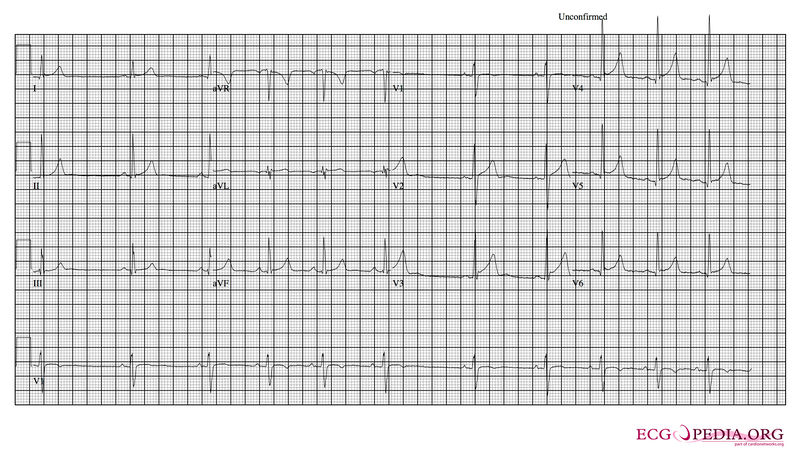 Sinus arrhythmia.jpg 800 × 464; 97 KB
-
 Hyperkalemia case3.jpg 794 × 381; 87 KB
-
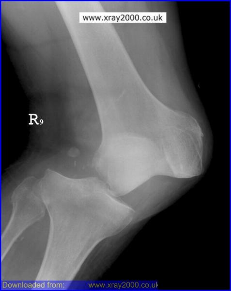 Knee dislocation 1.jpg 477 × 600; 21 KB
-
 Knee dislocation 2.jpg 160 × 160; 3 KB
-
 Knee dislocation 3.jpg 377 × 543; 59 KB
-
 Torsades de Pointes.png 763 × 411; 134 KB
-
 Gross.png 398 × 514; 457 KB
-
 Echo of asvd.jpg 404 × 372; 43 KB
-
 COA 2.jpg 502 × 146; 12 KB
-
 Echo.jpg 819 × 460; 37 KB
-
 TA 1.jpg 610 × 742; 47 KB
-
 Bicuspid1.JPG 306 × 223; 12 KB
-
 Aortic stenosis.JPG 283 × 273; 14 KB
-
 VSD types and blood flow.png 344 × 406; 222 KB
-
 CXRCOA.jpg 504 × 481; 26 KB


