Hiatus hernia
For patient information click here
| Hiatus hernia | |
 | |
|---|---|
| ICD-10 | K44, Q40.1 |
| ICD-9 | 553.3, 750.6 |
| OMIM | 142400 |
| DiseasesDB | 29116 |
| MeSH | D006551 |
|
Hiatus Hernia Microchapters |
|
Diagnosis |
|---|
|
Treatment |
|
Case Studies |
|
Hiatus hernia On the Web |
|
American Roentgen Ray Society Images of Hiatus hernia |
Editor-In-Chief: C. Michael Gibson, M.S., M.D. [1]
Overview
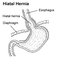
A hiatus hernia or hiatal hernia is the protrusion (or herniation) of the upper part of the stomach into the thorax through a tear or weakness in the diaphragm.
Symptoms
The symptoms include acid reflux, and pain, similar to heartburn, in the chest and upper stomach.
In most patients, hiatus hernias cause no symptoms. Sometimes patients experience heartburn and regurgitation, when stomach acid refluxes back into the esophagus.
Causes
The following are possible causes or contributing factors for having a hiatus hernia:
- Obesity
- Frequent coughing
- Straining with constipation
- Frequent bending over or heavy lifting
- Heredity
- Smoking
- Stress
Diagnosis

The diagnosis of a hiatus hernia is typically made through an upper GI series or endoscopy.
The imaging findings are
- On chest radiographs, a paraesophageal hernia may appear as a soft-tissue-opacity lesion posterior to the heart near the esophageal hiatus.
- CT helps verify migration of the stomach cranially through the hiatus. Sagittal and coronal reformatted images often help demonstrate the hernia and the hiatal defect.
Patient #1: Sliding hiatal hernia
Patient #2: Sliding hiatal hernia
Patient #3: Paraesophageal hernia
Types
There are two major kinds of hiatus hernia:
- The most common (95%) is the sliding hiatus hernia, where the gastroesophageal junction moves above the diaphragm together with some of the stomach.
- The second kind is rolling (or paraesophageal) hiatus hernia, when a part of the stomach herniates through the esophageal hiatus beside, and without movement of, the gastroesophageal junction. It is about 100 times less common than the first kind. [1]
A third kind is also sometimes described, and is a combination of the first and second kinds.
Treatment
In most cases, sufferers experience no discomfort and no treatment is required. However, when the hiatal hernia is large, or is of the paraesophageal type, it is likely to cause esophageal stricture and discomfort. Symptomatic patients should elevate the head of their beds and avoid lying down directly after meals until treatment is rendered. If the condition has been brought on by stress, stress reduction techniques may be prescribed, or if overweight, weight loss may be indicated. Medications that lower the lower esophageal sphincter (or LES) pressure should be avoided. Antisecretory drugs like proton pump inhibitors and H2 receptor blockers can be used to reduce acid secretion.
Where hernia symptoms are severe and chronic acid reflux is involved, surgery is sometimes recommended, as chronic reflux can severely injure the esophagus and even lead to esophageal cancer.
The surgical procedure used is called Nissen fundoplication. In fundoplication, the gastric fundus (upper part) of the stomach is wrapped, or plicated, around the inferior part of the esophagus, preventing herniation of the stomach through the hiatus in the diaphragm and the reflux of gastric acid. The procedure is now commonly performed laparoscopically. With proper patient selection, laparoscopic fundoplication has low complication rates and a quick recovery.[2]
Complications include gas bloat syndrome, dysphagia (trouble swallowing), dumping syndrome, excessive scarring, and rarely, achalasia. The procedure sometimes fails over time, requiring a second surgery to make repairs.
Complications
A hiatus hernia per se does not cause any symptoms. The condition promotes reflux of gastric contents (via its direct and indirect actions on the anti-reflux mechanism) and thus is associated with gastroesophageal reflux disease (GERD). In this way a hiatus hernia is associated with all the potential consequences of GERD - heartburn, esophagitis, Barrett's esophagus and esophageal cancer. However the risk attributable to the hiatus hernia is difficult to quantify, and at most is low.
Besides discomfort from GERD and dysphagia, hiatal hernias can have severe consequences for patients if not treated. While sliding hernias are primarily associated with gastroesophageal acid reflux, rolling hernias can strangulate a portion of the stomach above the diaphragm. This strangulation can result in esophageal or GI tract obstruction and the tissue even become ischemic and necrose.
Another severe complication, although very rare, is a large herniation that can restrict the inflation of a lung, causing pain and breathing problems.
Epidemiology
Hiatus hernias affect anywhere from 1 to 20% of the population. Of these, 9% are symptomatic, depending on the competence of the lower esophageal sphincter (LES). 95% of these are "sliding" hiatus hernias, in which the LES protrudes above the diaphragm along with the stomach, and only 5% are the "rolling" type (paraesophageal), in which the LES remains stationary but the stomach protrudes above the diaphragm. People of all ages can get this condition, but it is more common in older people.
Notes and references
- ↑ Lawrence, P. (1992). Essentials of General Surgery. Baltimore: Williams & Wilkins. p. 178. ISBN 0-683-04869-4.
- ↑ Lange CMDT 2006
External links
Template:Gastroenterology
Template:Congenital malformations and deformations of digestive system
de:Hiatushernie it:Ernia iatale he:בקע סרעפתי fi:Palleatyrä sv:Hiatusbråck


