Musculoskeletal problems of the foot
Editors-In-Chief: Robert G. Schwartz, M.D. [1], Piedmont Physical Medicine and Rehabilitation, P.A.; Michael Tollison, M.D. [2], Piedmont Orthopaedic Associates, Greenville, SC
Please Join in Editing This Page and Apply to be an Editor-In-Chief for this topic: There can be one or more than one Editor-In-Chief. You may also apply to be an Associate Editor-In-Chief of one of the subtopics below. Please mail us [3] to indicate your interest in serving either as an Editor-In-Chief of the entire topic or as an Associate Editor-In-Chief for a subtopic. Please be sure to attach your CV and or biographical sketch.
Overview
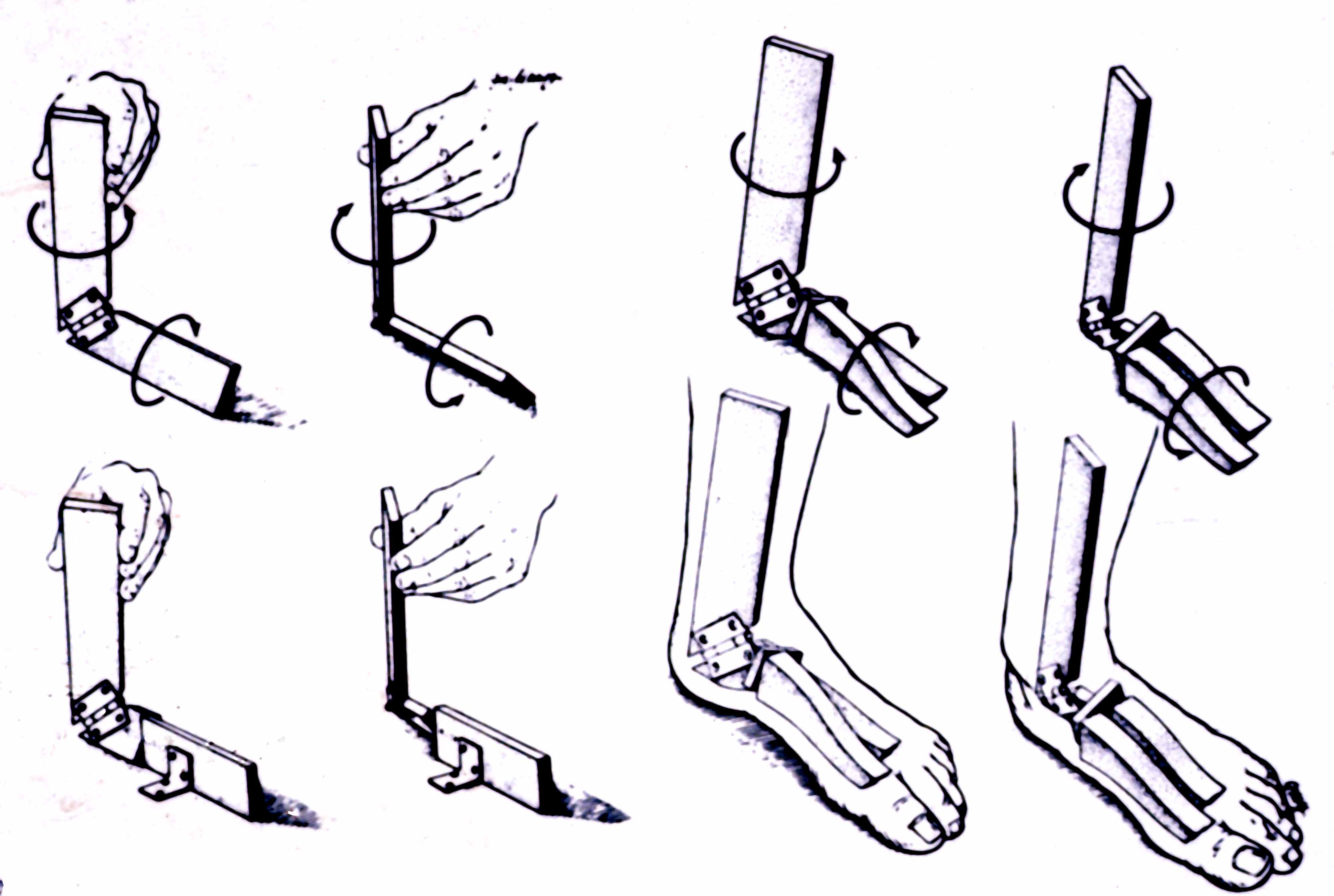
Foot and ankle pain is a very common condition. Anomalies range from osteoarthritis, ligamentous strain [4], nerve entrapment and neuropathy [5], to mechanical and postural induced abnormalities. Most cases respond to conservative care including physical therapy, orthotics, local injection and percutaneous tenotomy [6]. In more recalcitrant cases surgical intervention is required.
Associated Conditions
It is not at all uncommon for someone to present with foot or ankle pain even when the primary problem is coming from nerve root irritation in the back [7], venous reflux [8] or peripheral arterial disease [9]. It is therefor important to keep in mind that not all foot and ankle problems arise from the foot or ankle itself. If pain persists beyond a few weeks, it is wise to get a doctors opinion.
History and Symptoms
The history of present illness (the facts that surround the onset of pain) and the symptoms associated with the chief complaint, can provide valuable clues as to its source. Examples of several conditions are listed below.
Acute Traumatic Injury
- Fracture of Proximal Phalanx of Great Toe
- Usually via direct trauma or toe-stubbing injury
- Usually minimal displacement; can by treated conservatively
- Fracture of Metatarsals 1-4
- Usually via direct blow to top of foot
- Midfoot pain, inability to bear weight, direct tenderness
- Nondisplaced
- Can be treated conservatively
- Displaced
- Require surgical reduction
- Fracture of 5th Metatarsal (MT)
- Dancer’s Fracture
- Severe inversion injury--avulsion of bone from proximal metatarsal
- Occurs at site of peroneus brevis insertion
- Can be treated conservatively (immobilization)
- Jones’ Fracture
- Fracture of proximal tuberosity of base of metatarsal
- Transverse fracture of shaft
- Can be managed conservatively, but high rate nonunion
- Dancer’s Fracture
- Calcaneal Fracture
- Most commonly fractured tarsal bone
- Usually via vertical falls or twisting injuries
- Intra-articular Fractures
- All require orthopedics referral; unpredictable healing
- May be complicated by chronic joint pain, arthritis, nerve entrapment
- Extra-articular Fractures
- Most can be treated without surgery
- Ortho referral for displaced posterior process fractures (Achilles disruption)
- Ortho referral for nonunion of anterior process fracture
Nontraumatic
- Great toe
- Hallux valgus (bunion)
- Metatarso-phalangeal osteoarthritis (MTP OA)--painful swelling of dorsomedial aspect of 1st metatarsal head
- Hallux valgus deformity associated (toe angulates laterally)
- Hyperpronation (flat feet) and poor footwear contribute
- Arthritic flares or bursitis can occur with ongoing pressure at medial joint line
- Adventitial bursitis
- Inflammation of bursal sac over medial 1st MTP joint due to friction/pressure
- Dramatic erythema, edema and tenderness
- Gout (podagra):
- Dramatic inflammatory response to monosodium urate (MSU) crystal deposition
- Can also affect tenosynovial sheaths (enthesitis) and other small joints of foot
- Sesamoid disorders
- Two sesamoids (medial and lateral) articulate on plantar aspect of 1st metatarsal (MT)
- Inflammation or fracture can occur with chronic stress (runners, dancers)
- Localized pain and swelling at plantar aspect of 1st
- Hallux valgus (bunion)
- Forefoot
- Metatarsalgia
- Pain at any of the 2nd-5th metatarsal heads with weight-bearing
- Can be related to inflammatory deformity with subluxation of MT head (right anterior)
- Morton’s neuroma
- Chronic irritation of digital nerve running between metatarsal heads
- Most commonly occurs between 3rd and 4th toes
- Burning pain btwn toes; cramping; numbness along sides of 2 adjacent toes
- Typically associated with poorly padded shoes, improves with forefoot massage
- Metatarsal stress fracture
- Microfracture of metatarsal after prolonged walking/standing (“march fracture”)
- Usually 2nd or 3rd metatarsal
- Sudden onset of pain, often without history of trauma
- Military recruits, athletes, osteoporotic patients at risk
- Metatarsalgia
- Hindfoot – Plantar Region
- Plantar fasciitis
- One of most common causes adult foot pain
- Heel pain worse with initiation of walking/standing after inactivity
- Results from strain of plantar fascia after jumping, prolonged standing
- Predisposing factors
- Obesity
- Flat feet (pes planus) or high arches (pes cavus)
- Excessive pronation
- Short Achilles tendons
- Can be an inflammatory process associated with systemic disease (rheumatoid arthritis (RA), Reiter’s)
- Calcaneal spurs may coexist or develop due to inflammation (but usually asymptomatic)
- Infracalcaneal bursitis
- Inflammation of bursa beneath calcaneus
- Pain/ache in mid-plantar aspect of calcaneus
- Symptoms increase with duration of weight-bearing
- Calcaneal periostitis
- Bilateral pain along plantar and lateral aspects of heels
- Can be due to trauma
- Can be due to inflammatory disease (RA, psoriatic arthritis, ankylosing spondylitis, Reiter’s)
- May improve with treatment of underlying disease process
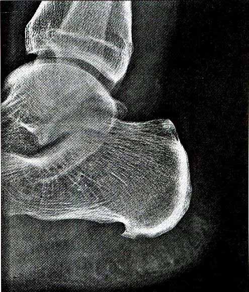
- Calcaneal spurs
- Bony outgrowths that develop on plantar tuberosity
- Usually asymptomatic
- Pain can occur if large (>1 cm) with apex angled downward--pain with weight-bearing
- Heel pad syndrome
- Irritation of fat pad due to trauma
- Pain localized to heel pad; plantar fascia not tender
- Most commonly in marathon runners
- Self-limited, resolves within 2-3 weeks
- Tarsal tunnel syndrome
- Posterior tibial nerve compressed in tarsal tunnel
- (Beneath flexor retinaculum inferoposterior to medial malleolus)
- Can occur via local trauma (sprain, fracture), repetitive hyperpronation
- Also via inflammatory disease (RA), bony prominences, pregnancy, hypoT4
- Paresthesias, plantar pain (medial/lateral plantar nerve distribution)
- Symptoms often nocturnal or after standing, relieved by foot/ankle movement
- Posterior tibial nerve compressed in tarsal tunnel
- Referred pain (subtalar arthritis, lumbosacral (LS) radiculopathy)
- Treat the underlying condition
- For example, see [RSD], [CRPS], [Low back pain]
- Plantar fasciitis
- Ligament Strain
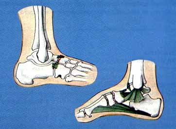
- Pain anywhere that ligament connects bone to bone
- May be associated with swelling and skin color changes
- Often due to an inciting event (such as a turned or twisted ankle)
- May also be due to normal wear and tear or other forgotten injury
- Ligament Strain
- Hindfoot – Posterior Region
- Achilles tendinitis
- Inflammation and microtears near Achilles tendon insertion
- Often associated with repetitive irritation (athletes)
- Bilateral involvement (in absence of quinolone) suggests Reiter’s or spondylitis
- Pain behind ankle with walking, standing, weight-bearing athletic activities
- Pain worse with activity--later stiffness and swelling
- Untreated cases--acute rupture (up to 10%) or chronic tendinitis
- Achilles tendon rupture
- Occurs after abrupt calf muscle contraction
- Typically occurs in patients > 30 years old with sporadic athletic activity
- Also associated with fluoroquinolone use
- Audible snap followed by severe pain in calf
- Partial rupture can occur without precipitating event
- Posterior tibial tenosynovitis
- Inflammation of tendon as it passes around the medial malleolus
- Exacerbants = ankle pronation, pes planus, obesity
- Can be associated w/tarsal tunnel syndrome, especially if significant pronation
- Pain and swelling at inner aspect of ankle, worse wtih walking
- Retrocalcaneal bursitis
- Inflammation of bursa between Achilles tendon and calcaneus
- Uncommon
- Pain behind ankle, increased with walking (plantar flexion)
- Pre-Achilles bursitis
- Inflammation of bursa between Achilles tendon (calcaneal insertion) and skin
- May resemble Achilles tendinitis, but less disabling; no significant risk tendon rupture
- Pain and localized swelling behind the heel
- Aggravated/caused by inappropriate shoes
- Ligament Strain
- Pain anywhere that ligament connects bone to bone
- May be associated with swelling and skin color changes
- Often due to an inciting event (such as a turned or twisted ankle)
- May also be due to normal wear and tear or other forgotten injury
- Achilles tendinitis
Presentation and Physical Exam
1st MTP Joint Conditions
- Hallux Valgus
- Valgus deformity of MTP joint
- Prominent metatarsal head, toe points laterally
- First and second toe may overlap if advanced
- Tenderness along medial joint line (or over whole joint if acute flare)
- Joint enlargement due to subluxation, osteophytes, edema
- Crepitation with passive movement of MTP joint
- +/- Pain at extremes of passive plantar/dorsiflexion of toe
- +/- Limited range of motion (ROM) (hallux rigidus)
- Valgus deformity of MTP joint
- Adventitial Bursitis of 1st MTP
- Erythema, swelling over medial aspect of MTP joint (focal area vs. full joint involvement with gout)
- Maximal tenderness over medial joint line
- Associated with hallux valgus deformity (increased friction with shoes) +/- resultant loss of ROM
- Mild pain with MTP flexion/extension (vs. gout with severe pain)
- Isometric toe flexion/extension against resistance painless (tendons spared)
- Gout
- Significant erythema, swelling and exquisite tenderness involving entire joint
- Severe pain with MTP flexion/extension
- Sesamoiditis or Fracture
- Localized tenderness/swelling on plantar palpation of MTP
Lesser MTP Forefoot Conditions
- Metatarsalgia
- Maximal tenderness at the MTP heads
- Plantar protrusion of metatarsal head(s) may be visible with patient lying prone
- Callus often present beneath involved metatarsal(s)
- Adjacent metatarsals may be hypermobile (shifting weight to involved metatarsal)
- Morton’s Neuroma
- Tenderness greatest in web space between MTP heads (vs. at MTP heads in metatarsalgia)
- Pain reproduced by squeezing MTP heads from sides (electric pain to ends of adjacent 2 toes)
- Click may be felt with squeeze + deep palpation in distal intermetatarsal space (Mulder’s sign)
- Passive ROM of MTP joints painless
- +/- Loss of sensation along inner aspects of 2 adjacent toes (advanced cases)
- Metatarsal Stress Fracture
- Localized tenderness over metatarsal shaft
- Dramatic dorsal foot swelling
- Pain when metatarsals squeezed from the sides
Hindfoot (Plantar) Conditions
- Plantar Fasciitis
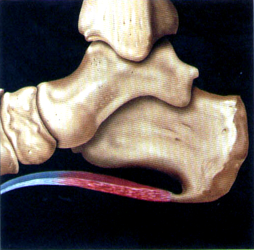
- Focal point tenderness at the calcaneal origin of the plantar fascia – pain can be increased by toe dorsiflexion (stretch fascia) during palpation of medial plantar surface ~1.5” distal to posterior heel
- Medial-to-lateral compression of calcaneus usually less painful than local fascial tenderness (if compression more painful, must rule out calcaneal stress fracture)
- +/- Limited foot dorsiflexion (nl = 25-30°) due to shortening of Achilles tendon
- +/- Associated pes planus/cavus
- Infracalcaneal Bursitis
- Point tenderness directly under center of calcaneus (plantar surface)
- +/- Localized warmth/swelling
- Calcaneal Periostitis
- Diffuse tenderness along plantar aspect of heel and midfoot bilateral and along lateral edges of heels
- Evaluation for signs underlying rheumatologic disease indicated
- Calcaneal Spurs
- Usually no specific findings; prominent spur palpable through skin may require intervention
- Heel Pad Syndrome
- Pain localized to heel pad, aggravated by squeezing pad from side to side
- Plantar fascia not tender; pain not exacerbated by toe dorsiflexion
- Tarsal Tunnel Syndrome
- Reproduction of symptoms with percussion or pressure over flexor retinaculum (Tinel’s sign)
- Ligament Strain
- Pain anywhere that ligament connects bone to bone
- Swelling and skin color changes may be present
- Weather sensitivity can exist
Hindfoot (Posterior) Conditions
- Achilles Tendinitis
- Tender, fusiform thickening of Achilles tendon with “cobblestone” texture
- Pain exacerbated with resisted plantar flexion and passive stretching in dorsiflexion
- Ankle ROM normal, though may be limited by pain in dorsiflexion
- Preserved calf muscle strength with no palpable defects in tendon
- Achilles Tendon Rupture
- Weakness--patient may be unable to stand up on toes (with full rupture)
- Thompson Test
- Patient kneels on chair with feet hanging over edge
- Squeeze of normal calf muscle foot plantar flexion
- Squeeze on side with tendon rupture--no foot response
- Crescent Sign
- Blood tracking in soft tissues can be seen beneath malleolus or in foot/toes
- Posterior Tibial Tenosynovitis
- Local tenderness/swelling inferior and posterior to medial malleolus
- Swelling may obliterate normal depression inferior to malleolus
- Pain exacerbated by resisted ankle inversion and plantar flexion
- Passive forced eversion may worsen pain (tendon stretch)
- Normal ankle ROM
- +/- Pes planus, pes cavus, or ankle pronation
- Retrocalcaneal Bursitis
- Local tenderness and swelling in soft-tissue space between Achilles tendon and calcaneus/talus
- Pain increased with forced extreme plantar flexion (compression of bursa)
- Resisted ankle plantar/dorsiflexion, inversion/eversion painless (no tendon involvement)
- Normal ankle ROM
- +/- Significant swelling
- Pre-Achilles Bursitis
- Local midline tenderness and swelling ~1” superior to heel pad, small area of involvement
- Passive stretch of Achilles tendon (dorsiflexion) painless or minimally painful
- Resisted plantar flexion painless or minimally painful (unlike Achilles tendinitis)
- Normal ankle ROM
- Ligament Strain
- Pain anywhere that ligament connects bone to bone
- Swelling and skin color changes may be present
- Weather sensitivity can exist
Management
Acute Traumatic Injury
- Fracture of Proximal Phalanx of Great Toe – nondisplaced
- Buddy tape the toe to adjacent toe
- Stiff shoes or a short-leg walking cast for 2 weeks
- Fracture of Lesser Toes – nondisplaced
- Buddy tape the toe to adjacent larger toe with cotton placed in toe web
- Wide toe-box shoes until healed
- Fracture of Metatarsals 1-4 – nondisplaced
- Ice, elevation, analgesia
- Short-leg walking cast for fractures of metatarsals 2-4
- First metatarsal fractures requires non-weightbearing casting for 2-3 weeks, then short-leg walking cast for 2-3 weeks more (total immobilization ~5 weeks)
- Fracture of 5th Metatarsal
- Dancer’s Fracture
- Short-leg walking cast
- Immobilization for 3-4 weeks to allow tendon reattachment
- Jones’ Fracture
- Bulky Jones dressing for 24-36 hours; no weightbearing
- Then short-leg walking cast for 3-4 weeks
- Transverse Fracture of Shaft
- Short-leg walking cast; at risk for nonunion despite immobilization
- Dancer’s Fracture
- Calcaneal Fracture – extra-articular
- Strict bedrest for 5-6 days with leg elevation (reduce swelling)
- Jones compression dressing for 2-3 days
- Short-leg walking cast
- Non-weightbearing ambulation only (crutches) until union seen on follw-up X-rays – usually takes weeks
- Gradual resumption of weightbearing thereafter
- Ligament Strain
- For the first 72 hours "ICE": ice, compression and elevation
- Range of Motion
- Depending upon the severity of the strain, reduced activity to non weight bearing for 4-6 weeks
- Use of a splint prn
- Ankle and foot intrinsic strengthening and balance exercises
Nontraumatic Injury
- Great Toe
- Hallux Valgus (bunion)
- Cotton or rubber spacer between 1st and 2nd toes
- Wide-toe-box shoes
- Felt ring or bunion shield to protect medial joint from shoe irritation
- Ice to side/top of toe for pain relief
- +/- nonsteriodal anti-inflammatory drugs (NSAIDs), elevation during flare
- Steroid injection (periarticular) at 4-6 weeks if above measures fail
- Podiatry/ortho referral for chronic cases (palliative bunionectomy)
- Adventitial Bursitis
- Wide-toe-box shoes
- Felt ring or bunion shield over medial aspect of joint
- Consider steroid injection for pain relief after rule out infection (caution in diabetic (DM) patients)
- NSAIDs often ineffective
- Gout (podagra)
- Ice, elevation, NSAIDs, +/- colchicine or prednisone taper
- Joint aspiration prone to confirm diagnosis and rule out infection
- Steroid injection (periarticular) if other treatment contraindicated
- Sesamoid Disorders
- Stiff-soled, low-heeled shoe with soft innersole – reduce stress on sesamoids
- Orthotics if above measures inadequate
- If sesamoid fracture, short-leg walking cast for 3-4 weeks, then stiff shoes
- Hallux Valgus (bunion)
- Forefoot
- Metatarsalgia
- Soft innersoles, molded shoes, or metatarsal bars to disperse weight from MT
- Surgery needed in some cases, e.g. metatarsal head resection in rheumatoid arthritis (RA)
- Morton’s Neuroma
- Wide-toe-box shoes
- Soft, padded insoles with cotton or rubber spacer between involved toes
- Nerve block
- NSAIDs often ineffective
- Steroid injection may be beneficial if no relief with above measures
- Surgical neurectomy if above fails – may cause permanent toe numbness
- Metatarsal Stress Fracture
- Wide-toe-box shoes (decrease medial/lateral pressure)
- Padded insoles, walking with shortened stride to reduce impact
- Restricted weightbearing (standing/walking) till pain much improved
- Short-leg walking cast if persistent symptoms
- Metatarsalgia
- Hindfoot – plantar region
- Plantar Fasciitis
- Padded arch supports, weight loss if obese
- Soft heel pads or heel cups may relieve pain
- Ice to heel, massage of heel with tennis ball or frozen water bottle
- Achilles tendon stretching exercises
- NSAIDs may have limited benefit (2-3 week course)
- Steroid injection along plantar fascia can provide short-term relief
- Judicious use of injections given risk heel pad atrophy and fascial rupture
- Short-leg walking cast for 4-8 weeks may be beneficial
- Percutaneous Tenotomy for recalcitrant cases{{Diagnostic musculoskeletal ultrasound]]
- Open surgery rarely indicated
- Infracalcaneal Bursitis
- Ice, massage, NSAIDs
- Soft heel pad or heel cup to reduce impact
- Calcaneal Periostitis
- NSAIDs, heel lifts, treat any underlying inflammatory conditions
- Calcaneal Spurs
- Rarely requires treatment; consider heel pad or custom orthotic
- Surgery if painful spur palpable beneath heel pad
- Heel Pad Syndrome
- Ice during acute phase
- Rubber heel cups or padded arch supports worn for 1-2 weeks
- Limited weight bearing during first few days (crutches if needed)
- Avoidance of hard surfaces
- Ankle ROM and Achilles tendon stretching exercises during recovery
- Tarsal Tunnel Syndrome
- Cushioned soles, arch supports; orthoses if significant pronation
- NSAIDs
- Steroid injection with variable response
- Nerve blocks can be helpful
- Surgery may be beneficial, especially if anatomic deformity, e.g. ganglion
- Ligament Strain
- Ankle and foot intrinsic strengthening exercises
- Balance exercises
- Therapeutic modalities
- Orthotics
- Prolotherapy
- Plantar Fasciitis
- Hindfoot – posterior region
- Achilles Tendinitis
- Crutches/non-weightbearing for 7-10 days if severe, acute symptoms
- +/- Short-leg walking cast or air cast for moderate/severe cases
- Ice +/- NSAIDs (3-4 week course)
- Daily gentle stretching in dorsiflexion after acute symptoms to improve
- Padded heel cups or heel lift; double socks to decrease friction over tendon
- Vigorous stretches (goal 30° painless dorsiflexion) 3-4 weeks after symptoms resolve
- Local injection with either steriod, Prolotherapy
- Persistent tendinitis requires ortho referral (may need surgery)
- Achilles Tendon Rupture
- Orthopedics referral
- Posterior Tibial Tenosynovitis
- Correct ankle pronation with arch supports or high top shoes
- Correct pes planus with arch supports
- Limit standing and walking; use Velcro pull-on ankle brace
- Ice +/- NSAID (4 week course)
- Persistent symptoms may require injection, rigid immobilization
- Ankle stretching exercises during recovery phase
- Local injection with either steriod or [Prolotherapy]
- Retrocalcaneal Bursitis
- Restriction of repetitive ankle motion (jogging, stair-climbing)
- Ice, NSAIDs, elevation
- Avoidance of high heels
- Padded heel cups, shortened walking stride
- +/- High top shoes or velcro ankle brace to control heel motion
- Steroid injection can be very effective
- Achilles tendon stretching exercises during recovery phase
- Pre-Achilles Bursitis
- Padded heel cups, double socks or felt ring to decrease heel friction
- Avoidance of rigid-backed shoes; shortened walking/running stride
- Ice for analgesia
- Injection + immobilization (air or walking cast) for severe/recurrent cases
- Achilles tendon stretching exercises
- Ligament Strain
- Ankle and foot intrinsic strengthening exercises
- Balance exercises
- Therapeutic modalities
- Orthotics
- Prolotherapy
- Achilles Tendinitis
References
Acknowledgements
The content on this page was first contributed by: Rebecca Cunningham, M.D.