Histoplasmosis physical examination
|
Histoplasmosis Microchapters |
|
Diagnosis |
|---|
|
Treatment |
|
Case Studies |
|
Histoplasmosis physical examination On the Web |
|
American Roentgen Ray Society Images of Histoplasmosis physical examination |
|
Risk calculators and risk factors for Histoplasmosis physical examination |
Editor-In-Chief: C. Michael Gibson, M.S., M.D. [1] Associate Editor(s)-in-Chief: Serge Korjian M.D.
Overview
Physical Examination
Physical examination findings vary in patients with histoplasma infection and it depends on stage of the disease and extent of the spread of infection. Patients with acute or chronic pulmonary histoplasmosis present with features similar to pneumonia. The following physical examination findings can be demonstrated:
General Appearance
Patient will appear ill with fever and dyspnea.
Skin
- Erythema nodosum
Lungs
In patients with acute or chronic pulmonary histoplasmosis occasional wheezing and rochi are a common finding.
Palpation
- Increased tactile fremitus
Percussion
- Dullness on percussion
Auscultation
- Decreased breath sounds
- Bronchial breath sounds
- Rhonchi
- Crackles, Rales
- Increased vocal fremitus
Disseminated Histoplasmosis
Patients with disseminated histoplasma infection have similar features as sepsis. The following physical examination findings can be demonstrated in a patient with disseminated infection:
Vital Signs
Sepsis is considered present if infection is highly suspected or proven and two or more of the following systemic inflammatory response syndrome (SIRS) criteria are met:[1][2]
- Heart rate > 90 beats per minute
- Temperature < 36 (96.8 °F) or > 38 °C (100.4 °F)
- Tachypnea > 20 breaths per minute or, on blood gas, a PaCO2 < 32 mm Hg
Skin
HEENT
- Cervical Lymphadenopathy
Extremities
- Decreased peripheral pulses
Neurologic
- Altered sensorium, lethargy, and coma
Gallery
-
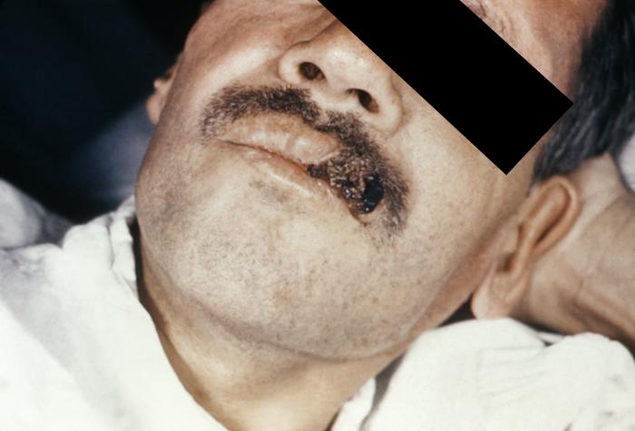
Skin lesion on the upper lip due to Histoplasma capsulatum infection.
-
Interior view of an elderly man’s oral cavity. A histoplasmosis infection induced inflammatory response on right inferior gingival tissues. From Public Health Image Library (PHIL). [3]
-
This image depicts the perianal region of a male patient afflicted with a disease known as histoplasmosis, which is caused by the fungal organism, Histoplasma capsulatum. From Public Health Image Library (PHIL). [3]
-
This image depicts an intraoral view, which reveals a lesion of the patient’s maxillary gingival mucosa that had been diagnosed as histoplasmosis, caused by the fungal pathogen, Histoplasma capsulatum. From Public Health Image Library (PHIL). [3]
References
- ↑ Dellinger RP, Levy MM, Carlet JM, Bion J, Parker MM, Jaeschke R, Reinhart K, Angus DC, Brun-Buisson C, Beale R, Calandra T, Dhainaut JF, Gerlach H, Harvey M, Marini JJ, Marshall J, Ranieri M, Ramsay G, Sevransky J, Thompson BT, Townsend S, Vender JS, Zimmerman JL, Vincent JL (2008). "Surviving Sepsis Campaign: international guidelines for management of severe sepsis and septic shock: 2008". Critical Care Medicine. 36 (1): 296–327. doi:10.1097/01.CCM.0000298158.12101.41. PMID 18158437. Retrieved 2012-09-16. Unknown parameter
|month=ignored (help) - ↑ Bone RC, Balk RA, Cerra FB, Dellinger RP, Fein AM, Knaus WA, Schein RM, Sibbald WJ. Definitions for sepsis and organ failure and guidelines for the use of innovative therapies in sepsis. The ACCP/SCCM Consensus Conference Committee. American College of Chest Physicians/Society of Critical Care Medicine. Chest. 1992 Jun;101(6):1644-55. PMID 1303622.
- ↑ 3.0 3.1 3.2 "Public Health Image Library (PHIL)".

![Interior view of an elderly man’s oral cavity. A histoplasmosis infection induced inflammatory response on right inferior gingival tissues. From Public Health Image Library (PHIL). [3]](/images/3/33/Histoplasmosis10.jpeg)
![This image depicts the perianal region of a male patient afflicted with a disease known as histoplasmosis, which is caused by the fungal organism, Histoplasma capsulatum. From Public Health Image Library (PHIL). [3]](/images/4/45/Histoplasmosis07.jpeg)
![This image depicts an intraoral view, which reveals a lesion of the patient’s maxillary gingival mucosa that had been diagnosed as histoplasmosis, caused by the fungal pathogen, Histoplasma capsulatum. From Public Health Image Library (PHIL). [3]](/images/d/d9/Histoplasmosis03.jpeg)