Empty sella syndrome
| Empty sella syndrome | |
| ICD-9 | 253.8 |
|---|---|
| DiseasesDB | 31523 |
| MeSH | D004652 |
For patient information click here
Editor-In-Chief: C. Michael Gibson, M.S., M.D. [1]
Please Take Over This Page and Apply to be Editor-In-Chief for this topic: There can be one or more than one Editor-In-Chief. You may also apply to be an Associate Editor-In-Chief of one of the subtopics below. Please mail us [2] to indicate your interest in serving either as an Editor-In-Chief of the entire topic or as an Associate Editor-In-Chief for a subtopic. Please be sure to attach your CV and or biographical sketch.
Overview
Empty sella syndrome (abbreviated ESS) is a disorder that involves the sella turcica, a bony structure at the base of the brain that surrounds and protects the pituitary gland. ESS is a condition that is often discovered during tests for pituitary disorders, when radiological imaging of the pituitary gland reveals a sella turica that appears to be empty.
Classification
There are two types of ESS: primary and secondary.
- Primary ESS happens when a small anatomical defect above the pituitary gland increases pressure in the sella turica and causes the gland to flatten out along the interior walls of the sella turica cavity. Primary ESS is associated with obesity and high blood pressure in women. The disorder sometimes results in a build-up of fluid pressure inside the skull and the pituitary gland may be smaller than usual.
- Secondary ESS is the result of the pituitary gland regressing within the cavity after an injury, surgery, or radiation therapy. Individuals with secondary ESS due to destruction of the pituitary gland have symptoms that reflect the loss of pituitary functions, such as the ceasing of menstrual periods, infertility, fatigue, and intolerance to stress and infection.
Associated conditions and diagnosis
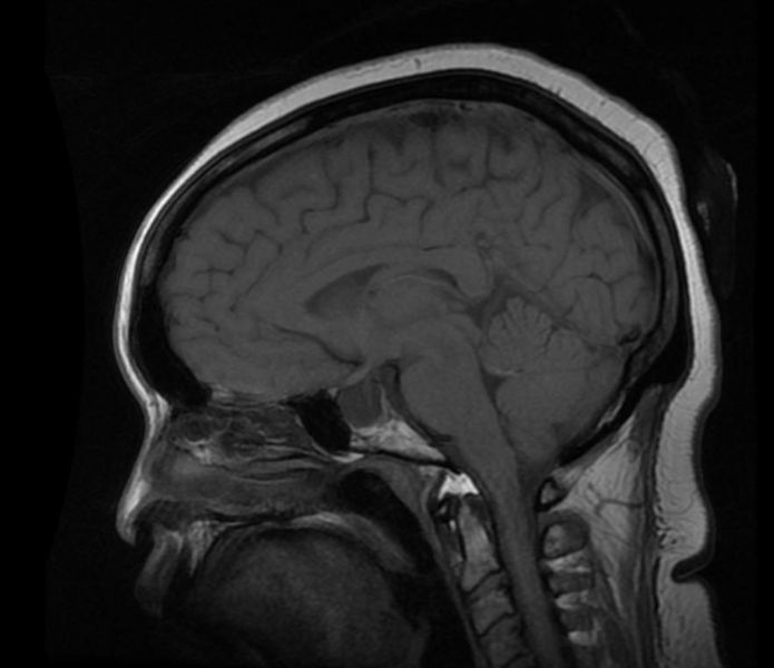
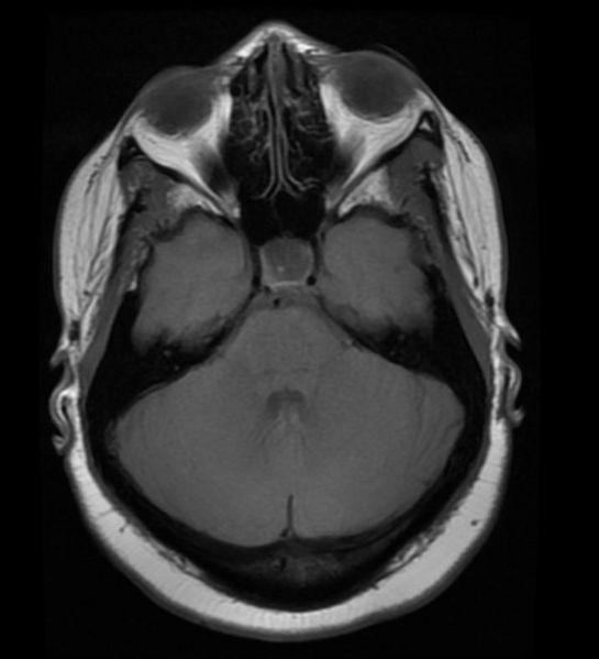
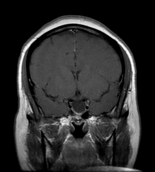
In children, ESS may be associated with early onset of puberty, growth hormone deficiency, pituitary tumors, or pituitary gland dysfunction. MRI scans are useful in evaluating ESS and differentiating it from other disorders that produce an enlarged sella.
Differential Diagnosis
- Autoimmune factors
- Cavernous sinus thrombosis
- Communicating hydrocephalus
- Congenital absence of the diaphragma sellae
- Diabetes Mellitus
- Familial
- Head trauma
- Increased intracranial pressure
- Radiation therapy complication
- Meningitis
- Pituitary adenoma
- Primary in middle-aged obese women
- Rupture of intrasellar or parasellar cyst
- Sheehan's Syndrome
- Surgery
- Vascular diseases
Diagnosis
Patient #1: MR images demonstrate an expanded and empty sella
Treatment
Unless the syndrome results in other medical problems, treatment for endocrine dysfunction associated with pituitary malfunction is symptomatic and supportive. In some cases, surgery may be needed.
Prognosis
ESS is not a life-threatening illness.


