Uploads by Rohan Bhimani
Jump to navigation
Jump to search
This special page shows all uploaded files.
| Date | Name | Thumbnail | Size | Description | Versions |
|---|---|---|---|---|---|
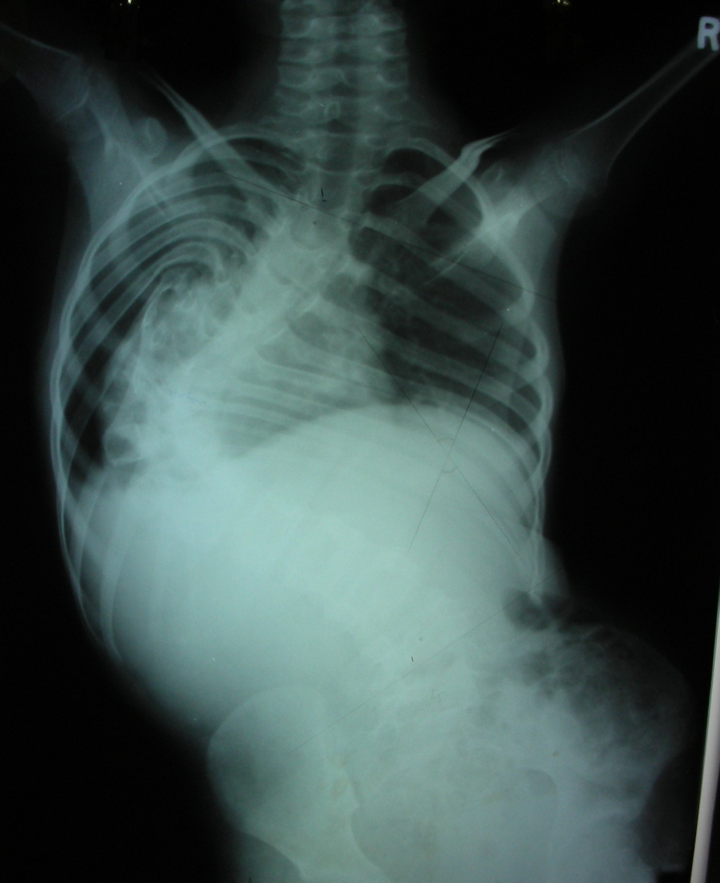
| 00:43, 21 January 2019 | Xray UBC.gif (file) |  |
375 KB | 1 | |
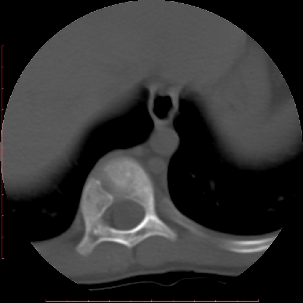
| 17:05, 18 January 2019 | NOF CT GIF.gif (file) |  |
187 KB | 1 | |
| 16:41, 18 January 2019 | NOF MRI GIF.gif (file) |  |
173 KB | 1 | |
| 16:14, 18 January 2019 | NOF xrays.gif (file) |  |
240 KB | 1 | |
| 17:10, 31 December 2018 | Histology osteoma.jpg (file) |  |
1.39 MB | 1 | |
| 17:01, 31 December 2018 | CT osteoma.gif (file) |  |
356 KB | 1 | |
| 16:52, 31 December 2018 | Xray osteoma.gif (file) |  |
335 KB | 1 | |
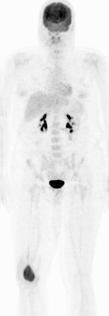
| 20:16, 27 December 2018 | Bone scan giant-cell-tumour-femur.jpg (file) |  |
15 KB | 1 | |
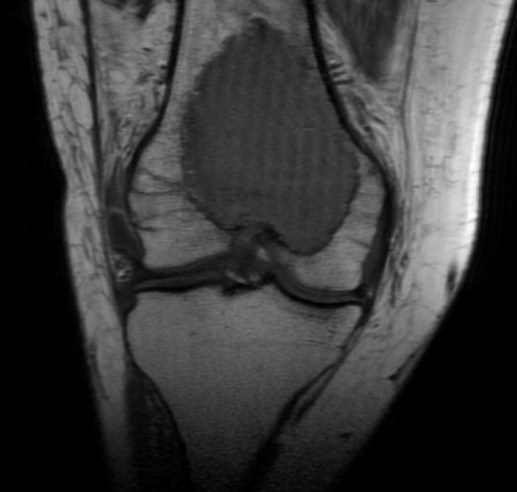
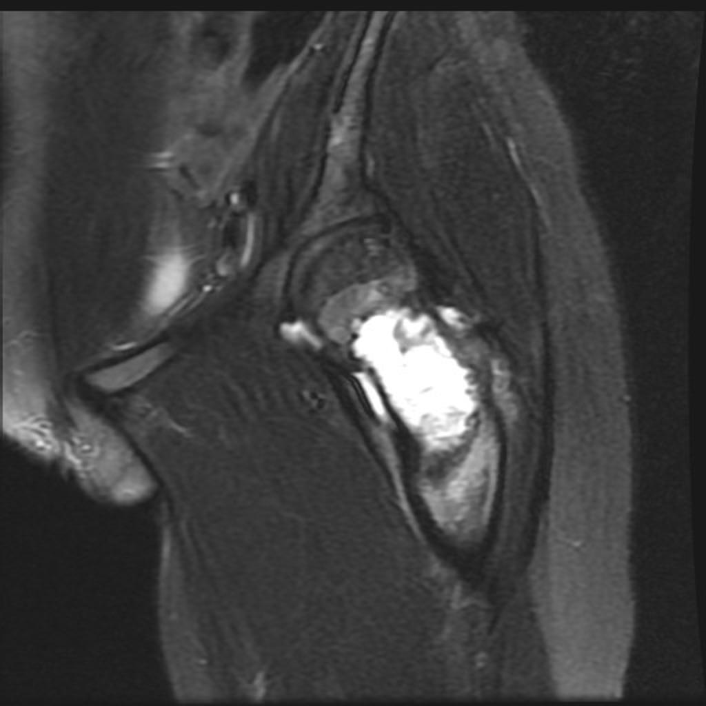
| 20:14, 27 December 2018 | MRI giant-cell-tumour-femur.jpg (file) |  |
57 KB | 1 | |
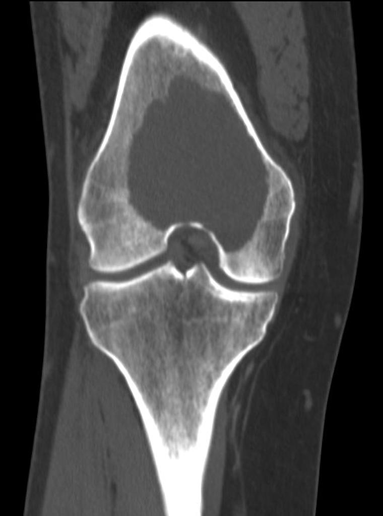
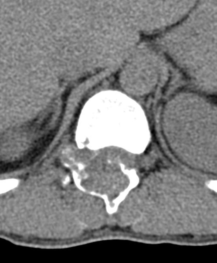
| 20:13, 27 December 2018 | CT giant-cell-tumour-femur .jpg (file) |  |
42 KB | 1 | |
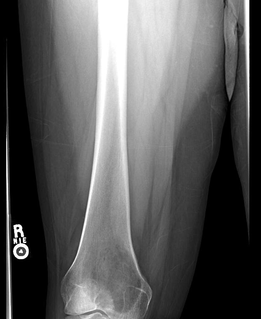
| 20:07, 27 December 2018 | Giant-cell-tumour-femur xray.jpg (file) |  |
53 KB | 1 | |
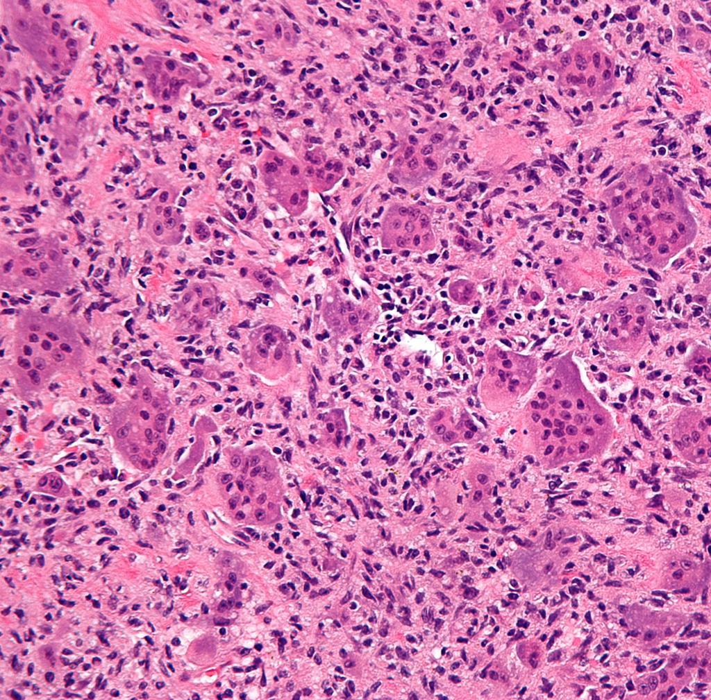
| 19:59, 27 December 2018 | Giant-cell-tumour-histology.jpg (file) |  |
281 KB | 1 | |
| 20:29, 21 December 2018 | CT scan aneurysmal-bone-cyst.jpg (file) |  |
61 KB | 1 | |
| 20:23, 21 December 2018 | MRI aneurysmal-bone-cyst.jpg (file) |  |
58 KB | 1 | |
| 20:19, 21 December 2018 | X ray aneurysmal-bone-cyst.jpg (file) |  |
67 KB | 1 | |
| 15:52, 21 December 2018 | Aneurysmal-bone-cyst pathology.jpg (file) |  |
454 KB | 1 | |
| 15:39, 20 December 2018 | MRI CHondroblastoma.jpg (file) |  |
52 KB | 1 | |
| 15:33, 20 December 2018 | Chondroblastoma CT scan.jpg (file) |  |
38 KB | 1 | |
| 15:26, 20 December 2018 | Xray chondroblastoma of tibia.jpg (file) |  |
52 KB | 1 | |
| 17:10, 17 December 2018 | Osteoblastoma Histology.jpg (file) |  |
6.62 MB | 1 | |
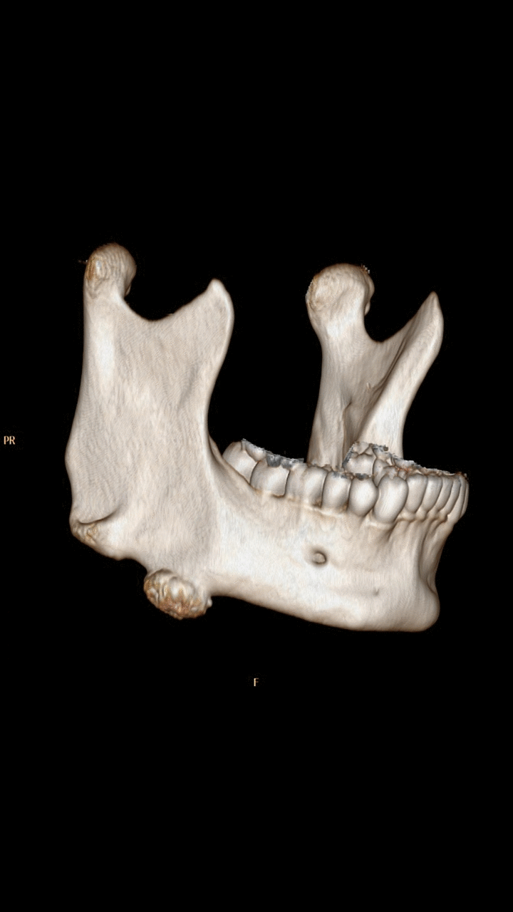
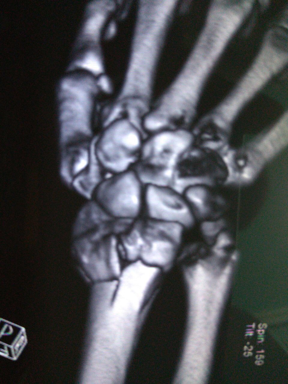
| 17:10, 13 December 2018 | Xray Wrist AP and Lat view.JPG (file) |  |
78 KB | 1 | |
| 16:35, 12 December 2018 | Dinner fork Deformity.jpg (file) |  |
232 KB | 1 | |
| 16:26, 12 December 2018 | Sign DRF.jpg (file) |  |
329 KB | 1 | |
| 21:55, 11 December 2018 | MRI- non-displaced-distal-radial-fracture-.jpg (file) |  |
82 KB | 1 | |
| 21:32, 11 December 2018 | CT scan DER VENTRAL.JPG (file) |  |
221 KB | 1 | |
| 21:25, 11 December 2018 | CT scan Lateral View with intraarticular step.JPG (file) |  |
98 KB | 1 | |
| 21:23, 11 December 2018 | CT scan wrist ventral view.JPG (file) |  |
221 KB | 1 | |
| 21:21, 11 December 2018 | CT scan Wrist dorsal view.JPG (file) |  |
233 KB | 1 | |
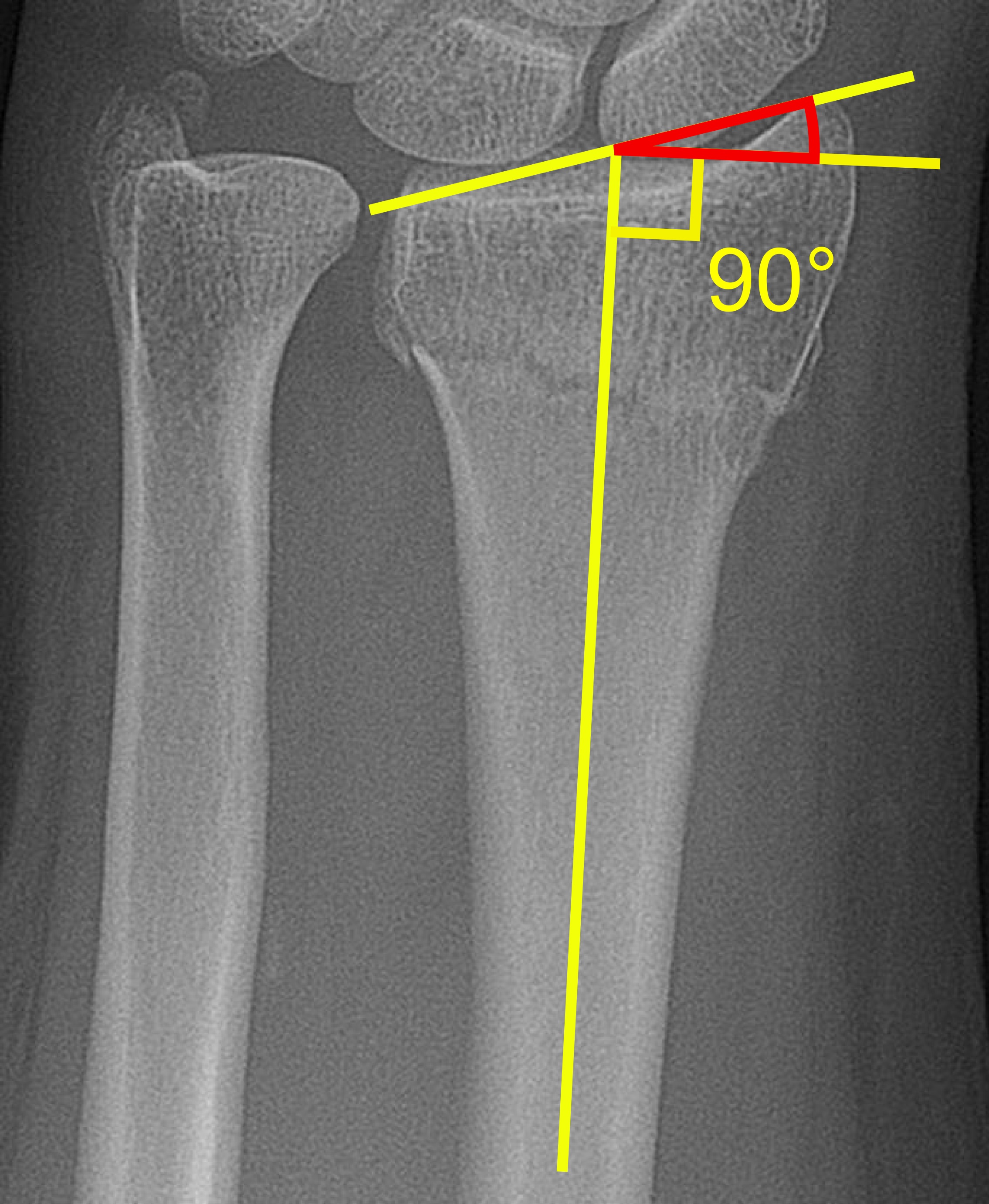
| 20:29, 11 December 2018 | Radial inclination of distal radius fracture.jpg (file) |  |
1.07 MB | 1 | |
| 20:23, 11 December 2018 | Dorsal tilt of distal radius fracture.jpg (file) |  |
1.24 MB | 1 | |
| 16:32, 11 December 2018 | Closed rduction of Distal radius fracture..png (file) |  |
175 KB | 1 | |
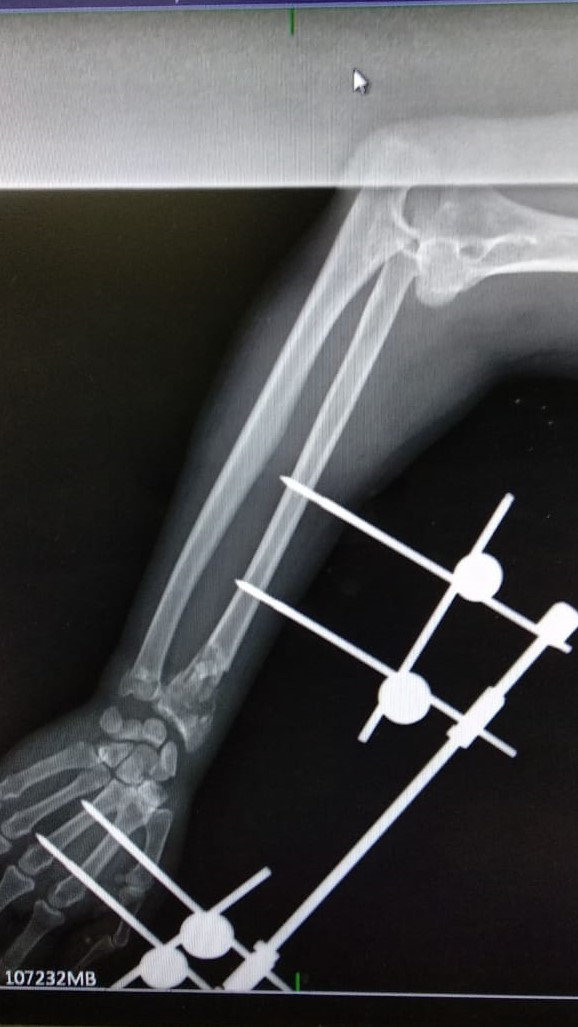
| 15:18, 11 December 2018 | External fixation for Distsal end radius.jpg (file) |  |
111 KB | 1 | |
| 15:13, 11 December 2018 | MUlti plate Radius.JPG (file) |  |
61 KB | 1 | |
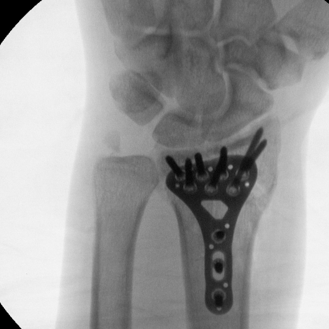
| 15:09, 11 December 2018 | Distal end radius Volar plate.jpg (file) |  |
176 KB | 1 | |
| 21:19, 7 December 2018 | Scoliosis complications.jpg (file) |  |
77 KB | 1 | |
| 21:17, 7 December 2018 | Scoliosis post-op.jpg (file) |  |
69 KB | 1 | |
| 18:59, 7 December 2018 | Boston brace.jpg (file) |  |
37 KB | 1 | |
| 17:40, 7 December 2018 | CTLSO -scoliosis.jpg (file) |  |
1.13 MB | 1 | |
| 23:49, 6 December 2018 | Adams forward bend test.jpg (file) |  |
51 KB | 1 | |
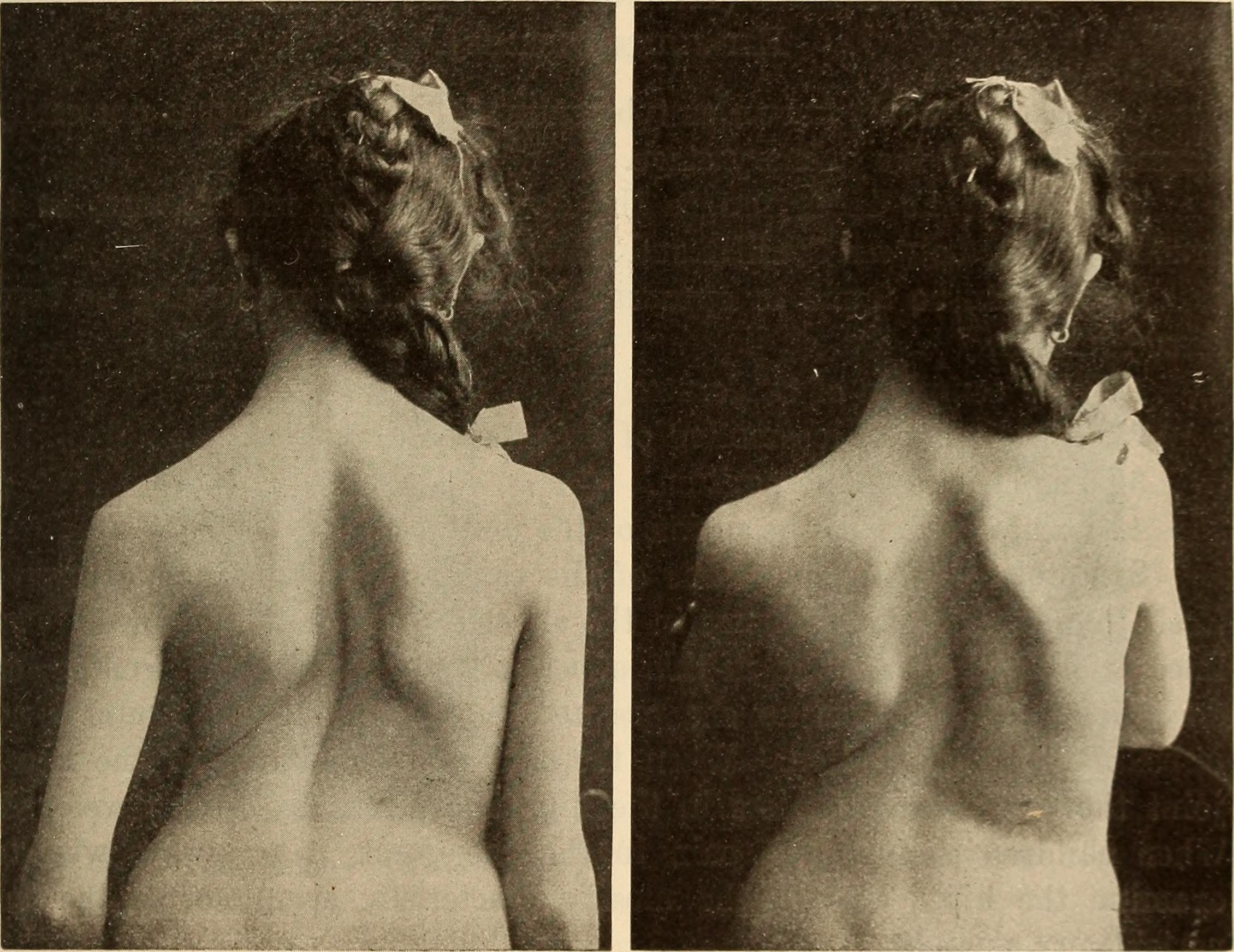
| 23:29, 6 December 2018 | Scoliosis physical exam.jpg (file) |  |
5.82 MB | 1 | |
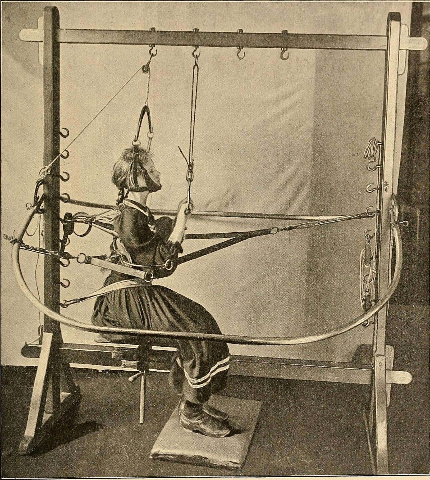
| 23:18, 6 December 2018 | Scoliosis Historical Treatment.jpg (file) |  |
773 KB | 1 | |
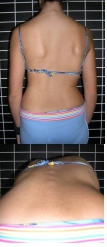
| 23:07, 6 December 2018 | Scoliosis clinical pic.jpg (file) |  |
510 KB | 1 | |
| 22:51, 6 December 2018 | Sep median nerve.gif (file) |  |
196 KB | 1 | |
| 17:49, 6 December 2018 | PET scan -Thoracic endplate osteomyeltisdiscitis.jpg (file) |  |
45 KB | Case courtesy of Dr Chris O'Donnell, Radiopaedia.org, rID: 18272 | 1 |
| 16:45, 6 December 2018 | MRI hemivertebra with congenital-scoliosis.jpg (file) |  |
126 KB | Coronal T2: L1 hemivertebra is causing congenital scoliosis. Case courtesy of Dr Ahmed Almuslim, Radiopaedia.org, rID: 6919 | 1 |
| 15:17, 6 December 2018 | CT scan Hemivertebra withcongenital-scoliosis.jpg (file) |  |
160 KB | Right supernumerary D10/D11 hemivertebra is noted associated with mild right dorsal scoliosis as well as mild focal kyphotic deformity. Source: Case courtesy of Dr Mohammad A. ElBeialy, Radiopaedia.org, rID: 41542 | 1 |
| 21:57, 5 December 2018 | Idiopathic Scoliosis.JPG (file) |  |
254 KB | Thoracolumbar scoliosis Source: DR. Rohan A. Bhimani | 1 |
| 21:44, 5 December 2018 | Hemivertebra-with-congenital-scoliosis.jpg (file) |  |
159 KB | Right supernumerary D10/D11 hemivertebra is noted associated with mild right dorsal scoliosis as well as mild focal kyphotic deformity. Source: Case courtesy of Dr Mohammad A. ElBeialy, Radiopaedia.org, rID: 41542 | 1 |
| 21:17, 5 December 2018 | Scoliosis coronal balance.jpeg (file) |  |
60 KB | The saggital and coronal balance are both within the normal range, the lines drawn form the middle of the C7 vertebral view pass through the midline at the level of the S1 on the coronal view, and within a 2 cm distance from the posterosuperior corner... | 1 |
| 15:37, 3 December 2018 | Dr. Rohan Bhimani.jpg (file) |  |
139 KB | 1 |