Pulmonary nodule chest x ray: Difference between revisions
Jump to navigation
Jump to search
No edit summary |
No edit summary |
||
| Line 9: | Line 9: | ||
[[Image:Malignant solitary pulmonary nodule 1.jpg|thumb|center|Malignant solitary pulmonary nodule]] | |||
Image: | |||
*The patient is a 67 year old woman with a solitary pulmonary nodule on a recent chest x-ray. A retrospective review of prior chest x-rays suggests that this is nodule is of recent origin. This lesion was felt to be too peripheral for reliable bronchial wash findings. | |||
*Concern over potential sampling error associated with needle biopsy prompted a referral for PET imaging to rule out a malignant process. | |||
<div align="left"> | <div align="left"> | ||
<gallery heights="200" widths="200"> | <gallery heights="200" widths="200"> | ||
Image:Pulmonary AVM as nodule | Image:Pulmonary AVM as nodule 1.jpg|Chest x-ray: A 32 year old woman. 1. Two pulmonary arteriovenous malformations consistent with the nodules seen on the recent chest film. There is breathing artifact on several of the images and other tiny AVMs cannot be excluded. 2. Cardiomegaly with right atrial and left atrial enlargement and hepatic congestion. | ||
Image:Pulmonary AVM as nodule | Image:Pulmonary AVM as nodule 2.jpg|Thorax CT | ||
</gallery> | </gallery> | ||
</div> | </div> | ||
==References== | ==References== | ||
Revision as of 14:41, 25 September 2012
|
Pulmonary Nodule Microchapters |
|
Diagnosis |
|---|
|
Treatment |
|
Case Studies |
|
Pulmonary nodule chest x ray On the Web |
|
American Roentgen Ray Society Images of Pulmonary nodule chest x ray |
|
Risk calculators and risk factors for Pulmonary nodule chest x ray |
Editor-In-Chief: C. Michael Gibson, M.S., M.D. [1]
Overview
Chest X Ray

- The patient is a 67 year old woman with a solitary pulmonary nodule on a recent chest x-ray. A retrospective review of prior chest x-rays suggests that this is nodule is of recent origin. This lesion was felt to be too peripheral for reliable bronchial wash findings.
- Concern over potential sampling error associated with needle biopsy prompted a referral for PET imaging to rule out a malignant process.
-
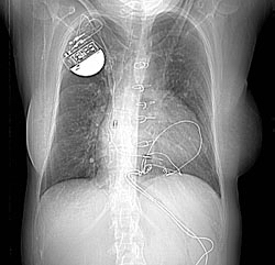
Chest x-ray: A 32 year old woman. 1. Two pulmonary arteriovenous malformations consistent with the nodules seen on the recent chest film. There is breathing artifact on several of the images and other tiny AVMs cannot be excluded. 2. Cardiomegaly with right atrial and left atrial enlargement and hepatic congestion.
-

Thorax CT

