Cirrhosis chest x ray: Difference between revisions
Megan Merlo (talk | contribs) No edit summary |
|||
| Line 2: | Line 2: | ||
{{Cirrhosis}} | {{Cirrhosis}} | ||
{{CMG}} {{AE}} {{VVS}} | {{CMG}} {{AE}} {{VVS}} | ||
{{PleaseHelp}} | |||
==Overview== | ==Overview== | ||
Chest x ray has a limited role in the diagnosis and management of cirrhosis, but can be helpful in identifying certain complications that can occur as a result of cirrhosis. | Chest x ray has a limited role in the diagnosis and management of cirrhosis, but can be helpful in identifying certain complications that can occur as a result of cirrhosis. | ||
== Chest X Ray == | ==Chest X Ray== | ||
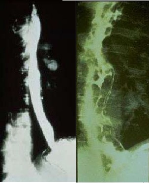
[[Image:Normal versus Abnormal Barium study of esophagus.jpg|thumb|left|200px|Normal versus Abnormal Barium study of esophagus with varices]] | [[Image:Normal versus Abnormal Barium study of esophagus.jpg|thumb|left|200px|Normal versus Abnormal Barium study of esophagus with varices]] | ||
| Line 14: | Line 17: | ||
==References== | ==References== | ||
{{reflist|2}} | {{reflist|2}} | ||
[[Category:Gastroenterology]] | |||
[[Category:Hepatology]] | |||
[[Category:Disease]] | |||
{{WS}} | |||
{{WH}} | {{WH}} | ||
Revision as of 14:58, 18 July 2016
|
Cirrhosis Microchapters |
|
Diagnosis |
|---|
|
Treatment |
|
Case studies |
|
Cirrhosis chest x ray On the Web |
|
American Roentgen Ray Society Images of Cirrhosis chest x ray |
Editor-In-Chief: C. Michael Gibson, M.S., M.D. [1] Associate Editor(s)-in-Chief: Vishnu Vardhan Serla M.B.B.S. [2]
Please help WikiDoc by adding content here. It's easy! Click here to learn about editing.
Overview
Chest x ray has a limited role in the diagnosis and management of cirrhosis, but can be helpful in identifying certain complications that can occur as a result of cirrhosis.
Chest X Ray

Chest x ray has a limited place in the diagnosis and management of patients with cirrhosis. It is used to screening for ascites, seeking evidence of bowel perforation in patients with suspected spontaneous bacterial peritonitis, and monitoring bowel distension in acutely ill patients admitted for treatment of decompensation or variceal hemorrhage. X ray may show elevation of the diaphragm from ascites. Gynecomastia may be appreciated. The azygous vein may be enlarged because of collateral flow and pleural effusions may occur from the presence of pleuroperitoneal fistulas.