Blastic NK cell lymphoma: Difference between revisions
No edit summary |
No edit summary |
||
| Line 8: | Line 8: | ||
==Historical Perspective== | ==Historical Perspective== | ||
*Blastic NK cell lymphoma was first discovered by Adachi, an American hematologist, in 1994 following an unusual presentation of cutaneous lymphoma that express CD4 and CD56 antigens but no other T cell and B cell antigens.<ref name="pmid7526680">{{cite journal| author=Adachi M, Maeda K, Takekawa M, Hinoda Y, Imai K, Sugiyama S et al.| title=High expression of CD56 (N-CAM) in a patient with cutaneous CD4-positive lymphoma. | journal=Am J Hematol | year= 1994 | volume= 47 | issue= 4 | pages= 278-82 | pmid=7526680 | doi= | pmc= | url=http://www.ncbi.nlm.nih.gov/entrez/eutils/elink.fcgi?dbfrom=pubmed&tool=sumsearch.org/cite&retmode=ref&cmd=prlinks&id=7526680 }} </ref> | *Blastic NK cell lymphoma was first discovered by Adachi, an American hematologist, in 1994 following an unusual presentation of cutaneous lymphoma that express CD4 and CD56 antigens but no other T cell and B cell antigens.<ref name="pmid7526680">{{cite journal| author=Adachi M, Maeda K, Takekawa M, Hinoda Y, Imai K, Sugiyama S et al.| title=High expression of CD56 (N-CAM) in a patient with cutaneous CD4-positive lymphoma. | journal=Am J Hematol | year= 1994 | volume= 47 | issue= 4 | pages= 278-82 | pmid=7526680 | doi= | pmc= | url=http://www.ncbi.nlm.nih.gov/entrez/eutils/elink.fcgi?dbfrom=pubmed&tool=sumsearch.org/cite&retmode=ref&cmd=prlinks&id=7526680 }} </ref> | ||
* Chaperot et al demonstrated that predendritic cells (plasmacytoid monocytes) and CD4/CD56 leukemic cells are similar in the production of alpha-interferon in response to influenza virus stimulation, which on maturation with interleukin 3 (IL-3) become powerful inducers for CD4 proliferation and Th-2 polarization. | * Chaperot et al demonstrated that predendritic cells (plasmacytoid monocytes) and CD4/CD56 leukemic cells are similar in the production of alpha-interferon in response to influenza virus stimulation, which on maturation with interleukin 3 (IL-3) become powerful inducers for CD4 proliferation and Th-2 polarization. .<ref name="pmid11342451">{{cite journal| author=Chaperot L, Bendriss N, Manches O, Gressin R, Maynadie M, Trimoreau F et al.| title=Identification of a leukemic counterpart of the plasmacytoid dendritic cells. | journal=Blood | year= 2001 | volume= 97 | issue= 10 | pages= 3210-7 | pmid=11342451 | doi= | pmc= | url=http://www.ncbi.nlm.nih.gov/entrez/eutils/elink.fcgi?dbfrom=pubmed&tool=sumsearch.org/cite&retmode=ref&cmd=prlinks&id=11342451 }} </ref> | ||
.<ref name="pmid11342451">{{cite journal| author=Chaperot L, Bendriss N, Manches O, Gressin R, Maynadie M, Trimoreau F et al.| title=Identification of a leukemic counterpart of the plasmacytoid dendritic cells. | journal=Blood | year= 2001 | volume= 97 | issue= 10 | pages= 3210-7 | pmid=11342451 | doi= | pmc= | url=http://www.ncbi.nlm.nih.gov/entrez/eutils/elink.fcgi?dbfrom=pubmed&tool=sumsearch.org/cite&retmode=ref&cmd=prlinks&id=11342451 }} </ref> | |||
* Petrella and Karube demonstrated that CD123, which is a marker of predendritic cells are expressed on the tumor cells in blastic NK cell lymphoma and proposed an oncogenic transformation | * Petrella and Karube demonstrated that CD123, which is a marker of predendritic cells are expressed on the tumor cells in blastic NK cell lymphoma and proposed an oncogenic transformation | ||
of NCAM-expressing plasmacytoid monocyte-like cells as cell of origin in CD4/CD56 blastic NK cell lymphoma. | of NCAM-expressing plasmacytoid monocyte-like cells as cell of origin in CD4/CD56 blastic NK cell lymphoma.<ref name="pmid15323148">{{cite journal| author=Petrella T, Meijer CJ, Dalac S, Willemze R, Maynadié M, Machet L et al.| title=TCL1 and CLA expression in agranular CD4/CD56 hematodermic neoplasms (blastic NK-cell lymphomas) and leukemia cutis. | journal=Am J Clin Pathol | year= 2004 | volume= 122 | issue= 2 | pages= 307-13 | pmid=15323148 | doi=10.1309/0QPP-AVTU-PCV9-UCLV | pmc= | url=http://www.ncbi.nlm.nih.gov/entrez/eutils/elink.fcgi?dbfrom=pubmed&tool=sumsearch.org/cite&retmode=ref&cmd=prlinks&id=15323148 }} </ref><ref name="pmid14508398">{{cite journal| author=Karube K, Ohshima K, Tsuchiya T, Yamaguchi T, Suefuji H, Suzumiya J et al.| title=Non-B, non-T neoplasms with lymphoblast morphology: further clarification and classification. | journal=Am J Surg Pathol | year= 2003 | volume= 27 | issue= 10 | pages= 1366-74 | pmid=14508398 | doi= | pmc= | url=http://www.ncbi.nlm.nih.gov/entrez/eutils/elink.fcgi?dbfrom=pubmed&tool=sumsearch.org/cite&retmode=ref&cmd=prlinks&id=14508398 }} </ref> | ||
==Pathophysiology== | ==Pathophysiology== | ||
*Blastic NK cell lymphoma is a type of [[lymphoma]]. It does not appear to be associated with [[Epstein Barr virus]].<ref name="pmid9192774">{{cite journal |author=Chan JK, Sin VC, Wong KF, ''et al'' |title=Nonnasal lymphoma expressing the natural killer cell marker CD56: a clinicopathologic study of 49 cases of an uncommon aggressive neoplasm |journal=Blood |volume=89 |issue=12 |pages=4501–13 |year=1997 |month=June |pmid=9192774 |doi= |url=http://www.bloodjournal.org/cgi/pmidlookup?view=long&pmid=9192774}}</ref> | *Blastic NK cell lymphoma is a type of [[lymphoma]]. It does not appear to be associated with [[Epstein Barr virus]].<ref name="pmid9192774">{{cite journal |author=Chan JK, Sin VC, Wong KF, ''et al'' |title=Nonnasal lymphoma expressing the natural killer cell marker CD56: a clinicopathologic study of 49 cases of an uncommon aggressive neoplasm |journal=Blood |volume=89 |issue=12 |pages=4501–13 |year=1997 |month=June |pmid=9192774 |doi= |url=http://www.bloodjournal.org/cgi/pmidlookup?view=long&pmid=9192774}}</ref> | ||
Revision as of 13:42, 11 May 2016
Editor-In-Chief: C. Michael Gibson, M.S., M.D. [1]
Synonyms and keywords: Agranular CD4+CD56+ hematodermic neoplasm; CD4+CD56+ hematodermic neoplasm; HDT; Blastic plasmacytoid dendritic cell neoplasm; BPDCN;
Overview
Blastic NK cell lymphoma was first discovered by Adachi, an American hematologist, in 1994 following an unusual presentation of cutaneous lymphoma that express CD4 and CD56 antigens but no other T cell and B cell antigens. Blastic NK cell lymphoma is a type of lymphoma. It does not appear to be associated with Epstein Barr virus.[1]
Historical Perspective
- Blastic NK cell lymphoma was first discovered by Adachi, an American hematologist, in 1994 following an unusual presentation of cutaneous lymphoma that express CD4 and CD56 antigens but no other T cell and B cell antigens.[2]
- Chaperot et al demonstrated that predendritic cells (plasmacytoid monocytes) and CD4/CD56 leukemic cells are similar in the production of alpha-interferon in response to influenza virus stimulation, which on maturation with interleukin 3 (IL-3) become powerful inducers for CD4 proliferation and Th-2 polarization. .[3]
- Petrella and Karube demonstrated that CD123, which is a marker of predendritic cells are expressed on the tumor cells in blastic NK cell lymphoma and proposed an oncogenic transformation
of NCAM-expressing plasmacytoid monocyte-like cells as cell of origin in CD4/CD56 blastic NK cell lymphoma.[4][5]
Pathophysiology
- Blastic NK cell lymphoma is a type of lymphoma. It does not appear to be associated with Epstein Barr virus.[1]
- Blastic NK cell lymphoma is derived from plasmacytoid type 2 dendritic cell precursors. These dendritic cell precursors are possibly related to a common myeloid/NK-cell precursor cell.[6]
- Blastic NK cell lymphoma is currently classified by World Health Organization Classification of Tumours of Haematopoietic and Lymphoid Tissues as an aggressive neoplasm derived from the precursors of plasmacytoid dendritic cells and categorically placed under the heading “Acute myeloid leukemia (AML) and related precursor neoplasms.”
- The deletion in 5q has been associated with the development of blastic NK cell lymphoma.
- Other genetic abnormalities associated with Blastic NK cell lymphoma are deletion of 12p, abnormalities of chromosome 13, deletion of 6q, and loss of chromosomes 15 and 9.
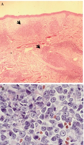
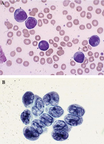
- On microscopic histopathological analysis, fine chromatin and scanty cytoplasm resembling lymphoblasts, or in some cases, myeloblasts, and may on occasion exhibit sub-membranous cytoplasmic vacuolations surrounding the nucleus are characteristic findings of blastic NK cell lymphoma.
- Tumor cells are invariably CD4+ and CD56+, and usually HLA-DR and CD45RA are positive as well. CD2 and CD34 are usually negative; and expression of TdT, CD7 and cytoplasmic CD3 is variable.[7]
-
Hematoxylin & Eosin stain of skin lesion biopsy. Low power view of leukemic infiltrate corresponding to the raised plaque (A, black arrows) and high power view of the malignant cells in the skin infiltrate
-
Large malignant-appearing cells, with agranular cytoplasm, cleaved nuclei and prominent neocleoli on peripheral blood smear using Wright stain (A) and similar blast cells present in cerebrospinal fluid
Differentiating Blastic NK cell lymphoma from other Diseases
- Blastic NK cell lymphoma must be differentiated from other malignancies such as:
- Acute myeloid leukemia
- Human T-cell lymphotropic virus 1 associated adult T-cell leukemia/lymphoma
- Cutaneous NK/T-cell lymphoma
- Primary and secondary cutaneous pleomorphic T-cell lymphomas
- Undifferentiated carcinoma
- Malignant melanoma.
Epidemiology and Demographics
- The prevalence of blastic NK cell lymphoma is unknown as it is an extremely rare disorder.[8]
Age
- Blastic NK cell lymphoma is more commonly observed amongmiddle-aged or elderly patients. The mean age at diagnosis is 66 years.[9]
Gender
- Males are more commonly affected with blastic NK cell lymphoma than female.
Natural History, Complications and Prognosis
- The majority of patients with Blastic NK cell lymphoma have an aggressive clinical course.
- Prognosis is generally poor, and the median survival rate is 15 months.
Diagnosis
Cells are positive for CD4 and CD56.[10][11]
Symptoms
- Symptoms of blastic NK cell lymphoma may include the following:
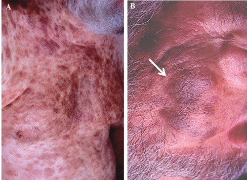
- Nodules, plaques and patches of variable sizes on skin
Physical Examination
- Physical examination may be remarkable for:
- Nodules, plaques and patches of variable sizes on skin

Laboratory Findings
- There are no specific laboratory findings associated with blastic NK cell lymphoma.
Imaging Findings
- There are no imaging study findings associated with blastic NK cell lymphoma
Other Diagnostic Studies
- Blastic NK cell lymphoma may also be diagnosed using immunochemistry.
- Immunohistochemical staining is positive for r CD4 and CD56, with variable positivity for CD43, TdT, and CD68.
Treatment
Medical Therapy
- The mainstay of therapy for blastic NK cell lymphoma is chemotherapy with CHOP or COP-like regimens.
- Immunomodulation or immunotherapy with IL-3 or with antiCD123 antibody is also being considered for chemotherapy-resistant patients.
Prevention
- There are no primary preventive measures available for blastic NK cell lymphoma.
References
- ↑ 1.0 1.1 Chan JK, Sin VC, Wong KF; et al. (1997). "Nonnasal lymphoma expressing the natural killer cell marker CD56: a clinicopathologic study of 49 cases of an uncommon aggressive neoplasm". Blood. 89 (12): 4501–13. PMID 9192774. Unknown parameter
|month=ignored (help) - ↑ Adachi M, Maeda K, Takekawa M, Hinoda Y, Imai K, Sugiyama S; et al. (1994). "High expression of CD56 (N-CAM) in a patient with cutaneous CD4-positive lymphoma". Am J Hematol. 47 (4): 278–82. PMID 7526680.
- ↑ Chaperot L, Bendriss N, Manches O, Gressin R, Maynadie M, Trimoreau F; et al. (2001). "Identification of a leukemic counterpart of the plasmacytoid dendritic cells". Blood. 97 (10): 3210–7. PMID 11342451.
- ↑ Petrella T, Meijer CJ, Dalac S, Willemze R, Maynadié M, Machet L; et al. (2004). "TCL1 and CLA expression in agranular CD4/CD56 hematodermic neoplasms (blastic NK-cell lymphomas) and leukemia cutis". Am J Clin Pathol. 122 (2): 307–13. doi:10.1309/0QPP-AVTU-PCV9-UCLV. PMID 15323148.
- ↑ Karube K, Ohshima K, Tsuchiya T, Yamaguchi T, Suefuji H, Suzumiya J; et al. (2003). "Non-B, non-T neoplasms with lymphoblast morphology: further clarification and classification". Am J Surg Pathol. 27 (10): 1366–74. PMID 14508398.
- ↑ Willemze R, Jaffe ES, Burg G, Cerroni L, Berti E, Swerdlow SH; et al. (2005). "WHO-EORTC classification for cutaneous lymphomas". Blood. 105 (10): 3768–85. doi:10.1182/blood-2004-09-3502. PMID 15692063.
- ↑ Petrella T, Bagot M, Willemze R, Beylot-Barry M, Vergier B, Delaunay M; et al. (2005). "Blastic NK-cell lymphomas (agranular CD4+CD56+ hematodermic neoplasms): a review". Am J Clin Pathol. 123 (5): 662–75. PMID 15981806.
- ↑ 8.0 8.1 Saeed H, Awasthi M, Al-Qaisi A, Massarweh S (2014). "Blastic plasmacytoid dendritic cell neoplasm with extensive cutaneous and central nervous system involvement". Rare Tumors. 6 (4): 5474. doi:10.4081/rt.2014.5474. PMC 4274438. PMID 25568744.
- ↑ Hou, Steve; Jaworski, Joseph; Swami, Vanlila; Heintzelman, Rebecca; Cusack, Carrie; Chung, Christina; Peck, Jeremy; Fanelli, Matthew; Styler, Michael; Rizk, Sanaa (2015). "Blastic plasmacytoid dendritic cell neoplasm with absolute monocytosis at presentation". Pathology and Laboratory Medicine International: 7. doi:10.2147/PLMI.S71492. ISSN 1179-2698.
- ↑ Ng AP, Lade S, Rutherford T, McCormack C, Prince HM, Westerman DA (2006). "Primary cutaneous CD4+/CD56+ hematodermic neoplasm (blastic NK-cell lymphoma): a report of five cases". Haematologica. 91 (1): 143–4. PMID 16434387. Unknown parameter
|month=ignored (help) - ↑ Kim Y, Kang MS, Kim CW, Sung R, Ko YH (2005). "CD4+CD56+ lineage negative hematopoietic neoplasm: so called blastic NK cell lymphoma". J. Korean Med. Sci. 20 (2): 319–24. PMID 15832009. Unknown parameter
|month=ignored (help)

