Rhabdomyosarcoma CT: Difference between revisions
Jump to navigation
Jump to search
No edit summary |
(→CT) |
||
| Line 4: | Line 4: | ||
==Overview== | ==Overview== | ||
==CT== | ==CT== | ||
On CT scan, | On CT scan, rhabdomyosarocma is characterized by: | ||
*Soft tissue density. | *Soft tissue density. | ||
*Some enhancement with contrast. | *Some enhancement with contrast. | ||
Revision as of 14:31, 8 September 2015
|
Rhabdomyosarcoma Microchapters |
|
Diagnosis |
|---|
|
Treatment |
|
Case Studies |
|
Rhabdomyosarcoma CT On the Web |
|
American Roentgen Ray Society Images of Rhabdomyosarcoma CT |
Editor-In-Chief: C. Michael Gibson, M.S., M.D. [1]
Overview
CT
On CT scan, rhabdomyosarocma is characterized by:
- Soft tissue density.
- Some enhancement with contrast.
- Adjacent bony destruction seen in over 20% of cases.
Rhabdomyosarcoma of biliary tract
- CT may show a heterogenous or hypo-attenuating mass with biliary ductal dilatation.
Rhabdomyosarcoma of the orbit
- Rhabdomyosarcomas are typically homogeneous soft tissue masses isodense to normal muscle.
- The mass may extend into the eyelid or through bone into the paranasal sinuses (especially the ethmoid sinus) and superiorly into the anterior cranial fossa.
Cardiac rhabdomyosarcoma
- CT may show a smooth or irregular low-attenuation mass in a cardiac chamber.
-
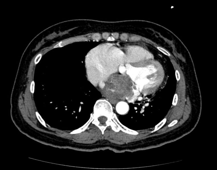
Cardiac Rhabdomyosarcoma Image courtesy of RadsWiki and copylefted
-
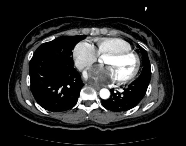
Cardiac Rhabdomyosarcoma Image courtesy of RadsWiki and copylefted
-
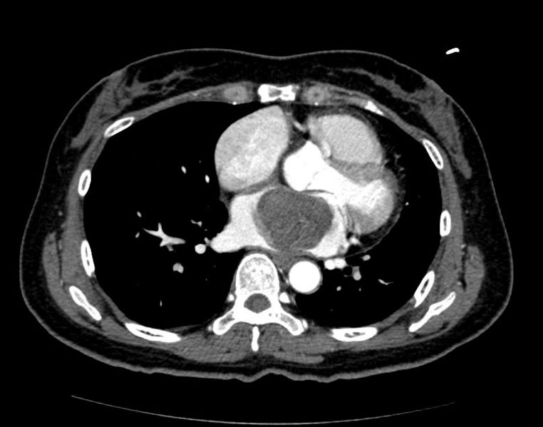
Cardiac Rhabdomyosarcoma Image courtesy of RadsWiki and copylefted


